
Abstract
Lithium batteries have been widely used to power up implantable medical devices such as pacemakers that are often designed to treat, diagnose, and prevent different diseases. However, due to their limited capacity and lifetime, patients have to undergo a surgical procedure to replace the discharged battery. Recently, nanogenerators have been emerged and are broadly accepted since they can convert tiny biomechanical forces, such as heartbeats, into electrical energy. This study aims to manufacture a biocompatible and high-performance piezoelectric energy harvester (PEH) that is capable to be charged by the energy received from the heartbeat and store the generated voltage. In this research, a hybrid structure of poly (vinylidene fluoride) (PVDF) coupling with polyamide-11 (PA-11) was fabricated using dual electrospinning to enhance the piezoelectric properties of the intended PEH. The piezoelectric test results show an acceptable increase in nanofibers’ piezoelectric sensitivity from 62.87 mV/N to 75.75 mV/N by adding 25% (v/v) of PA-11 to PVDF, indicating the synergistic effect of PVDF and PA-11. The specimen PVDF (75% v/v)-PA-11 (25% v/v) also showed the highest mechanical strength and consequently is suggested as the optimum sample. To further enhance the efficacy and sensitivity of PEH to convert the small mechanical forces into an acceptable voltage, 15% (w/w) of barium titanate (BaTiO3) nanoparticles were added to the hybrid structure. The crystallinity and mechanical strength were noticeably increased by incorporating BaTiO3 nanoparticles into the fibrous structure, leading to a piezoelectric sensitivity of 107.52 mV/N. This result lays the groundwork for producing an effective piezoelectric patch that could be used as pacemaker batteries.
Keywords
Introduction
Heart failure is a condition that causes inadequacy in pumping blood. In cases of severe heart failure, the use of pacemakers with coordination in the heart muscle contraction helps relieve the symptoms of the heart failure and often increases the life expectancy of patients.1–3 Pacemakers are devices that produce impulses and direct them to the heart. Any electrical impulse that comes out of the heart leads to heart stimulation and contraction. 4 Within the past decades, lithium batteries have been widely used to power these devices. Yet, due to their limited capacity and lifespan of 10–15 years, patients usually suffer from painful and risky replacement surgeries.5,6 Wireless power transmission technology, in which energy is transmitted through wiring inside and outside of the body by electromagnetic and ultrasonic waves, has relatively low capability, high equipment costs, and the potential for radiation that cause trouble for the patient. 7 Besides, in a thermoelectric generator, the temperature gradient must be high enough, which is not possible inside the human body. 8 Therefore, new techniques must be developed to provide effective and sustainable resources. 9 As an emerging energy converter that can transform very small mechanical/vibration energies in nature such as low-frequency motion, airflow, animal movement, and the vibrations from the heartbeats into electrical energy, nanogenerator has attracted a lot of attention, especially in recent years.9–10
Among the available smart materials, piezoelectric materials are often used due to their wide bandwidth, fast electromechanical response, low energy requirements, and high production forces.11–12 The piezoelectric nanogenerator has the potential to integrate with devices implanted in the body to produce a self-charging system. They can also be used in animal bodies to extract energies from the motion of internal organs such as heart, lungs, and diaphragm. Compared to other energy technologies, the nanogenerator can keep these devices charged for a longer period as it continuously converts mechanical motions to electricity. 13 However, available piezoelectric nanogenerators might not fit this purpose as the mechanical energies naturally present in the body are very small and the existing nanogenerators are not sufficiently sensitive to convert those mechanical energies into electricity. Thus, developing a high-performance piezoelectric nanogenerator is of high crucial importance and great interest. Many researchers have been exploiting new materials and fabrication processes to fabricate piezoelectric nanogenerators with enhanced efficiency. Hwang et al., 14 have developed flexible piezoelectric thin films made of Pb (Zr x and Ti1‐x), O3 (PZT), and (1‐x) Pb (Mg1/3Nb2/3) O3–xPbTiO3 (PMN‐PT). This patch was able to convert a slight movement into an electrical signal and could be used as a power generator or sensitive mechanical nanosensor. The electric power gathered from the bending motion of a flexible thin film was decent to stimulate the heart muscle. This technical improvement may extend the flexible thin‐film harvesters as a permanent energy source in self‐powered biomedical devices to substitute or support conventional embedded batteries. 14 It was also found in another study, the flexible lead zirconate titanate mechanical harvester with a biocompatible encapsulation layer could be utilized to capture energy directly from bovine heart movement. During cardiac motions of contraction/relaxation, the flexible energy harvester and storage system maintained conformal contact on the heart without any destruction. The in vivo energy harvester attached to the right ventricle (RV) of a bovine heart produced an open‐circuit voltage of 3–5 V from the organ’s movements. This strongly suggests a possibility to provide self‐powered electric energy for implantable biomedical devices. 15 Yuan et al. 16 fabricated 0.5Ba (Zr0.2Ti0.8) O3‐0.5 (Ba0.7Ca0.3) TiO3 (BZT‐BCT) nanowires (NWs) by electrospinning method. Their results showed that the lead‐free BZT‐BCT NWs had a high piezoelectric coefficient. They subcutaneously implanted the nanogenerator into a rabbit’s back to evaluate the in vivo biocompatibility and its piezoelectric properties. Their experiments further revealed that by periodically pressing the back of the rabbit, the nanogenerator could give an output current of 0.13 nA. 16
Realization of the proposed piezoelectric patch requires its integration with the pacemaker and in vivo implantation. So far, ceramic-based piezoelectric nanogenerators have been mostly exploited to harvest cardiac biomechanical energies. In spite of their high sensitivity and enhanced performance, they can cause toxicity both to the patients and the environment. Recently, some researchers have been inclined to use biocompatible polymers. Polyvinylidene fluoride (PVDF), poly (trifluoroethylene) (PTrFE), and polyamide-11 (Nylon-11) are polymers with the highest piezoelectric properties with a piezoelectric strain coefficient (d31) of 24 pC/N, 12 pC/N, and 8 pC/N, respectively. 17 Hydrogen and fluorine atoms, placing symmetrically at the top and bottom of the PVDF chain, increase the polarity and dielectric properties of its crystal structure. 18 Among the flexible piezoelectric polymers, PVDF has the higher piezoelectric stress and strain coefficient (d33 and d31) together with good mechanical flexibility, excellent biocompatibility, and acceptable chemical stability, which makes it to be considered as one of the most promising candidates for the fabrication of desired piezoelectric patch. It is worth noting that although the piezoelectric coefficient of ceramics and minerals is higher than that of polymers, they are not appropriate in terms of sensor constructions. In other words, those materials are less flexible, can cause toxicity, and their piezoelectric stress voltage constant (g31) indicating their sensitivity to mechanical load is lower than polymers.19–21 As a result, it is recommended not to directly use ceramics to manufacture samples. Rather, ceramic nanoparticles can be used as enhancers to improve the piezoelectric properties as well as mechanical resistance of the polymers. 22 Possessing high surface area, high efficiency, and sustainability have made nanoparticles receive considerable attention in recent years. 23 To produce a highly sensitive harvester, the piezoelectric properties of PVDF are required to be improved by utilizing the hybrid strategy of composing with ceramic nanoparticles. According to previous studies, among piezoelectric nanoparticles, barium titanate (BaTiO3) has a higher piezoelectric coefficient than the other lead-free ceramics, such as zinc oxide (ZnO), though is considered to be less biocompatible. Therefore, it should be utilized in small quantities as it can harm the adjacent cells after implantation. 24 Sahu et al. fabricated a self-powered piezoelectric nanogenerator by adding BaTiO3 particles into PVDF film that could increase both the polarization and the dielectric coefficient of the composite film. The synthesized nanogenerator consisting of PVDF and BaTiO3 (10 wt %) presented an electrical current of 38 nA, the output voltage of 7.2 V, and the power density of 0.8 µw/cm2. 25
Since most nanoparticles are not biocompatible and cannot be used in a large number, a novel strategy is recommended here to address this issue. To reduce the amount of nanoparticles incorporated in the fibers, and at the same time keep the piezoelectric response at its high level, the dual electrospinning of PVDF with a biocompatible piezoelectric polymer has been suggested. It has been reported that conjugated polymer-based samples usually surpass the patches using one polymer as conjugated polymers encompass collective properties that make them sensitive to tiny vibrations.26,27 At room temperature, odd-numbered polyamides have lower piezoelectric coefficients than PVDF, while their significant piezoelectric, ferroelectric and thermal properties will be achieved by reaching the temperature above their glass transition point. As a matter of fact, piezoelectric polyamides are widely used since heating and cooling of polyamide films would increase their polarization stability. 28 In this case, the maximum piezoelectric strain coefficient (d31) of PA-11 was measured to be 14 pC/N that is comparable with the one reported for PVDF . 29 In accordance with the findings of Su et al., the piezoelectric coefficient of PVDF was increased noticeably at ambient and human body temperatures when it was hybridized with PA-11 that is a non-biodegradable polymer with a natural origin. 30 As a result, bilateral electrospinning of PVDF with PA-11 is recommended here. In this way, only a small amount of BaTiO3 nanoparticles can lead to the formation of efficient electrospun composite fibers with a desirable electrical response.
The main objective of this study is to design and fabricate a fibrous piezoelectric energy harvester (PEH) that has the potential to convert the mechanical forces generated by heartbeats into electrical energies, which can be used to charge the pacemakers. Since the minimum output voltage of the lithium batteries is around 2.2 V, the intended energy harvester must generate a similar voltage in response to heartbeats. Consequently, it is suggested that the dual electrospinning of PVDF with the optimized volumetric ratio of PA-11 will create an efficient structure with acceptable electrical power. This composite material can easily incorporate BaTiO3 nanoparticles to form an electrospun mat with even larger piezoelectric properties that could ultimately power up medical implants such as pacemakers.
Methods and materials
Materials
Poly (vinylidene fluoride) (PVDF) powder with a molecular weight of 534,000 (g/mol) was purchased from Arkema, France. Polyamide-11 (PA-11) industrial-grade granules were used with a molecular weight of 183.30 (g/mol). The solvents used in this work were N, N-dimethyl formamide (DMF), acetone dichloromethane, and formic acid, all purchased from Merck Chem Company and used without any further purification. Titanium isopropoxide, barium hydroxide-octahydrate, diethanolamine, ammonia solution (25% w/v), hydrogen peroxide, and ethanol, which were utilized for BaTiO3 synthesis and hydroxylation, were also purchased from the same company.
Methods
In this paper, the following steps have been taken to fabricate a PEH with enhanced properties. First, BaTiO3 nanoparticles were synthesized and hydroxylated. Afterward, conjugate electrospinning of PVDF with PA-11 with different volume ratios was conducted to form hybrid structures. Then, the optimal volume ratio of the components was defined by comparing their piezoelectric properties. Finally, the nanoparticles were distributed in the hybrid fibrous structure with the optimal volume ratio, and its piezoelectric properties were investigated.
BaTiO3 nanoparticles synthesis and hydroxylation
First, for the nanoparticle’s synthesis, 10 mL of ethanol was added to 7.105 g of titanium isopropoxide. Then, 3.5 mL of ammonia solution (25% w/v) was mixed with the titanium precursor solution by the magnetic stirrer for about 20 min. The synthesis was continued by the incorporation of 11.82 g and 12 mL of barium hydroxide-octahydrate and preheated DI water, respectively. At this stage, for controlling the nanoparticle’s size, 2.5 g of diethanolamine was added to the solution. Finally, nanoparticles were created in an oven at 200°C for about 16 h. The product was washed several times with DI water and ethanol, followed by drying at 80°C for 24 h. Then, to hydroxylate the synthesized nanoparticles, 200 mg of fabricated BaTiO3 nanoparticles were placed into a container on which the reflux tube was placed. Then 80 mL of hydrogen peroxide was added to them. After that, the container was placed in the oil bath to control the solution’s temperature at 104°C for 4 h. When the solution heated up and boiled, its vapors entered the reflux and were condensed by the cold water passing through the reflux and returned to the container. After 4 h, the container was cooled subsequently. The resulting product was washed and centrifuged seven times with deionized water. The nanoparticles were utterly dried in an oven at 80°C for 24 h.
Fabrication of the polymeric hybrid fibers
Volume ratios of PVDF and PA-11 in three different samples.

BaTiO3 incorporation into hybrid polymeric structure
To produce the final sample, BaTiO3 nanoparticles (15% w/w) were added to the PVDF polymeric solution. To be more concise, 0.18g of BaTiO3 nanoparticles were homogenized properly in 2 mL of DMF. At the same time, the PVDF solution was prepared by dissolving PVDF powder in DMF/acetone (1:2). Then, two solutions were mixed at 50°C for about 15 min. For conjugate electrospinning, a syringe loaded with PA-11 was placed at one side, while another syringe containing PVDF and BaTiO3 nanoparticles was placed in the opposite side. PVDF/BaTiO3 and PA-11 solutions were electrospun with the mass flow rate of 1.2 mL/h and 0.2 mL/h, respectively. Voltages equal to 15 kV were applied for both syringes (Figure 1). Schematic illustration of the PEH fabrication steps.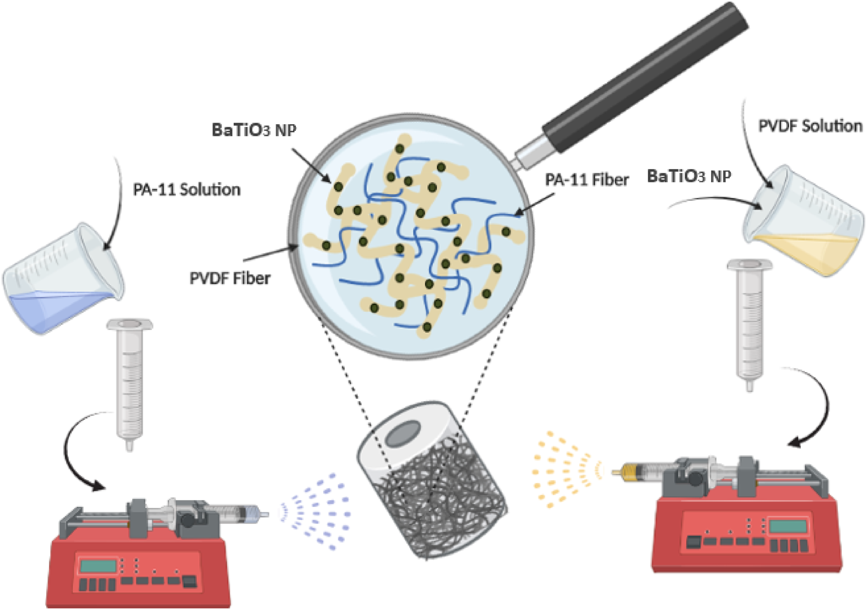
Characterization
Morphological characterization
Scanning electron microscopy (SEM, model: MIRA3, TESCAN Co.) was utilized to characterize the size and morphology of the synthesized nanoparticles as well as the microstructure of electrospun fibers. By applying the sputter coater (Pooshesh hayeh nanosakhtar, model: DSR1), the small pieces of samples were platinum coated. The fiber’s diameters were also measured using ImageJ.
Physiochemical characterization
The crystallinity percentage of the microfibers with or without nanoparticles was analyzed through Fourier transform infrared (FTIR) spectroscopy (PerkinElmer, model: Spectrum 2). Applicable piezoelectric properties of PVDF are contributed to one of its crystalline polymorph, β - phase. With the aid of equation (1), the percentage of β—crystalline phase (F (β)) can be calculated through the FTIR spectrum. To measure this parameter, the absorption peaks in 840 cm−1 and 764 cm−1, relating to α and β phase, respectively, must be determined at first .
31

To characterize the synthesized BaTiO3 nanoparticles and to investigate their impacts on microfiber’s crystalline structure, an X-ray diffraction analysis (XRD) (PANalytical, model: X’ Pert PRO MPD) was performed. Another test used to analyze the synthesized nanoparticles is the elemental analysis performed by X-ray scattering spectroscopy (EDX) conducted with the same SEM machine. This test shows the percentage of constituents in the material. To show that PA-11 and PVDF have piezoelectric properties, it is necessary to subject them to a mechanical load and measure their output currents or voltages. So far, a lot of research has been done on the piezoelectric property of PVDF,32,33 but the piezoelectric property of PA-11 has been proposed more theoretically, and less experimental study has been done regarding it.34–36 To evaluate the efficiency and piezoelectric properties of the samples in converting mechanical forces, measuring electrical response is the most practical method. To perform the piezoelectric test, a self-designed mechanical–electronic evaluation system (PiezoTester) was used to measure the output voltages of the samples.
37
This device applied the intended force to the samples in a cyclic manner to stimulate the heartbeat. Through a particular oscilloscope, the output voltage was shown on the screen. By enveloping the fibers with two aluminum foils acting as electrodes for sensing and measuring the external charges and putting two wires on them, which were fixed with adhesive tapes, the PEH was produced. Finally, the fabricated PEH was placed in the PiezoTester to fulfill this experiment (Figure 2). In addition to this analysis, longitudinal tensile tests and the fiber’s Young’s modulus could be applied to indirectly investigate the piezoelectric properties. To elaborate this, if a fibrous sample has a higher Young’s Modulus, it could transfer the applied forces more easily to the other parts. The better the force transmitted along the fibers, the better the polymer chains' alignment and, consequently, the dipoles could be achieved. Hence, more electrical charges would accumulate on the fiber’s surfaces leading to a higher output voltage. This test was performed with the Instron Tensile Testing machine (Hounsfield, model: H10KS) at room temperature. Schematic illustration of the PiezoTester and the fabricated PEH layers.
Biocompatibility characterization
The cytotoxicity of the fibers was assessed by MTT assay. First, L929 cells with the density of 1x10⁴ cells per well were seeded in 48-well plates and incubated at 37°C for 24 h. Afterward, 1 mg of the fibers was added to each well containing 500 μl of the cell culture medium. In this experiment, the wells that did not have any fibers and simply encompassed the cell culture medium were defined as the control groups. These 48 well-plates were incubated for 24, 48, and 72 h. For evaluating the cell viability, MTT (3-(4, 5-dimethyldiazol-2-yl)-2, 5-diphenyl tetrazolium bromide) assay was conducted. In brief, cell culture media were discarded, and 25 μl MTT solution with 225 μl of the fresh medium was added into each well, followed by 3-h incubation. After removing the supernatant, the remnant formazan was dissolved by MTT solvent. Next, the plate was wrapped in foil and shook for 15 min. For reading the absorbance of the solution, they were transferred to the 96-well plate, and an absorbance microplate reader (BioTech, model: ELx800) was used to read the optical density at 570 nm wavelength.
Statistical analysis
All the experiments were repeated three times (n=3) for each sample, and their results are expressed as mean ± standard deviations (SD). The significant difference was considered at p<0.05.
Results and Discussion
Analysis of hybrid PVDF/PA-11 fibers
Morphological characterization
SEM micrographs are used (Figure 3) to evaluate the morphology and structure of PVDF and PA-11 electrospun fibers. As shown in Figure 3, the PA-11 fiber diameters are less than PVDF fibers due to their lower concentration (4% w/v) compared to that of PVDF (24% w/v) and spinning flow rate (0.5 mL/h to 1.2 mL/h). Figure 3(c) also represents the physical entanglements caused by bilateral electrospinning of PA-11 and PVDF. This entanglement would affect the crystallinity, mechanical strength, and piezoelectricity of the fibers. This claim would be corroborated by conducting further experiments. SEM images of the electrospun a) PVDF, b) PA-11, and c) PVDF (75%)–PA-11 (25%) fibers with fiber diameter distributions.
Crystallinity characterization and piezoelectric sensitivity
Considering Figure 4 and equation (1), the β-phase percentage of the samples was calculated, and the results are illustrated in Table 2. In this equation, A is the absorption of the peaks. Regarding the FTIR spectrum of the samples and the results represented in Table 2, the percentage of the β-crystalline phase of PVDF is higher than the PA-11 percentage, which is related to the higher piezoelectric coefficient discussed in the previous sections. By increasing the volumetric ratio of PA-11 in samples, a rising trend is observed due to the synergistic effect of these two semi-crystalline polymers, which results in more crystalline hybridization compounds. As a result, the β-crystalline phase percentage of PVDF was increased from 59.38% to 69.2% when the PA-11 volumetric ratio of 34% (v/v) was achieved. The achieved β-crystalline phase of PVDF/PA-11 hybrid structure can be compared with the results of Kalani et al.
38
In their study, the β-crystalline phase was measured for the samples containing PVDF with different percentages of BaTiO3. For the sample incorporated with 25% (w/w) BaTiO3 nanoparticles, the corresponded β-phase fraction reached 68%, which is lower than the best result obtained in the current study (69.20%). It is worth noting that in the present study, the PVDF’s crystallinity was improved simply by its hybridization with a biocompatible polymer. However, Kalani et al. used a large amount of ceramic-based nanoparticles (25% (w/w) BaTiO3) to increase the crystalline structure of their electrospun fibers, which could adversely affect the mat’s flexibility and reduce its biocompatibility.
38
FTIR spectrum of the samples. The β-crystalline phase percentage of the samples.

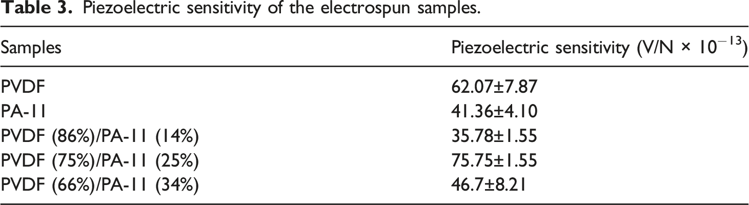
To test the piezoelectric properties of the fabricated patches, the setup device exerted a cyclic impact on the samples. A force equals to 1860 N was applied to all samples with a period of 50 milliseconds. The oscilloscope screen image representing the amount of the cyclic force and the output voltage of PVDF is shown in Figure 5. The yellow diagram is related to the mechanical force, and the red diagram indicates the cyclic output voltage. When this force was applied, the polymer chains and consequently the dipoles were aligned, creating a voltage difference between the upper and lower surfaces of the fibers. Table 3 shows the piezoelectric sensitivity, generated output voltage per 1 N of cyclic force, for all the samples. As expected, the output voltage of PVDF was higher than that of PA-11 due to the more significant piezoelectric coefficient of the PVDF. For hybrid fibers, when PVDF was co-spun with a small percentage (14% v/v) of PA-11, the output voltage decreased as the spun fibers did not purely consist of PVDF and were co-spun with a material having a lower piezoelectric coefficient. However, further increasing the volume ratio of PA-11 resulted in an upward trend for the output voltages. PA-11, like PVDF, has a semi-crystalline structure, and when co-spun with PVDF, it could enhance its crystalline structure. The stronger the crystalline structure, the higher the β-phase would be, and consequently, the piezoelectric sensitivity is expected to increase. The piezoelectric sensitivity of PVDF was increased from 62.07 mV.N−1–75.75 mV.N−1 when it was hybridized with 25% (v/v) of PA-11. Nevertheless, the piezoelectric sensitivity declined when this volumetric ratio reached 34% (v/v). This unexpected behavior should be evaluated through mechanical investigations, which is explained in the following sections. As shown, PVDF (75%)–PA-11 (25%) has the highest piezoelectric sensitivity among the samples and can be suggested as the optimum sample. This amount of piezoelectric sensitivity can be compared with the results reported by Sorayani et al.,
37
in which a PVDF/ZnO pressure sensor was tested with the same PiezoTester. The highest sensitivity reported by its authors was 2.18 mV.N−1, which is much lower than the best result here (75.75 mV.N−1). It means that the synergistic effect of these two semi-crystalline polymers surpasses the addition of a ceramic-based nanoparticle with a higher piezoelectric coefficient in improving the piezoelectric sensitivity. To accurately select the optimum specimen in terms of both output voltage and mechanical strength, these samples must be subjected to the tensile test in addition to the piezoelectric analysis. Output signals of PVDF in the piezoelectric test. The yellow diagram shows the cyclic mechanical force applied to the fibers and the red diagram shows the amount of the output voltage (Time/div = 50 ms/div, Volt/div sample = 1 V/div, and Volt/div Load cell = 1 V/div). Piezoelectric sensitivity of the electrospun samples.
Mechanical properties
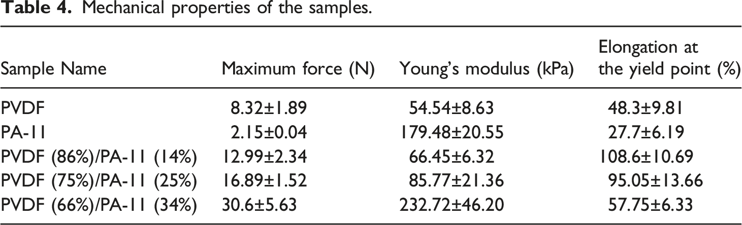
Mechanical properties of the samples.
Analysis of hybrid nanocomposite fibers
BaTiO3 nanoparticles characterization
To characterize the synthesized nanoparticles, XRD analysis was conducted, and the result is shown in Figure 6(a). The location of BaTiO3 matched with JCPDS Card (No. 83–1880) and illustrates the tetragonal phase information of BaTiO3 without impurity. By exploiting Debye–Scherrer formula, the average crystalline size of BaTiO3 nanoparticles was calculated to be about 24 nm, and the lattice parameters were found to be a = 4.15 A°, b = 4.15 A°, and c = 2.97 A°.
43
The peak with the highest intensity is observed at 2θ=32.17°. The presence of the peaks at 2θ = 22.21°, 32.17°, 38.90°, 45.60°, 50.81°, 56.13°, 65.87°, 70.32°, 74.79°, and 79.01° is a good confirmation of the BaTiO3 formation.
44
Other peaks are possibly attributed to the presence of BaCO3, produced by the reaction between barium hydroxide and CO2. BaTiO3 and BaCO3 characteristic peaks are distinguished in the XRD spectrum.45,46 As shown in Figure 6(b), only three peaks contribute to Ba, Ti, and O with a mass percentage of 64.5, 18.2, and 17.4, respectively. The observations mentioned above are found to be in accordance with BaTiO3 nanoparticle production. Since no peak corresponding to other elements is observed, it can be concluded that no by-products were formed during its synthesis. Since smaller particles could better transfer piezoelectric properties to the polymeric bulk, this synthesis aimed to produce particles at the nanometer scales. Barium titanate nanoparticles analysis. (A) XRD spectrum of the synthesized barium titanate. (B) EDX spectroscopy of the synthesized nanoparticle.
It is stated that covalent bonds forming between piezoelectric nanoparticles and polymers can help transfer this property up to 10 times more to the bulk of polymers. In fact, according to Kim et al., to distribute BaTiO3 nanoparticles well in the bulk of the polymer, chemical bonding was performed between BaTiO3 and PVDF. Because there are no functional groups in the PVDF structure, covalent bonds cannot be formed solely by the nanoparticles.
47
As Almadhoun et al. demonstrated, by incorporation of hydroxyl groups on BaTiO3 nanoparticles, hydrogen bonds could be formed between those functional groups and the fluorine atoms of PVDF.
48
This hydrogen interaction can improve the distribution of nanoparticles, which may enhance the piezoelectric properties. Figure 7 shows the FTIR spectrum of the modified BaTiO3 nanoparticles. The three primary absorption peaks are observed at 556.61 cm−1, 1041.74 cm−1, and 3415.39 cm−1. The peak at 556.61 cm−1 corresponds to the vibration of the Ti-O bond in BaTiO3, while the peak at 1041.74 cm−1 represents the tensile vibrations of -CO32-, which indicates the presence of barium carbonate (BaCO3) contamination in BaTiO3 powder. It has also been noted that the surface changes using hydrogen peroxide leave the contamination unchanged. The wide absorption peak at 3415.39 cm−1 represents the tensile state of the surface -OH groups on the particles. Compared to the unchanged BaTiO3 nanoparticles available in the references,49–51 BaTiO3-OH particles show a more intense adsorption peak for hydroxyl groups, meaning that the surface hydroxyl groups for these particles are denser and more concentrated, which is shown in this Figure. FTIR spectrum of the hydroxylated nanoparticles.
According to SEM images (Figure 8), it is observed that synthesized particles had spherical shapes with uniform morphologies. Moreover, no cluster is observed, and the particles are entirely separated with a diameter less than 100 nm. For further assessment, the diameters of the synthesized nanoparticles were measured using ImageJ software and the particle’s size distribution histogram is illustrated in Figure 8(c). The diagram shows the nanopowders have narrow size distribution ranges with the average diameter of 97.93 nm. Since a vast majority of them have a diameter less than 100 nm, the synthesis process leads to the formation of 100-nm grade nanopowders with a narrow-size distribution. Morphology of the synthesized barium titanate at different scales. A) 500 nm, b) 200 nm, and c) nanoparticle’s size distribution histogram.
Morphological observation
Figure 9 shows the PVDF fibers' morphology and structure incorporated with BaTiO3 nanoparticles. The addition of nanoparticles would affect the electrospinning conditions mentioned in the previous parts. To produce a uniform nanocomposite structure encompassing 15% w/w BaTiO3, the electrospinning mass flow rate, applied voltage, and nozzle-to-collector distance was optimized. As it was shown, the nanoparticles were distributed very well through the fibers, which would produce a uniform structure with steady crystallinity and piezoelectric properties.
31
This facilitates the detection of nanoparticles within the fibrous structure and shows that the particles are evenly distributed. Electrospun fibers containing 15% (w/w) BaTiO3 at different scales a) 500 nm, b) 1 μm, and c) 5 μm.
Crystallinity and piezoelectric considerations
Considering the FTIR spectrum of PVDF fibers shown in Figure 10, the β-crystalline phase percentage of PVDF was increased from 59.38% to 71.65% due to the incorporation of BaTiO3 nanoparticles (15% w/w). As previously mentioned, increasing the β-crystalline phase percentage directly enhanced the piezoelectricity of the microfibers. To investigate the influence of adding BaTiO3 nanoparticles on the β-crystalline phase of the fibers, XRD analysis was performed, and the results for the two samples of PVDF and PVDF–BaTiO3 are shown in Figure 11. As shown, a peak at 2θ=32° emerged in the spectrum of the PVDF–BaTiO3, which can match the synthesized BaTiO3 peaks reported in the previous sections. Furthermore, no shift in the BaTiO3 peak position was observed due to the presence of PVDF fibers. Therefore, this evidence would prove the presence of the well-distributed BaTiO3 nanoparticles in the PVDF fibers without any extra phase formation. To measure the degree of crystallinity, the ratio of the areas of the crystalline peaks to the areas under the scattering curve must be calculated.
52
From the XRD spectrum, it is observed that the crystallinity degree of PVDF was increased from 40.73% to 54.71% by incorporation of 15% (w/w) BaTiO3 nanoparticles into the hybrid structure. Additionally, the high intense peak was found to be close to the β-crystalline phase position of PVDF. Consequently, regarding the previous studies,
53
it can be predicted that these nanoparticles would strengthen the fibrous crystalline structure followed by the enhancement of piezoelectric properties. FTIR spectrum of PVDF and PVDF–BaTiO3. XRD spectrum of PVDF and PVDF–BaTiO3.

To prove the results obtained from the FTIR analysis, the piezoelectric test was also performed. By adding 15% (w/w) BaTiO3 nanoparticles to the PVDF-PA-11 hybrid fibrous structure, the piezoelectric sensitivity reached 107.52 mV/N, which was noticeably higher than the sample containing PVDF and PA-11 with optimal volumetric ratio (PVDF (75% v/v)–PA-11 (25% v/v)). It is also much higher than that of PVDF when it is exclusively composited with 25% (w/w) of BaTiO3. The PVDF/BaTiO3 pressure sensor was tested with the same PiezoTester and its piezoelectric sensitivity was reported to be 6 mV.N−1. 38 In this case, the prominent role of PA-11 in enhancing the piezoelectric response can be highlighted. Finally, since the heart systolic force was calculated to be approximately 10–20 N in various conditions. 42 by applying an equal force on the final PEH with the highest piezoelectric sensitivity, the output voltage of 1.07V–2.15 V would be generated. These voltage values are almost acceptable since the minimum voltage required for the pacemaker lithium batteries is 2.2 V. As a result, this patch would be a promising substitute for lithium batteries due to its capability of self-charging.
In vitro biocompatibility evaluations
The cytotoxicity of the optimum sample was studied by MTT assay. The viability of the L929 cells compared with that of the control groups for 24, 48, and 72 h was found to be approximately 81%, 96%, and 70%, respectively. As shown in Figure 12, (a) fixed concentration of (PVDF (75% v/v)–BaTiO3 (15% w/w))/PA-11 (25% v/v) fibers not only had a subtle influence on the growth of the cells but also enhanced the cell proliferation after 48 h in comparison with the first day of incubation. This result can be compared with the achievements of Azimi et al.,
27
in which the cell viability of mouse embryonic fibroblasts (MEF) cultured on PVDF/ZnO/rGO piezoelectric nanogenerator was investigated. In their study, the MTT assay revealed about 80% of cells were viable after 48 h, which is lower than the results presented here (96%).
27
However, after 72 h, a slight reduction in the cell viability was observed. This result is in accordance with previous studies claiming that the slight viability reduction might be contributed to the potential toxicity of mineral nanoparticles.
16
Regarding the former findings, it is stated that 10%–15% (w/w) is the appropriate percentage of mineral nanoparticles that can be incorporated into fibrous structures. This amount of nanomaterials not only could dramatically enhance the physicochemical characteristics but also would not have a devastating impact on the adjacent cells.
47
Accordingly, the current study shows that the fabricated PEH consisting of 15% (w/w) of BaTiO3 had the desired biocompatibility and subsequently could be implemented in the body. Cell viability of the (PVDF (75% v/v)–BaTiO3 (15% w/w))/PA-11(25% v/v) fibers.
Conclusion
In this research, we successfully manufactured a robust and novel sensitive piezoelectric patch as an efficient energy harvester with improved properties. In contrast to classical rigid energy harvesters that are typically made of lead-based ceramics and cause harm to the patients, the designed polymeric fibrous patch is flexible and can be easily placed on the heart’s lateral wall to efficiently harvest electricity from the cardiac motions. The PEH comprising of composite fibers of PVDF and PA-11 incorporated with BaTiO3 nanoparticles was optimized toward achieving large power electricity. It has been shown the conjugate electrospinning of PVDF with PA-11 would increase PVDF’s piezoelectric response due to the physical entanglement of their polymeric chains. Several experiments have been done to optimize the volumetric ratio of these piezoelectric polymers. Finally, the sample consisted of PVDF (75% v/v) and PA-11 (25% v/v) were found to be optimum with the piezoelectric sensitivity of 75.75 mV/N. Furthermore, to enhance the piezoelectric sensitivity, 15% (w/w) BaTiO3 nanoparticles were distributed evenly in the bulk of the hybrid fibers. Consequently, the crystallinity and mechanical strength were dramatically increased, causing the piezoelectric sensitivity to be obviously raised from 75.75 mV/N to 107.52 mV/N. As a result, the proposed PEH could be possibly charged by biomechanical forces like heartbeats or diaphragm movements to empower the pacemakers. Overall, the developed hybrid fibrous structures could be leveraged as PEH for numerous applications such as self-powered cardiac pacemakers.