
Abstract
Gelatin nanofibers containing Chromolaena odorata (C. odorata) crude extract were fabricated via electrospinning process to study the effect of C. odorata concentration on their size and properties for biomedical applications, especially for the purpose of wound healing and covering. C. odorata extracts, 0–20% weight by volume (w/v), in 20% (w/v) gelatin solutions, were spun at an operating voltage of 8 kV and flow rate of 0.5 mL/h. The distance between the needle tip and collector was fixed at 10 cm. The average diameters of the electro-spun fibers obtained did not exhibit significant difference when the concentration of the C. odorata extract was equal to or less than 10% (w/v). This was true in the case of both cross-linked and uncross-linked fibers with 0.04% (w/w) glutaraldehyde. The average diameters of the fibers obtained increased significantly when the concentration of the C. odorata extract was increased from 10% to 20% (w/v). All the uncross-linked fibers had smaller average diameters than those of the cross-linked fibers under all conditions. An increase in the C. odorata content of the fibers provided more hydrophilicity to the fiber surfaces. The novel finding from this study was that the gelatin fibers fabricated with C. odorata extract exhibited 180 times greater water adsorption than those of the commercial healing and covering films used presently. Moreover, all the fibers collected showed excellent antimicrobial activity against the Gram-positive bacterium, Staphylococcus aureus, but not against the Gram-negative bacterium, Escherichia coli.
Introduction
Appropriate medical treatment is the first important step in wound healing. However, efficient wound covering materials with excellent properties are additionally essential to accelerate wound healing. It is very critical to protect and keep the wound clean from any factors that can disturb healing, especially microbes. Wound covering materials must show significant advantages at least in term of blood coagulation, liquid adsorption, antimicrobial activity, cell growth, pain reduction, and flexibility. Various forms of materials such as thin film, hydrogel, nanoparticles, and nanofiber mat can be used as, or as part of, the covering materials depending on the wound type [1–6]. Each design with a different fabricated substance provides individual advantages and disadvantages.
Gelatin, chitosan, and cellulose acetate, for example, are typically used as base polymers for biomaterial fabrication because they exhibit good biocompatibility and biodegradability. However, gelatin is more widely used owing to its great functional and filmogenic properties and low cost [7–19]. To enhance the properties of gelatin-based biomaterials, the addition of some chemicals to the polymer solution is a convenient and practical alternative.
Chromolaena odorata or Siam weed is known as one of the growth inhibitors of agricultural crops especially young plants, because it is a rapidly growing invasive weed. This pest is generally found all over Thailand. Despite its disadvantages, it recognizably possesses some properties that are advantageous in medical treatment, such as anti-inflammatory, hepatotropic, and antimicrobial properties, wound and burn healing, as well as fever relief and usability in diabetes medication [20–23]. Alkaloids, essential oils, anisic acid, and a variety of flavonoids reported to be found in C. odorata crude extract [24] lead to its excellent properties, which render it suitable for use in traditional medical therapy.
In addition to the effect of the base polymer and additive, the suitable form of the material used affects its properties. Nanofibers are outstanding owing to their high porosity and large specific surface area, which offer significant potential for use in many applications of biomedical relevance, such as in wound healing and covering materials [25–27]. The process of electrospinning is usually employed in the synthesis of fibers of the micrometer to nanometer scale. A viscous polymer solution is fed into the system, extracted from the needle tip at a specific flow rate and operating voltage, and converted into non-woven fibers, which are collected at the collector.
In this study, crude extract from the leaves of Siam weed was used in the process of spinning together with gelatin. The diameter, surface appearance, hydrophilicity, water adsorption, and antimicrobial activity of the nanofibers obtained were examined corresponding to the crude extract concentration. The above mentioned properties are of initial concern and importance in the manufacture of wound healing and covering elements. It is presumed that this low-cost prototype of wound healing and covering materials, containing a substance extracted from an invasive and practically useless plant, can demonstrate better or equal wound healing and protection abilities compared with the currently used commercial alternatives.
Experiment
Materials
Gelatin powder (bioreagent grade), glacial acetic acid (reagent grade), ethanol (reagent grade), and glutaraldehyde (GTA, 25% solution) were purchased from Sigma (USA), Merck (Germany), Chemi (Thailand), and Panreac (Germany), respectively. All the chemicals were used as received without further purification. Fresh Siam weed leaves were collected from around the farmlands near Rajamangala University of Technology Thanyaburi, Pathum Thani, Thailand.
C. odorata crude extract preparation
The 10 topmost fresh leaves of the Siam weed plants were collected and rinsed with water to remove all dirt before they were dried at room temperature for 4–5 h. All the leaves were cut into small pieces and then soaked in 95% ethanol for two days. The solution was subsequently filtered using a filter cloth and filter paper (No. 1). The C. odorata crude extract was extracted by a rotary evaporator with a rotation rate of 140 r/min at 45°C. The crude extract obtained was kept in a glass bottle wrapped with aluminum foil and stored at 4°C until use.
C. odorata-gelatin nanofiber fabrication
Details of a similar experiment for nanofiber fabrication have been described in existing literature [25–28]. Briefly, 20% (w/v) gelatin solution was prepared by dissolving the gelatin powder in 60% (v/v) acetic acid at 40°C and using a magnetic stirrer for 2 h at 700 r/min to render the mixture homogeneous. Specific amounts of the C. odorata extract were mixed with the gelatin solution obtained in different ratios to yield various concentrations of the running solution for the process of electrospinning (0%, 5%, 10%, 15%, 20%, and 25% (w/v)). Each solution was divided into two parts. The first part of the solution was ready for use in nanofiber fabrication. The second part of each solution was used for crosslinking with GTA (0.04% by weight) before being used for electrospinning process. All the solutions with or without GTA, were used to fabricate nanofiber mats at a fixed flow rate of 0.5 mL/h, operating voltage of 8 kV, operating time of 2 h, and distance between the needle tip and collector of 10 cm. Aluminum foil was used to cover the collector before the fibers were fabricated. All the nanofiber mats synthesized were dried in an oven at 40°C for 1 h and kept in a de-humidifier before further characterization for morphological structure, wetting property, water uptake, and antimicrobial activity.
Characterization
Morphological structure
The morphologies of the nanofibers synthesized were observed using a scanning electron microscope (SEM, JSM-5410LV, JEOL, Japan). The fiber size was determined using ImageJ software. The average fiber size and standard derivation of both uncross-linked and cross-linked fibers with GTA were calculated based on 10 locations of each fiber for at least 10 fibers under each condition.
Wetting property
Owing to the solubility of uncross-linked fibers, only the cross-linked fibers were examined for their wettability, defined by the contact angle between the fiber mat surface and the water droplet. The contact angles of the fibers prepared were evaluated using a contact angle meter (DM-CE2, Kyowa, Japan) via the sessile drop method. At least 10 random areas of each fiber mats were selected to determine the average contact angle.
Water uptake
A simple method was used to test the cross-linked fibers under all conditions for their ability to uptake water. Briefly, the nanofiber mats were cut into 1 cm × 1 cm specimens and dried at 40°C to determine the dried mass (md). The mats were then soaked in deionized water for 1 h. The wet mass (mw) of each fiber mat was observed right after soaking. All the samples tested were dried again to confirm that there was nothing dissolved in the water during the experimental process. The water uptake ability can be demonstrated by the percentage of water uptake, which can be calculated using equation (1).

Antimicrobial activity
Escherichia coli (E. coli) and Staphylococcus aureus (S. aureus) were the two important pathogens chosen as the representatives of Gram-negative and Gram-positive bacteria, respectively, to test the antimicrobial activity (AATCC 100 method) of the nanofiber mats fabricated under all conditions because both bacteria are rapidly growing microorganisms commonly found in hospital-acquired infections [29–32]. E. coli (ATCC 25922) and S. aureus (ATCC 25923) were used in the experiments. The bactericidal activity with 24 h of exposure time using a nanofiber mat in a culture medium at 37°C was computed based on the percentage of inhibition in the viable cell number computed by equation (2)

Results and discussion
Morphological structure
Pure gelatin nanofibers and gelatin nanofibers containing C. odorata crude extract in different concentrations were successfully fabricated via electrospinning process using the experimental set-up provided in the Experiment section except for the gelatin solution with 25% C. odorata crude extract owing to its viscosity. The morphology of each nanofiber obtained corresponding to the different ratios of C. odorata crude extract are shown in Table 1. All the fibers obtained (without GTA) exhibited smooth surfaces with small diameters of 269.94–668.96 nm.
Morphologies of fabricated nanofibers.

Glutaraldehyde, which is the crosslink chemical, in gelatin solution increases the viscosity of the solution. Consequently, all the fibers prepared without GTA were smaller in size compared with those prepared with GTA via electrospinning process at the same operating voltage and flow rate. In both the fiber types, i.e., with and without GTA, the average diameter of the fibers synthesized increased slightly as the concentration of the C. odorata crude extract increased from 0% to 10% in solution. A noticeable change in the average diameter size was observed at a concentration of 15% C. odorata crude extract in solution. At a concentration of approximately 15–20%, the effect of C. odorata crude extract concentration in solution on the average diameter was evidently dramatic, as shown in Figure 1. At that concentration, the viscosity of the total solution was slightly higher than that of solutions with lower concentrations of C. odorata crude extract. However, to validate this concept, a solution with 25% C. odorata crude extract cannot be spun into fibers at the fixed experimental voltage and flow rate, as previously mentioned.

Change in average diameter of nanofibers prepared with different concentrations of C. odorata crude extract in gelatin solution via electrospinning process at fixed flow rate of 0.5 mL/h, operating voltage of 8 kV, operating time of 2 h, and distance between needle tip and collector of 10 cm.
Wetting property
The cross-linked nanofibers were examined for their wetting property through observations of the contact angle between the nanofiber mat surface and water droplet using the contact angle meter. The fibers fabricated under all conditions were hydrophilic, based on the contact angles examined. However, the gelatin nanofibers without C. odorata crude extract showed the highest contact angle and were less hydrophilic than the others. The nanofiber with greater content of C. odorata crude extract demonstrated a smaller contact angle, as seen in Figure 2, owing to the presence of more hydrophilic compounds, including flavonoid from the crude extract [33, 34], in the starting materials used for its fabrication.

Measured contact angle between cross-linked nanofiber mat surface and water droplet for every fabrication condition at different concentrations of C. odorata crude extract.
Water uptake
The effect of fiber diameter on the water adsorption properties is less reported in the literature. However, it is believed that the surface property and porosity of materials are normally the two main factors controlling their water uptake ability. Materials with more hydrophobic surfaces and less porosity absorb less water than those with more hydrophilicity and porosity. However, materials with more hydrophobic surfaces could adsorb more water if they have high porosity, while materials with more hydrophilic surfaces could adsorb more water despite their low porosity. This depends on which factor is the dominant one.
The water uptake of the materials could be affected by the concentration of the C. odorata crude extract in the solution prepared for nanofiber fabrication. All the C. odorata crude extract-containing nanofibers obtained, which exhibited more hydrophilicity as previously mentioned in the section above, adsorbed more water than the pure gelatin nanofiber. However, the percentage of water uptake of the fabricated nanofiber decreased with increase in the concentration of C. odorata crude extract, as shown in Figure 3. This result, together with the wetting property data, implied that porosity decreased with increase in the concentration of C. odorata crude extract in solution for nanofiber fabrication. This corresponds to the results obtained in the Morphological structure subsection, i.e., an increased amount of C. odorata crude extract affects larger nanofibers, thereby reducing porosity and water absorption capability. This confirms that porosity should be a dominant factor in the process of water absorption in this study. The percentage of water uptake of the nanofibers fabricated in this study is approximately 180 times higher than those of the commercial healing and covering films reported in the literature [35]. With the increased water adsorption ability of wound healing materials, it is easier to provide an optimal healing environment for wound dressing because the excess exudate is removed via adsorption to the materials [36,37].
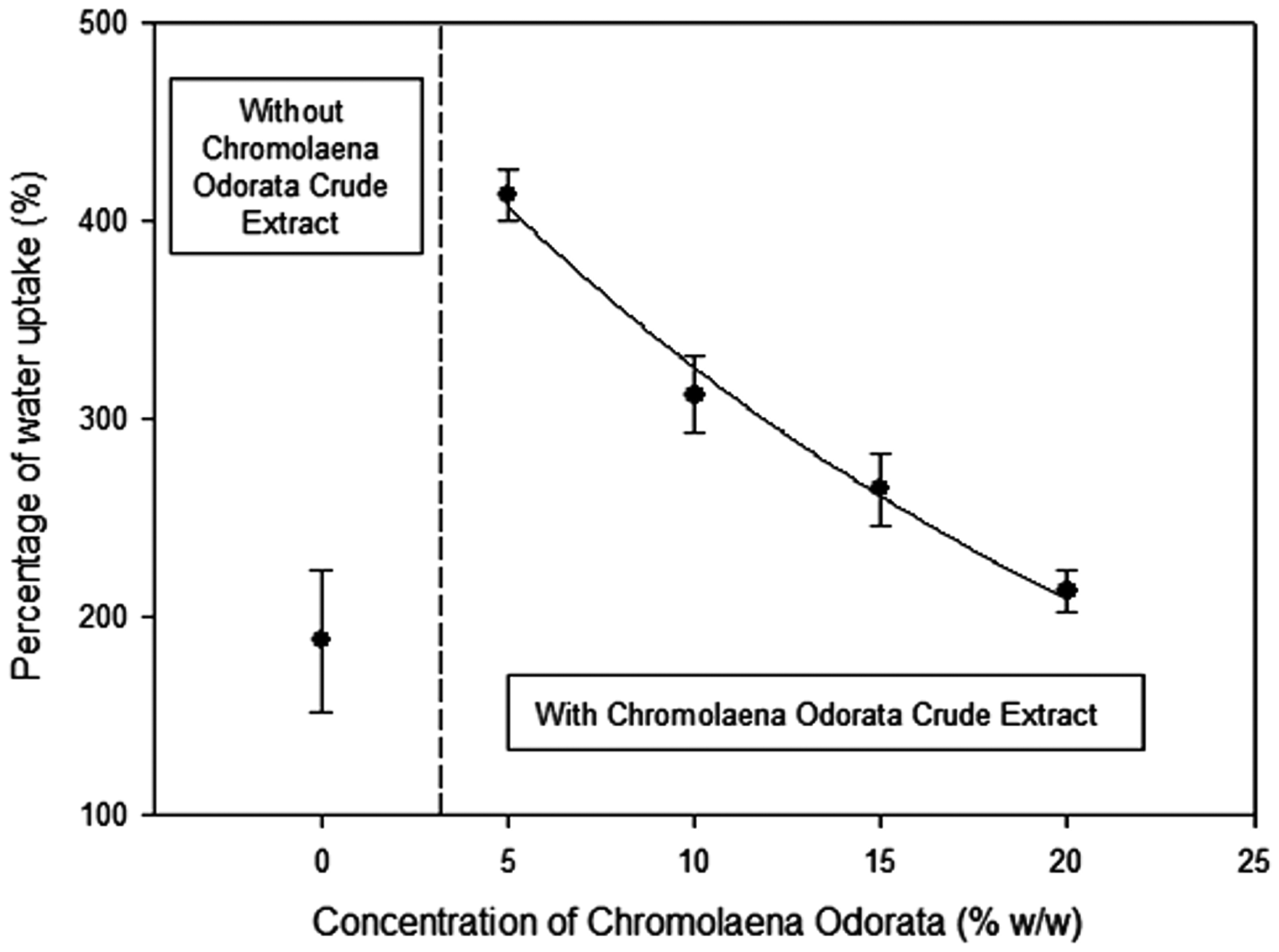
Percentage of water uptake of nanofibers fabricated with different concentrations of C. odorata crude extract.
Antimicrobial activity
The microbial activity of the nanofibers fabricated with 0–25% of C. odorata crude extract were investigated with S. aureus (ATCC 25923) and E. coli (ATCC 25922), which were the representative Gram-positive and Gram-negative bacteria, respectively. After 24 h of exposure time of the bacteria to nanofiber mats in the culture medium at 37°C, the percentage of inhibition of each bacterium was computed. The results are showed in Table 2.
Antimicrobial testing of fabricated nanofibers with S. aureus and E. coli.

NI: no inhibition in log CFU/mL.
Excellent inhibition of S. aureus by the nanofibers fabricated was found in the case of all the fibers containing C. odorata crude extract. In contrast, no inhibition of E. coli was noted for the nanofibers from all fabrication conditions although the cell wall of E. coli is thinner than that of S. aureus. Nevertheless, the difference between the cell wall structures and compositions of these bacteria might be the reason for the difference in their susceptibility to nanofibers containing C. odorata crude extract. In Gram-negative bacteria, the lipopolysaccharide of the outer membrane offers the ability to resist hydrophobic compounds [38–40]. The results here likely reflect that this antimicrobial activity was performed by some active agents from the C. odorata crude extract, such as alkaloids, essential oils, anisic acid, or a variety of flavonoids. These compounds might be able to specifically form at, or penetrate, the cell walls of S. aureus, thereby inhibiting the growth of the bacteria and destroying the microorganisms in the final stage. This implies that the nanofibers containing C. odorata crude extract are suitable for use in medical applications related to redness, pain, warmth, or swelling from abscesses. In vivo wound healing study and cytotoxicity examination are planned to be carried out in the next step of study. However, the C. odorata crude extract did not exhibit cytotoxicity when tested against T. vaginalis and B. hominis, and this is supported by Vital and Rivera [41].
Conclusions
Gelatin nanofibers containing C. odorata crude extract were successfully fabricated via electrospinning process and characterized based on initial physical and medical properties related to the C. odorata content. The size and hydrophilicity of the fibers increased slightly with the concentration of C. odorata whereas the percentage of water uptake decreased. The water uptake of the nanofibers synthesized was the novel finding from this study because its value was almost 180 times those of the commercial healing and covering films used presently. Another uniqueness of the nanofibers obtained was that they showed excellent antimicrobial activity against the Gram-positive bacterium, S. aureus, with 100% inhibition. A few other properties of the nanofiber mats, such as tenacity, extensibility, and biofilm infection, are our additional areas of interest for future research.
Footnotes
Acknowledgements
The authors appreciate the support provided by the Smart Materials Research Unit, Division of Physics, Faculty of Science and Technology, RMUTT, and the Department of Microbiology, Faculty of Science, Chulalongkorn University, Thailand.
Declaration of conflicting interests
The author(s) declared no potential conflicts of interest with respect to the research, authorship, and/or publication of this article.
Funding
The author(s) disclosed receipt of the following financial support for the research, authorship, and/or publication of this article: This work was funded by Rajamangala University of Technology Thanyaburi (RMUTT, grant number 256109A1650009) and partly supported by Thailand Toray Science Foundation, the 23rd Science & Technology Research Grant.