
Abstract
AOPAN nanofibers were prepared by electrospinning and amidoxime modification, subsequently, HEMA was used as the monomers for surface grafting via atom transfer radical polymerization, followed by coordination with Cu(II) ions, thereafter, the nanofibers AOPAN-poly(HEMA)-Cu(II) were explored as the novel support for laccase immobilization. Scanning electron microscopy was used to visualize the morphology of the nanofibers, and Fourier transform infrared spectroscopy was used to provide information on the surface chemistry of nanofibers. At the same time, the optimization of immobilization conditions and the relative properties of the immobilized laccase were also studied in this paper. The study showed the largest amount of immobilized laccase while the reaction time of atom transfer radical polymerization was 4 h. The immobilized laccase showed a better stability resistance to temperature and pH change, and the initial activity of immobilized laccase retained (60.3 ± 3.1)%, that of the free laccase retained only (21.3 ± 2.1)% when stored at 4℃ for 24 days. Immobilized laccase maintained its initial activity after 10 repeated batches of 64.5%.
Introduction
Enzyme is a protein-based biocatalyst with high efficiency, excellent selectivity and mild reaction conditions. Laccase is a polyphenol oxidase, one of the important wood fiber degrading enzymes, and belong to the blue oxidase family [1]. Laccase is a natural environment-friendly enzyme which can catalyze many aromatic compounds, especially polyphenol. Laccase has been widely utilized in field of wastewater treatment, biobleaching, aromatic compound conversion, biosensor construction and so on [2,3]. However, the extensive use of enzymes has been limited due to the poor stability and high cost, immobilization of enzymes is an effective way to solve these problems [4–6]. Immobilization methods include the physical adsorption, the embedding, the covalent binding, the coordination binding and the cross-linking [7,8]. Among these, coordination binding seems to have some advantages over the other methods, such as less possibility of inactivation and more stability of the immobilized laccase.
Atom transfer radical polymerization (ATRP) is by far the most promising living/controlled free radical polymerization, which has many advantages, such as it may be obtained with narrow molecular weight distribution, controlled molecular weight and different polymer structure, polymerization of various monomers, reaction conditions are mild and easy to control [9]. The ATRP method has often been adopted for making linear polymer chains/brushes with controlled molecular lengths/weights since the propagation centers do not undergo chain termination and/or chain transfer during polymerization, and thus the molecular weights/lengths increase linearly with the conversion of monomers [10–12].
Most of the transition metal ions such as Cu2+, Ni2+, Zn2+ and Fe3+ can form stable complexes with electron-rich compounds and coordinate molecules containing O, N and S, e.g. hydroxyl groups (–OH), amine groups (–NH2) and thiol groups (–SH) [13–16]. Immobilization of enzymes on a metal-chelated matrix is based on multipoint interactions between chelated metal ions on the support and the active sites such as the indole group of tryptophan, the imidazole group of histidine and the thiol group of cysteine [17–19].
In recent years, electrospun nanofibers has been used in many fields such as filtration/separation, tissue engineering, drug delivery, sensor/detector, composite materials and enzyme immobilization due to large surface area, high porosity, short regeneration rate and easy elution. In this study, AOPAN-poly(HEMA) nanofibers were prepared by grafting HEMA onto the surface of nanofibers by ATRP technology. Then, Cu(II) ions were incorporated into the hydroxyl groups of the AOPAN-poly(HEMA) nanofibers via metal chelation. Hereafter, laccase was immobilized onto the metal-chelated nanofibers, the relative properties of the immobilized laccase and free laccase were also studied, such as the quantity/activity, stability and reusability of immobilized laccase were evaluated.
Experimental
Materials
Laccase (EC 1.10.3.2) was a fungal laccase from Ganoderma lucidum, purchased from Sigma. Poyacrylonitrile (PAN, Mw = 90,000 g/mol), N,N-dimethylformamide (DMF), hydroxylamine hydrochloride, CuCl, CH3COOH, CH3COONa, H3PO4, CH3CH2OH, Coomassie brilliant blue (G250), CuSO4·5H2O, 2-methoxyphenol, tetrahydrofuran (THF), 2-bromoisobutyryl bromide (2-BIB), triethylamine (TEA), 1,1,4,7,10,10-hexamethyltriethyl- enetetramine(HMTETA), 2-hydroxyethyl methacrylate (HEMA) were purchased from Aladdin Chemical Reagent Co. Ltd (Shanghai, China) and used without further purification.
Preparation of AOPAN nanofibers
Prior to electrospinning, the PAN powder was first dried at 45℃ for 3 h, and then the PAN powder was dissolved in DMF solvent, a solution of 11 wt.% PAN in DMF was prepared at room temperature. The solution was then placed in syringes (content of 10 mL) with a blunt needle (the nozzle diameter was about 0.7 mm), and the solution flow rate was controlled by a micro-infusion pump (JZB-1800D, Changsha, China). The high-voltage supplier (DW-P503-4AC, Tianjin, China) was used to connect the grounded collector and metal needles for forming electrostatic fields [19]. During electrospinning, a positive high voltage of 18 kV was applied to the needle, solution flow rate was 0.5 mL/h, and collecting distance between the syringe needle tip and the grounded collector was 17 cm. PAN nanofibers were covered with an electrical ground covering film on the aluminum foil cover collecting roller. The membrane was dried at 45℃ for 3 h after electrospinning.
Then, the PAN nanofibers (0.1 g, dry weight) were accurately weighed. These nanofibers were added to 500 mL 0.1 mol/L hydroxylamine hydrochloride aqueous solutions. The pH of the reaction solution was adjusted to 7 by adding sodium carbonate solution. The reaction was carried out at 65℃ for 2 h. After the completion of reaction, the PAN nanofibers taken from reaction medium were washed with distilled water and dried at 40℃ in a vacuum oven. At the same time, the quality of nanofibers was also accurately weighed, and conversion rates of amidoxime were calculated according to equation (1) [20].

According to the experimental results, the conversion rates of amidoxime were 18.1% after 2 h.
Surface modification of the AOPAN nanofibers
AOPAN nanofibers were first immersed in 20 ml of THF for 10 min. Then, the AOPAN nanofibers were placed into a mixture of 70 µL of 10 mM TEA, 63 µL of 10 mM 2-BIB, and 50 mL of THF for 3 h at 35℃. As shown in Figure 1, 2-BIB would react with amidoxime groups on the surface of electrospun AOPAN nanofibers, while TEA would neutralize the byproduct of HBr. After completion of the reaction, the nanofibers were taken out and stored in THF.
Schematic showing the grafting of poly(HEMA) on the surface of an AOPAN nanofiber via the ATRP method. Schematic of laccase immobilized on the metal-chelated AOPAN-poly(HEMA) nanofibers [4].

First, 24 mL of HEMA and a mixture of 400 µL of HMTETA and 24 mL of DMF were deoxygenated through three freeze-pump-thaw cycles by using liquid nitrogen, then 100 mg of CuCl was added into the HMTETA/DMF mixture inside a vacuum glovebox followed by being magnetically stirred for 2 h. Subsequently, the initiated AOPAN nanofibers and HEMA were placed into the mixture of HMTETA/DMF/CuCl, and the system was kept in the glovebox at 25℃ for a predetermined time to complete the ATRP reaction of HEMA. Finally, the poly(HEMA)-modified nanofibers were rinsed by ethanol and dried in air.
According to the experimental results, the grafting rate of HEMA monomer in AOPAN-poly(HEMA) nanofibers was 19.7%.
Immobilization of laccase on AOPAN-poly(HEMA) nanofibers
Prior to immobilization, 0.1 g AOPAN-poly(HEMA) nanofibers was added to 40 mL of CuSO4 solution whose concentration was 100 mmol/L, the flack was shaken at 25℃ for 24 h (120 r/min), then rinsed with deionized water. The AOPAN-poly(HEMA)-Cu(II) nanofibers were dried in air and then dried at 35℃ in a vacuum over couch [21].
Then, the AOPAN-poly(HEMA)-Cu(II) nanofibers were immersed in 40 mL, 3 g/L laccase solution (pH = 4), and at 4℃ for 12 h. Finally, the modified nanofibers were removed from the solution and rinsed with the same HAc–NaAc buffer solution until no soluble protein was detected. And the immobilized laccase was stored in a HAc–NaAc buffer solution at 4℃.
In this experiment, the amount of laccase immobilization on the AOPAN-poly(HEMA)-Cu(II) nanofibers was determined by the method of Bradford. The fixed amount of laccase was determined by measuring the content of laccase in the buffer solution used for washing the immobilized enzyme nanofibers and the immobilized enzyme nanofibers before and after the immobilization, as shown in equation (2) [22].

Activity assays of free laccase and immobilized laccase
In order to test the respective activities of the free and immobilized laccase, 0.3 mL of laccase solution (3 g/L, pH = 4) or 0.01 g of AOPAN-poly(HEMA)-Cu(II) nanofibers immobilized laccase were mixed with 8.7 mL of 10 mmol/L 2-methoxyphenol solution (pH = 4). The system was kept at 50℃ for 30 min, and five replicates were tested for each sample. The activities of the free laccase or immobilized laccase were determined spectrophotometrically by measuring the decrease of absorbance at 465 nm, as a consequence of 2-methoxyphenol consumption the specific activity of enzyme was calculated by using the following equation [23].

And the relative activity of enzyme was then calculated by using equation (4).

Characterization of electrospun nanofibers
Scanning electron microscopy (SEM) (Hitachi S4800 field emission) SEM was used to examine the morphology of different nanofibers [25]. Prior to SEM examination, the specimens were sputtering gold to avoid charge accumulation. FT-IR spectrometer (Nicolet nexus 470) was used to investigate the functional groups on the surface of the nanofibers. Samples of PAN, AOPAN and AOPAN-poly(HEMA) were sliced into pieces and mixed with KBr.
Effect of ATRP reaction time
Various ATRP reaction times were studied to control the length/thickness of grafted polymer chains/brushes on the electrospun AOPAN nanofibers. To determine the optimal reaction times, the amounts of immobilized laccase on the final AOPAN-poly(HEMA)-Cu(II) with different ATRP times were measured according to ‘Immobilization of laccase on AOPAN-poly(HEMA) nanofibers’ section.
Evaluation of immobilized laccase
Temperature and pH dependence of free and immobilized laccase
In order to assess the temperature dependence, free and immobilized laccase were, separately, mixed with 2-methoxyphenol solution (pH = 4) first. The activities were then measured in the temperature range from 30 to 70℃. To evaluate the pH dependence, the activities of free and immobilized laccase were determined at different pH values (from 2.0 to 7.0). The relative activity under various conditions was calculated, while the highest activity was 100%.
Storage stability of immobilized laccase.
In order to analysis storage stability, the free and immobilized laccase were stored at 4℃ in HAc–NaAc (pH = 4) for 20 days, then the residual activity was calculated.
Reusability of immobilized laccase.
To examine the reusability of immobilized laccase, the activity of immobilized laccase was tested 10 times within 24 h. Before each test, the nanofibers with immobilized laccase were rinsed with HAc–NaAc solution (pH = 4) to remove any residual substrate, and the activity of immobilized laccase was then tested in a fresh reaction medium.
Results and discussion
Morphological structure of different nanofibers
Scanning electron microscopy (SEM) was used to observe the surface morphology of PAN, AOPAN and AOPAN-poly(HEMA) nanofibers, as shown in Figure 3. Electrospun PAN nanofibers and AOPAN nanofibers were formed with randomly oriented fiber membranes. The electrospun PAN nanofibers have good morphology with uniform diameter. Compared with PAN nanofibers, the diameter of AOPAN nanofibers was almost unchanged, and the fiber structure was not obviously deformed, however, after amidoxime modification, the surface of the nanofibers slightly roughened as shown in Figure 3(a) and (b). The AOPAN-poly(HEMA) nanofibers grafted by ATRP still maintain good fiber morphology, but the average diameter of the modified nanofibers was increased as shown in Figure 3(c).
SEM images of (a) PAN nanofibers, (b) AOPAN nanofibers and (c) AOPAN-poly(HEMA) nanofibers.
FT-IR analysis of different nanofibers
The FT-IR spectroscopy was generally used to qualitatively determine functional groups in a wide range of samples. In this paper, FT-IR was employed to study chemical differences among PAN, AOPAN and AOPAN-poly(HEMA) nanofibers, and the results of FT-IR spectra are presented in Figure 4.
FT-IR spectras of (a) PAN nanofibers, (b) AOPAN nanofibers, (c)AOPAN-poly(HEMA) nanofibers.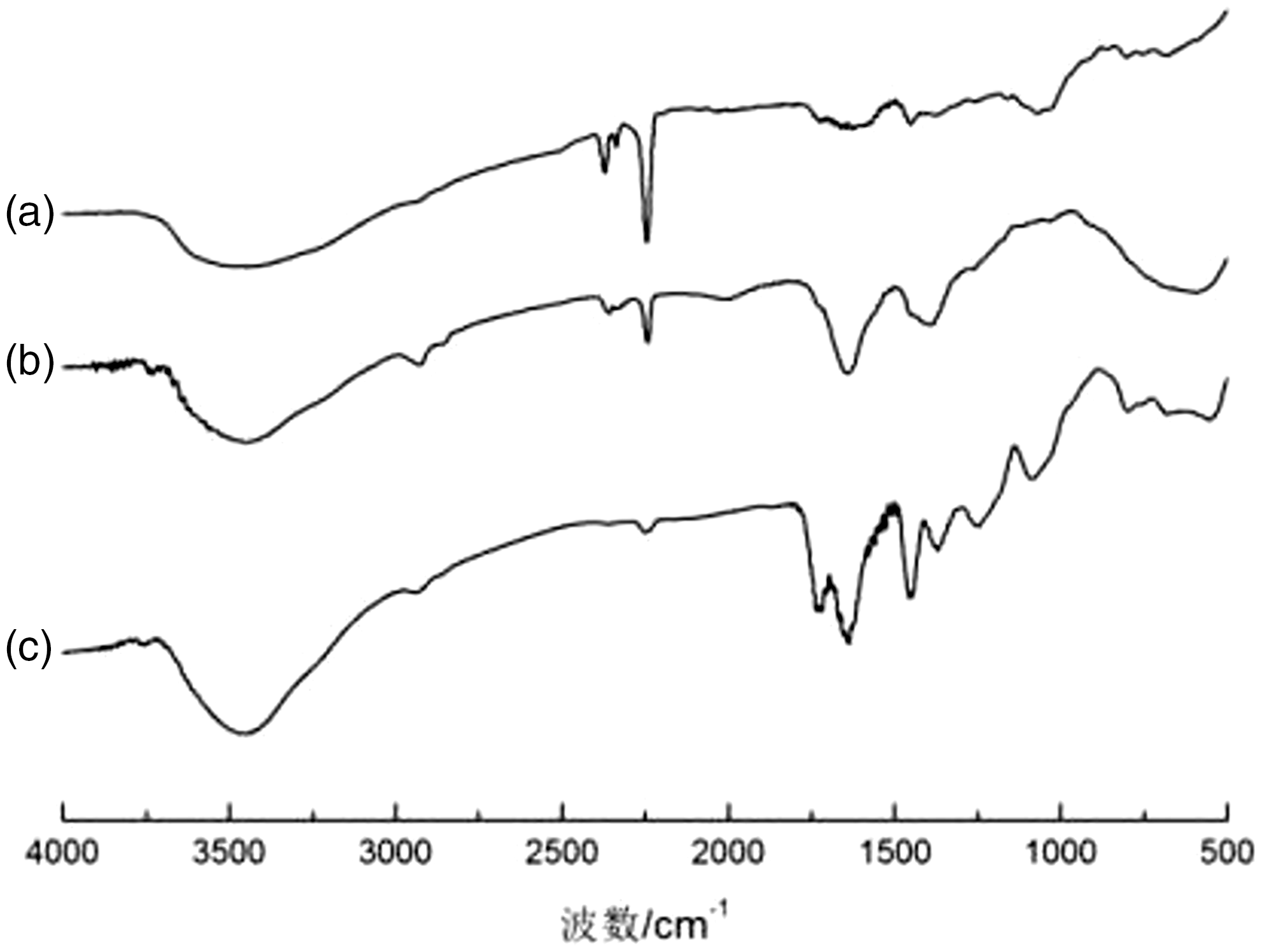
The spectra in Figure 4(a) had a characteristic band at 2247 cm−1, a directive of C≡N stretching in PAN. Compared with PAN nanofibers, the FTIR spectra of the AOPAN nanofibers showed some differences. The spectra Figure 4(b) had bands around 3650–3150, 1638 cm−1, an indication of N–H, O–H and C = N stretching vibration peak in AOPAN nanofibers. Simultaneously, the characteristic absorption peak of C ≡ N stretching at 2247 cm−1 was weakened obviously. It can be concluded that, some of the cyano on the PAN nanofibers have been successfully converted to amine oxime groups after modified by amidoxime modification. The spectra in Figure 4(c) acquired from the AOPAN-poly(HEMA) nanofibers had a band centered at 1727 cm−1, this band could be attributed to the stretching motion of cyano groups in HEMA, indicating that poly(HEMA) chains/brushes were generated on the surface of AOPAN nanofibers.
Effect of ATRP reaction time
Different ATRP reaction time caused different length/thickness of grafted polymer chains/brushes on the electrospun AOPAN nanofibers. In this work, AOPAN nanofibers were placed into the grafting reaction system in the glovebox at 25℃ for 1, 2, 4, 6, 8, 12 h, then the AOPAN-poly(HEMA)-Cu(II) nanofibers were used to immobilized laccase, and the result is shown in Figure 5.
Amount of immobilized laccase versus reaction time of ATRP for AOPAN-poly(HEMA) nanofibers.
As depicted in Figure 5, the amount of laccase immobilization increased gradually with the increased ATRP reaction time, and the biggest amount of the laccase immobilization was 87 ± 4.3 mg/g, while the ATRP reaction time was 4 h. As time goes by, the amount of immobilized laccase began to decreased gradually. This is because that, at the beginning of the ATRP reaction, the length/thickness of grafted polymer chains/brushes on the AOPAN nanofibers increased gradually, simultaneously, the binding sites with laccase also increased, so the amount of laccase immobilization showed a grown trend. With the reaction continued, the length/thickness of the grafted polymer chains/brushes continued to grow too long so that it cannot completely extend the reaction solution; therefore, the amount of immobilized laccase began to decline.
Temperature and pH dependence of free and immobilized laccase
The effect of temperature and pH value on relative activity of free and immobilized laccase is depicted in Figure 6. With the increase of ATRP reaction temperature, the relative activity of laccase would become higher. However, while the reaction temperature was further increased, the relative activity of laccase would be lower. The highest relative activities of laccase under both free and immobilized conditions were discovered at 50℃ and 45℃, respectively; moreover, the immobilized laccase normally had higher relative activities than the free laccase in the entire temperature range from 30 to 70℃. The higher relative activity would primarily be attributed to the increased structural stability of immobilized laccase molecules, while the multipoint interactions between laccase molecules and functional surface on the supports might provide the further protection against inactivation at a higher temperature. The optimum pH of the free and immobilized laccase can be easily identified at 4 and 4.5 from Figure 6(b). In addition, the immobilized laccase shown lower sensitivity on the pH value, and the corresponding residual activities of immobilized laccase were generally higher than that of free laccase.
Effect of pH (a) and temperature (b) on the activity of free and immobilized laccase.
Storage stability of immobilized laccase
As shown in Figure 7, the relative activity of free laccase and immobilized laccase were (21.3 ± 2.1)% and (60.3 ± 3.1)% after being stored at 4℃ in HAc-NaAc (100 mM, pH = 4) for 24 days, when the corresponding initial activities were set as 100%. The reduction of enzyme activity is a time-dependent natural phenomenon; however, the degree of enzyme activity reduction could be mitigated considerably through immobilization. The immobilized enzyme molecules could better retain their conformational structure; therefore, the inactivation upon long-term storage would be mitigated, thus ameliorating the storage stability of immobilized laccase.
Storage stability of free and immobilized laccase.
Reusability of immobilized laccase
Unlike free laccase, the residual activities of AOPAN-poly(HEMA) nanofibers immobilized laccase remained 63.4 ± 3.1% of their initial activities upon reuse 10 times (the nanofibers with immobilized laccase were washed with HAc-NaAc after each time) as shown in Figure 8. It is well known that the reusability of an enzyme is among the major factors/concerns for many practical applications. The results indicated that the AOPAN-poly(HEMA) nanofibers immobilized laccase exhibited the excellent performance on reusability and has a great value in the practical applications.
Residual activity of the immobilized laccase.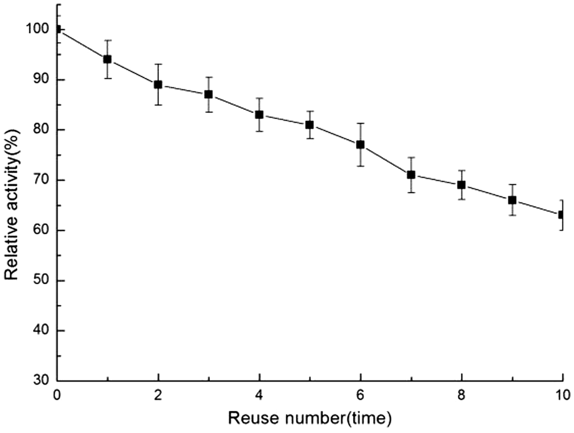
Conclusion
In this document, the electrospun AOPAN nanofibers was surface-grafted with HEMA via the ATRP reaction for high-density immobilization of laccase. The largest amounts of laccase immobilization were 87 ± 4.3 mg/g, while the ATRP reaction time for poly(HEMA) was determined at 4 h. And the immobilized laccase exhibited significantly higher resistance to changes in pH (tested from 2 to 7) and temperature (tested from 30 to 70℃) than free laccase. In addition, the storage stability of immobilized laccase was obviously better than free one. The functional nanofibers-immobilized laccase also has excellent reusability compared with the free laccase. These results confirm that the immobilized laccase had a high affinity with the support and gained more stable character compared to free one, which demonstrated the modification of AOPAN-poly(HEMA) nanofibers could be used as a promising material for immobilizing a wide range of bioactive molecules.
Footnotes
Declaration of Conflicting Interests
The author(s) declared no potential conflicts of interest with respect to the research, authorship, and/or publication of this article.
Funding
The author(s) disclosed receipt of the following financial support for the research, authorship, and/or publication of this article: This research was supported by the National Natural Science Foundation of China (Grant No. 21377004), the Projects of International Cooperation of Anhui Province (Grant No. 1604b0602024), and the Natural Science Foundation of Anhui Provincial Education Department (Grant No. KJ2016SD04). The authors would also acknowledge the Department of Education in Anhui Province of China (Grant No. 2015LJRCTD001).