
Abstract
This study introduced trimethylolpropane trimethacrylate into ultra-high molecular weight polyethylene fibers through supercritical CO2 pretreatment before the fibers were irradiated under an electron beam. Significant differences, emerging in the ultra-high molecular weight polyethylene fibers’ gel content, mechanical properties, and creep property according to their different irradiation doses, were studied through one-way analysis of variance. Regression equations were established between the irradiation dose and the gel content, breaking strength, elongation at break, and creep rate by regression analysis. A reasonable irradiation dosage range was determined after a verification experiment and the impact trends were analyzed; additionally, the sensitized irradiation crosslinking mechanism of ultra-high molecular weight polyethylene fibers was preliminarily examined. Then the surface morphology, chemical structures, thermal properties, and crystal properties of treated ultra-high molecular weight polyethylene fibers were measured. The results showed that as the irradiation dose increased, the gel content first rose and then declined; the breaking strength decreased continuously; the elongation at break increased at first and then decreased; and the creep rate originally fell and then rose before finally declining slowly. Electron beam irradiation had a significant etching effect on the fibers’ surface, and both the melting point and crystallinity decreased slightly.
Keywords
Introduction
Ultra-high molecular weight polyethylene (UHMWPE) fiber has excellent properties such as high tensile strength, high specific modulus, low density, high work of fracture, and good workability, which have led to its wide use in manufacturing marine vessel ropes, military protective materials, and aerospace materials [1,2]. However, because of its simple planar zigzag structure, lacking of hydrogen bonding between UHMWPE fiber macromolecules, and relatively small intermolecular forces, UHMWPE macromolecules are prone to slipping, which results in intermolecular creep [3–5]. This defect greatly limits the applications of UHMWPE fiber in many areas.
Many modifications to UHMWPE fiber are being explored at home and abroad, especially various irradiation methods, such as gamma ray irradiation, ultraviolet (UV) beam irradiation, and electron beam irradiation. Zhao et al. [6] studied the effects of gamma ray irradiation on UHMWPE fibers and concluded that after irradiation the gel fraction decreased by chain scission, while the tensile strength had similar stress–strain curves both in a vacuum and in air; the elongation at break decreased more obviously in air, while the crystallinity changed little between a vacuum and air. Zhang et al. [7] investigated the influence of sunlight-simulated UV beam irradiation on UHMWPE fibers and demonstrated that UV irradiation dramatically degraded the tensile properties of UHMWPE fibers while slightly increasing crystallinity. Wang et al. [8] studied the surface of modified UHMWPE fibers by high-energy UV initiated grafting reactions and acrylamide groups were grafted onto UHMWPE chains, as well as the initiating and grafting mechanism of the reactions and discussed the influence of important factors such as the crystallinity of UHMWPE fibers, the concentration of the initiating reagent, the grafting time, and the concentration of the grafting monomer. Mahboubeh and Nadereh [9] optimized the irradiation conditions of UHMWPE/PET composite fibers by Taguchi design using high-energy γ-ray and electron beam irradiation, but the results indicated that the higher irradiation doses had inflicted great damage on the mechanical properties of irradiated composite fibers.
In electron beam irradiation crosslinking, the polymer’s free radicals are produced by irradiation and they combine with each other to form crosslinks; then, regional network structures gradually form as the crosslinks increase, and the process ultimately results in the formation of a three-dimensional network structure [10,11]. At present, the literature regarding the crosslinking modification of UHMWPE fibers by electron beam irradiation focuses on simple electron beam irradiation, but higher irradiation doses greatly compromise the excellent properties of the fibers [12]. Lower irradiation doses can generate higher gel content after the introduction of crosslinking agent into UHMWPE fibers [13–15], greatly reducing the damage to the fibers’ properties.
No studies have investigated the possibility of first introducing a multifunctional monomer into UHMWPE fibers through supercritical CO2 pretreatment [16–18] and then irradiating the fibers under an electron beam to achieve the purpose of crosslinking modification. Trimethylolpropane trimethacrylate (TMPTMA), a tri-functional monomer with high reactivity containing three C=C double bonds and C=O double bonds between ortho-double bonds, is sensitive to high-energy irradiation and is a candidate monomer for use in this method. This paper set the optimum supercritical CO2 pretreatment parameters at a pressure of 30 MPa, a temperature of 80℃, and a time of 50 min, according to our previous experiment results. Under these conditions, TMPTMA was introduced into UHMWPE fibers, after which active points of UHMWPE were interconnected by electron beam irradiation in order to change the straight chain structure into a three-dimensional network structure [19]. This method effectively prevented relative slippage between macromolecules and also improved the irradiated crosslinking effects. The anticreep property was improved while maintaining the basic mechanical properties of UHMWPE fibers.
Experimental
Materials and chemicals
The UHMWPE fiber used in this study was supplied by Hunan Zhongtai Special Equipment Co., Ltd (Hunan, China). The TMPTMA used as an irradiation sensitizer was purchased from Shanghai Fangruida Co., Ltd (Shanghai, China). Xylene and acetone were provided by Shanghai Gouyao Chemical Reagent Co., Ltd (Shanghai, China).
Process treatment
Supercritical carbon dioxide pretreatment
The pretreatment of UHMWPE fibers was carried out using the supercritical CO2 treatment apparatus (SFE-500-2-CLO, China). The pretreatment procedure proceeded as follows. First, the UHMWPE multifilament fibers of about 40 m with the monofilament fineness of 1.85 dtex were wound on the hollow stainless steel pipes with the external and internal diameter of 3 and 1 cm, respectively, and the height of 10 cm, using a winding machine and dried at 50℃ to constant weight after ultrasonic cleaning. Afterward, the sample holder and TMPTMA were put into the supercritical CO2 apparatus for 50 min at a pressure of 30 MPa and a temperature of 80℃. After pretreatment, the samples were washed in distilled water in order to remove the TMPTMA oligomers and monomers, and after drying they were sealed in plastic bags for the next processing step.
Electron beam irradiation
The UHMWPE fibers pretreated in supercritical CO2 fluid were irradiated at room temperature using an ESS-010-1 electron accelerator from Shanghai Beam Irradiation Technology Co., Ltd, with 10 MeV energy, 12 kW power, and 10 kGy/s dose rate. Irradiation doses of 5, 30, 60, 100, and 150 kGy under air atmosphere were selected. After irradiation, the fiber samples were put into a dryer for further experiments.
Property testing and characterization
Gel content measurement.
The gel content of UHMWPE fibers was measured according to the international standard of ASTM D2765-2006 [20,21]. A mesh bag measuring 50 mm × 25 mm folded and using 200 mesh stainless steel was weighed, and about 0.3 g of the dried fiber samples was put into the mesh bag and weighed before being immersed in xylene solvent. Extraction proceeded for 48 h at a temperature around 138℃ of the boiling point of xylene. Then the sample mesh bags were taken out and dried at 100℃ for 10 h until they achieved a constant weight. The percentage gel content was calculated using the following formula (1)

Mechanical properties test
The breaking strength and elongation at break were measured using a XQ-1 C electronic single-fiber strength tester (Shanghai, China). The gauge length and drawing speed were kept at 20 mm and 10 mm/min, respectively. Each experiment was carried out 50 times, and average values were obtained using the results from the 50 measurements.
Creep property test
The creep rate of the UHMWPE fibers was determined using a XQ-1 C electronic single-fiber strength tester (Shanghai, China). The test procedure was carried out at a gauge length of 20 mm, drawing speed of 10 mm/min, creep time of 20 min, and creep load of 20% of breaking strength. The average of 10 measurements was used as the result.
Infrared spectroscopy FT-IR test
Infrared spectroscopy analysis was performed on a NEXUS-670 spectrometer (Nicolet, America) using the attenuated total reflection method at room temperature. The infrared spectrum obtained was the result of 64 scans at a range of 500–4000 cm−1 and a resolution of 4 cm−1.
Scanning electron microscope (SEM) test
The surface morphology of UHMWPE fibers before and after the different treatments was observed using a Quanta-250 environmental SEM (FEI, Czech). The accelerated voltage and magnification were 10 kV and 5000, respectively.
X-ray diffraction (XRD) test
XRD measurement was carried out using a D/Max-B X-ray diffractometer (Rigaku, Japan) and analyzed by MDI/JADE 6 software. The XRD patterns were obtained at room temperature via Cu Kα radiation, with a tube voltage of 40 kV and a tube current of 50 mA, measured over a range of 2θ values between 6° and 36° at a scanning speed of 2°/min. The crystallinity of the fiber samples was calculated using the peak separation program.
Differential scanning calorimetry (DSC) test
The thermal property analysis was tested by DSC using a DSC822 instrument (Mettler Toledo, Switzerland) at a heating rate of 10℃/min, from 25℃ up to 180℃ under a nitrogen atmosphere. The melting endothermic curve was recorded during the heating process, and the melting point of the fiber samples was obtained based on the melting peak.
Results and discussion
Supercritical CO2 pretreatment
Penetration rate characterization
This study set the conditions for supercritical CO2 pretreatment of UHMWPE fibers at a pressure of 30 MPa, temperature of 80℃, and time of 50 min. The penetration rate of TMPTMA, 4.08%, was calculated according to the following formula (2)

FT-IR analysis
Figure 1 depicts the FT-IR spectra of the UHMWPE fibers before and after supercritical CO2 pretreatment. The spectra of all samples demonstrated strong characteristic absorption peaks at 716, 1472, 2848, and 2915 cm−1 of CH2 stretching vibration [22]. In contrast to the unpretreated sample, the pretreated sample has an absorption peak in the range of 1000–1300 cm−1, corresponding to the C–O stretching vibration absorption peak. Meanwhile, a new peak appeared corresponding to the C=C stretching vibration absorption peak, which can be mainly attributed to TMPTMA. The spectra changes indicate that supercritical CO2 pretreatment introduces new functional groups into UHMWPE fibers. Therefore, based on the penetration rate of 4.08% calculated using formula (2), we can draw the conclusion that TMPTMA permeated into the UHMWPE fibers.
FT-IR spectra of UHMWPE fibers, untreated and treated with supercritical CO2: 0# without any treatment; 1# supercritical CO2 treatment without irradiation. FT-IR: Fourier transform infrared; UHMWPE: ultra-high molecular weight polyethylene.
Single factor experimental design
The four response variables—namely the gel content, breaking strength, elongation at break, and creep rate—were tested between each mean in order to determine whether significant differences exist at five different levels of irradiation, 5, 30, 60, 100, and 150 kGy. Five samples were selected randomly, and one-way ANOVA was used to make the determination of significant differences. Regression relationships were separately established between the irradiation dose and each of the four response variables through regression analysis, and a reasonable irradiation dosage range was determined after the verification experiments.
Equal variance test
Figure 2 shows 95% Bonferroni confidence intervals of the standard deviations. It separately illustrates the single 95% confidence interval of the four response variables’ standard deviations at the five different irradiation doses. Overlaps among the five groups of standard deviation intervals for each response variable indicate that no significant difference exists between the standard deviations of the response variables. The overlapping intervals of gel content, breaking strength, elongation at break, and creep rate are (0.91, 2.59), (0.57, 0.86), (0.024, 0.074), and (0.009, 0.011), respectively. Meanwhile, the equal variance test of P values for the four response variables produced results of 0.988, 0.511, 0.992, and 0.604, respectively; these values are all greater than 0.05, indicating that the variance data for each of the five groups corresponding to the four response variables are equal. Therefore, the one-way ANOVA can be carried out as follows.
95% Bonferroni confidence intervals of the standard deviations: (a) gel content, (b) breaking strength, (c) elongation at break, and (d) creep rate.
One-way ANOVA
Figure 3 shows 95% Tukey confidence intervals of the mean values’ differences, and it separately shows all pairwise comparisons of mean values for the four response variables under the five different irradiation doses. Table 1 shows the results of one-way ANOVA. The results include the degree of freedom, adjusted sum of squares, adjusted mean of squares, variance ratio, and P value. As Figure 3 and Table 1 demonstrate, no pairwise interval contained the zero point of the Tukey confidence interval for each response variable; furthermore, the ANOVA result of P = 0.000 (<0.05) for each response variable indicates that the mean values of the groups corresponding to the four response variables had significant differences at the five different levels of irradiation. Next, the regression analysis will be discussed.
95% Tukey confidence intervals of the mean values’ differences: (a) gel content, (b) breaking strength, (c) elongation at break, and (d) creep rate. One-way ANOVA results. ANOVA: analysis of variance; DF: degree of freedom; F: variance ratio; MS: adjusted mean of squares; SS: adjusted sum of squares; P: significance level.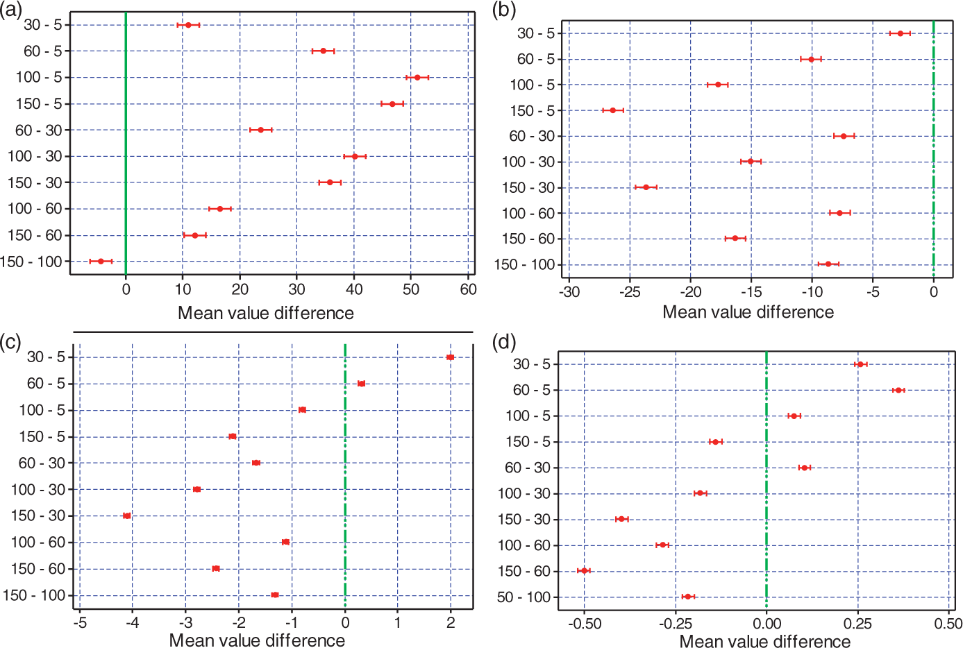

Regression analysis
Figure 4 shows the regression plot between each response variable and irradiation dose. R-Sq represents the percentage of the regression model error to the total error; the higher the R-Sq value, the better the regression model fits with the data. R-Sq (adj) represents the adjusted R-Sq; the closer the R-Sq and R-Sq (adj) values, the more reliable the regression model. P represents the significance level, and P values below 0.05 indicate a significant correlation. Equations (3), (4), (5), and (6) represent the regression relationships between the irradiation dose and each of the four response variables, where X and Y are the values of the irradiation dose and each response variable, respectively
Regression plot between response variables and irradiation dose: (a) gel content, (b) breaking strength, (c) elongation at break, and (d) creep rate.


According to the regression results, the R-Sq and R-Sq (adj) values for gel content, breaking strength, elongation at break, and creep rate are, respectively, 97.0 and 96.8%, 99.3 and 99.2%, 70.5 and 72.8%, and 81.1 and 79.4%, as shown in Figure 4(a) to (d). The differences between the two terms of each response variable are very small, at 0.2, 0.1, 2.2, and 1.7%. Furthermore, P = 0.000 (<0.05) for each response variable’s regression equation, indicating that the four regression models are reliable and the four regression equations are valid.
Fitting charts and regression equations show the linear regression relationship between irradiation dose and breaking strength and the quadratic regression relationships between irradiation dose and the other three response variables. Each regression model fits well with the measured data, such that it can accurately reflect the relationship between irradiation dose and the response variable. Equation (4) shows a negative correlation of −0.1884 between irradiation dose and breaking strength, indicating that the breaking strength decreased continuously as irradiation dose increased within the scope of the test. According to equations (3), (5), and (6), the gel content, elongation at break, and creep rate reached their maximum values of 67.781, 5.7559, and 1.1425%, respectively, at doses of 125, 42, and 65 kGy, respectively, which compared to their maximum values of 70.921, 6.752, and 1.228%, respectively, at doses of 125, 30, and 60 kGy, respectively, according to measured data. In order to determine the accuracy of the prediction model further, the following verification experiment should be carried out.
Regression verification
Level design table of irradiation dosage.

represents the gel content, elongation at breaking and creep rate reach their maximum at the maximum absolute value points of 125, 42 and 65 kGy according to equations (3), (5) and (6), respectively.
Figure 5 displays the regression validation diagrams for each response variable and irradiation dose. The diagrams separately depict the tendencies of both the fitting data and measured data for each response variable as the irradiation dose increases. The fitting curve and measured curve were very close for each response variable, producing similar changing trends with increasing irradiation dose; this consistency indicates that the verification results are within the range of normal prediction. In summary, the results of both regression analyses and verification experiments indicate that the four regression models are correct and the prediction results are credible. The reasonable range of irradiation dose is therefore 5–200 kGy. Given this finding, the effects of irradiation dose on UHMWPE fibers can be further researched within the reasonable dosage range.
Regression validation diagram between response variables and irradiation dose: (a) gel content, (b) breaking strength, (c) elongation at break, and (d) creep rate.
Effects of electron beam irradiation
Influence of irradiation dose on gel content
Figure 6 clearly illustrates the tendency of the gel content to change with increasing irradiation dose. The gel content increased rapidly and then decreased gradually as the irradiation dose increased. As shown in Figure 6, the curve can be divided into two parts. A significant increase in gel content is observed as the irradiation dose changes from 5 to 125 kGy, a range that can be considered to represent the first part of the curve. The maximum value of the gel content reaches 70.921% at the dose of 125 kGy, which can be called the “rapid increase area.” The gel content then decreases slightly to 43.496% as the dose rises from 125 to 200 kGy, a range that can be considered to represent the second part of the curve called the “postirradiation effect zone.”
Effects of irradiation dose on UHMWPE fibers. UHMWPE: ultra-high molecular weight polyethylene.
These changes can be explained by the theory that irradiation crosslinking and irradiation cracking exist at the same time during the irradiation process. The irradiation crosslinking reaction plays a leading role in the preirradiation process, and free radicals are generated by the introduction of TMPTMA. This significantly promotes the irradiation crosslinking of the UHMWPE fibers. As a result, the gel content increases rapidly in the “rapid increase area.”
With the irradiation dose increasing to a certain extent, the irradiation cracking leads to a sharp decrease in molecular weight, which has a dominant effect on the irradiation. Furthermore, TMPTMA has a much higher sensitivity to irradiation than UHMWPE fiber macromolecules, such that it basically becomes exhausted at low irradiation doses due to its self-polymerization coupled with limited content. Therefore, the TMPTMA’s effect of promoting crosslinking at high irradiation doses becomes gradually inconspicuous, with the result that the gel content mainly depended on the cracking to crosslinking ratio of the UHMWPE fibers at the moment. Therefore, the gel content shows a slight downward trend in the “postirradiation effect zone.”
Influence of irradiation dose on mechanical properties
Figure 6 also shows the relationships between the irradiation dose and the breaking strength and elongation at break, respectively, of the UHMWPE fibers. The results indicate that the breaking strength decreases drastically with increasing dosage, while the elongation at break reaches its maximum value at the dose of 30 kGy, then declines as the dose rises from 30 to 200 kGy. The breaking strength and elongation at break experience their minimum values simultaneously at the dose of 200 kGy.
UHMWPE fiber macromolecules have an ordered structure characterized by high crystallinity and high degree of orientation before irradiation. As the irradiation dose increases, the macromolecular chain scission of UHMWPE fibers also increases, giving rise to a sharp decline in molecular weight and leading to a substantial reduction in the breaking strength [23].
Within a certain range of irradiation dose, the presence of TMPTMA promotes the formation of crosslinks and reduces slips between molecular chains that lead to increased elongation at break. However, as the irradiation dose increases beyond a certain point, a large number of molecular chains have already broken, while at the same time the sensitization effect of TMPTMA decreases significantly as its content gradually decreases, resulting in a dramatic loss of elongation at break.
Influence of irradiation dose on creep rate
Figure 6 displays the changes in creep rate with irradiation dose as well. The creep rate increases first and then decreases as the irradiation dose rises from 5 to 200 kGy. It declines to 0.866% at the 5 kGy dose, compared to 1.461% before irradiation, and then reaches its maximum value of 1.228% when the dose rises to 60 kGy. It reaches its minimum value of 0.209% at the dose of 200 kGy.
When the irradiation dose is lower than 5 kGy, TMPTMA plays a significant role in promoting crosslinking due to its higher concentration. Crosslinking reactions occur mainly between TMPTMA and macromolecular chains of UHMWPE fibers, such that a rapid increase in three-dimensional network structures leads to a significant reduction in the creep rate. As the dose increases, three-dimensional network structures form between macromolecular chains at a reduced rate due to the decreasing TMPTMA content and its self-polymerization. Meanwhile, cracking reactions gradually replace crosslinking reactions, resulting in a slight increase in the creep rate. By the time the dose rises to 200 kGy, the TMPTMA has mostly been consumed, and the probability of crosslinking reactions between macromolecular chains and TMPTMA decreases accordingly. At the same time, a large number of molecular chain scissions occur, resulting in increased crosslinking points between the macromolecular chains. Therefore, crosslinking reactions between macromolecular UHMWPE chains increase, and intermolecular slippage becomes relatively ease. As a result, the creep rate exhibits a significant decrease.
SEM analysis
Figure 7 shows the surface morphology of UHMWPE fibers before and after different treatments. In Figure 7(a), many obvious dot embossments appeared on the untreated fibers’ surface, along with longitudinal grooves of different sizes with relatively smooth edges, probably due to the spinning process. In Figure 7(b), the surface smoothness, uniform thickness, and longitudinal deformation of the fibers pretreated with supercritical CO2 did not change significantly; however, the surface roughness of the irradiated fibers increased after supercritical CO2 pretreatment, and obvious grooves and cracks emerged. No significant differences could be observed clearly at doses of 5 and 150 kGy, as shown in Figure 7(c) and (d), respectively. These results indicate that supercritical CO2 pretreatment did not have a great impact on the surface morphology of UHMWPE fibers, but electron beam irradiation created a significant etching effect on the fibers’ surface, indirectly leading to the decrease of breaking strength to some extent.
SEM photos of UHMWPE fibers: (a) without any treatment, (b) supercritical CO2 treatment without irradiation, (c) irradiated at the dose of 5 kGy after supercritical CO2 treatment, and (d) irradiated at the dose of 150 kGy after supercritical CO2 treatment. SEM: scanning electron microscope; UHMWPE: ultra-high molecular weight polyethylene.
XRD analysis
Figure 8 shows the diffractogram for untreated and treated UHMWPE fibers. The XRD pattern reveals two strong diffraction peaks at 2θ around 21.6° and 24.1°, which are characterized by 110 and 200 crystallographic planes of the monoclinic phase, respectively [24]. It also shows no significant changes in the shapes of the diffraction curves, which suggests that supercritical CO2 pretreatment and electron beam irradiation at doses less than 150 kGy do not change the crystal form of UHMWPE fibers.
XRD patterns of UHMWPE fibers with different irradiation doses. UHMWPE: ultra-high molecular weight polyethylene; XRD: X-ray diffraction.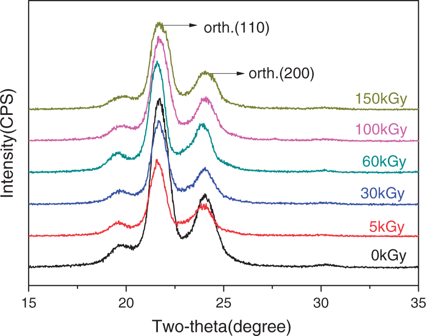
The crystallinity of UHMWPE fibers under different treatments.

UHMWPE: ultra-high molecular weight polyethylene.
XRD peak analysis results of UHMWPE fibers with different irradiation doses.

FWHM: full width half maximum; UHMWPE: ultra-high molecular weight polyethylene; XRD: X-ray diffraction.
One explanation for this phenomenon is that the introduction of TMPTMA improves the density of free radicals and increases the rate of crosslinking reactions between molecular chains during irradiation, thus restraining the recrystallization of macromolecular chains in the crystalline region. On the other hand, as the irradiation dose increases, the molecule chain packing density decreases, resulting in slight changes in interplanar spacing and crystallite dimension. These structural changes of fibers treated by the two methods led to the changes of breaking strength, elongation at break, and creep rate as well.
DSC analysis
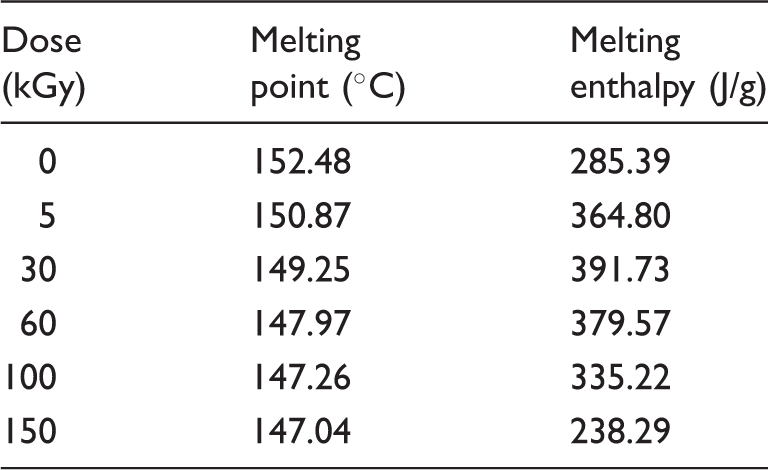
Figure 9 shows the DSC curves for untreated and treated UHMWPE fibers. In both Figure 9 and Table 5, the melting peaks move to the lower temperature region as the irradiation dose increases. At the same time, the melting enthalpies increase at first and then decrease. This occurs because irradiated crosslinking plays a dominant role in a certain dosage range, leading to a slight increase in the melting enthalpy values; however, irradiated cracking intensifies as the irradiation dose increases, resulting in declining melting enthalpy and decreasing melting point.
DSC curves of UHMWPE fibers with different irradiation doses. DSC: differential scanning calorimetry; UHMWPE: ultra-high molecular weight polyethylene. DSC data of UHMWPE fibers with different irradiation doses. DSC: differential scanning calorimetry; UHMWPE: ultra-high molecular weight polyethylene.
Discussion of sensitized irradiation crosslinking mechanism
Based on previous research [25–28], this study summarizes the sensitized irradiation crosslinking mechanism of UHMWPE fibers according to experimental observations and conclusions. The results explain the promoting effect of TMPTMA on the irradiation crosslinking of UHMWPE fibers through irradiation crosslinking reaction equations.
Typically, multifunctional monomers containing multiple C=C double bonds are more sensitive to high-energy radiation. During the irradiation process, they can increase the kinetic chain length of the crosslinking reaction and promote the formation of crosslinking networks in order to enhance the effect of irradiation crosslinking. However, multifunctional monomers in UHMWPE disperse in the form of clumps of aggregates because of their relatively poor compatibility with UHMWPE. At first, because of their self-polymerization, monomer aggregates form monomer polymers that contain a branched structure or a three-dimensional network structure during irradiation; meanwhile, free radicals are generated from the monomer aggregates and the adjacent macromolecular chains. At the beginning of irradiation, the number of free radicals is relatively low, but a series of chain reactions can easily be induced as long as a single free radical is present. Furthermore, the monomer aggregates inevitably come into contact with the macromolecular chains during the irradiation process, resulting in chain transfer that produces macromolecular free radicals, while C–C crosslinking bonds form among macromolecules coupling together. Ultimately, a crosslinking network forms between monomer polymers and macromolecules. This process can be represented as follows


Based on the above analysis of the sensitized irradiation crosslinking mechanism, this study introduced TMPTMA into UHMWPE fibers through supercritical CO2 pretreatment. The main crosslinking reactions under electron beam irradiation are shown in Figure 10.
Schematic illustration of irradiation crosslinking reactions.
Because the C=C double bond in TMPTMA is more sensitive to irradiation than the C–H single bond in UHMWPE, more free radicals are generated in the early irradiation process from the monomer aggregates. Therefore, the greater the probability of the chain transfer, the more crosslinking networks form between monomer polymers (TMPTMA) and macromolecules (UHMWPE). As the irradiation dose increases, the content of C=C double bonds contained in monomer aggregates of TMPTMA decreases gradually; at the same time, the TMPTMA becomes exhausted at low irradiation doses because of its self-polymerization and limited content. As a result, the TMPTMA’s effect of promoting crosslinking becomes gradually unnoticeable at high irradiation doses. As the irradiation dose increases further, the double bonds contained in TMPTMA monomers become depleted, especially on the surface of monomer aggregates, leaving the TMPTMA with no promoting effect on UHMWPE fibers. At that point, the crosslinking reactions become identical to those of crosslinking without monomer polymers. This process can be verified from the changed curve of the gel content of irradiated UHMWPE fibers, as shown in Figure 6.
Conclusion
In this study, TMPTMA was effectively introduced into UHMWPE fibers through supercritical CO2 pretreatment, providing a crosslinking bridge for electron beam irradiation and promoting crosslinking reactions. TMPTMA played an important role in promoting the effect of irradiated crosslinking.
Significant differences emerged in gel content, breaking strength, elongation at break, and creep rate among UHMWPE fibers at five different dose levels of irradiation. The regression models fit well with the measured data, as demonstrated by regression plots obtained through regression analysis, and the relationships between each variable and the irradiation dose were reflected accurately.
The performance of UHMWPE fibers changed to varying degrees after supercritical CO2 pretreatment and electron beam irradiation. As the irradiation dose increased, the gel content first rose, then declined; the breaking strength continuously decreased; the elongation at break increased at first and then decreased; and the creep rate originally decreased and then rose before finally declining slowly. The surface morphology of UHMWPE fibers changed obviously after irradiation. The crystal form did not change, but the interplanar spacing and crystallite dimension showed different degrees of change, and the crystallinity decreased slightly. The melting enthalpies increased at first and then decreased, while the melting point decreased slightly.
The promoting effect of multifunctional monomers on the irradiation crosslinking of UHMWPE fibers functions mainly through the transfer of macromolecular chains, which results in the formation of crosslinking networks between monomer polymers and macromolecules. This process describes the sensitized irradiation crosslinking mechanism of UHMWPE fibers in the presence of TMPTMA.
Footnotes
Declaration of Conflicting Interests
The author(s) declared no potential conflicts of interest with respect to the research, authorship, and/or publication of this article.
Funding
The author(s) received no financial support for the research, authorship, and/or publication of this article.