
Abstract
To enhance the anti-ultraviolent properties of technical jute fabrics, the enzymatic surface coating with the in-situ produced phenolic polymers of polyhydric phenols was investigated in this study. Firstly, the laccase-mediated polymerization of the five polyhydric phenols (catechol, resorcinol, hydroquinone, pyrogallol and phloroglucinol) was analyzed by FT-IR. Catechol and pyrogallol were polymerized together by laccase with ether bonds linked. On the contrary, the units of resorcinol, hydroquinone and phloroglucinol in their enzymatically formed polymers concatenated to each other by C-C bonds. Then, the coated jute fabrics were characterized in terms of X-ray photoelectron spectroscopy and scanning electron microscopy. The increasing of the C/O ratio on the jute fabric surface after the coating treatments supported the achievement of the enzymatic coating on jute fabrics via the in-situ polymerization of phenolic compounds and the grafting reaction of polyphenols with lignins on the surface. The sequence of the coating extent by using various polyhydric phenols was proved to be catechol, pyrogallol, resorcinol, phloroglucinol and hydroquinone in order from rich to poor according to the O-C-O component of cellulose in the C1s spectra of jute fabrics and the scanning electron microscopy photographs of jute surfaces. Lastly, the ultraviolent protection factor and the ultraviolent resistance of the coated jute fabrics were measured. The ultraviolent protective performance of jute fabrics after the coating treatments depended both on the coating amount and the chemical structure of the coated polymers. Among the tested polyhydric phenols, the polymerization of catechol obtained the best coating for ultraviolent protection. Different polyhydric phenols employed for the enzymatic coating showed different trends in ultraviolent protection factor of jute fabrics with the increasing of incubation time. The jute fabrics coated with in-situ-generated polycatechols or polyresorcinols had excellent ultraviolent resistances.
Introduction
In recent decades, the destruction of ozone layer in air due to wanton discharge of chlorofluorocarbons in production and living has gained wide concern. Nowadays, the surface of earth receives more ultraviolent (UV) rays than that in the past. As a consequence, human beings as well as lifeless substrates are suffering more challenges of UV radiation. Despite the elimination of air pollution has to be carried out, textiles with UV protective properties are also needed for preventing human beings or vulnerable materials from UV lights [1–6].
Jute fiber is a widely used textile material in home and industry [7–9]. Jute fabrics can be utilized in technical textiles outdoors, such as packing bags, ropes and textile coverings. The anti-UV performance of jute products can enhance their working life and maintain the mechanical properties for a longer time under sun light. In order to increase the UV resistance of technical jute textiles, chemical modifications including surface coating have been employed generally [10–13]. Nowadays, the use of enzymes as important biotechnological catalysts has emerged in the processing of lignocellulosic polymers [14–19]. Enzymatic processes have numerous advantages such as specificity, low cost to effectness, eco-friendly nature and working in mild conditions as compared to the conventional methods. Laccases (EC 1.10.3.2, benzenediol: oxygen oxidoreductase) are multi-copper-containing zymoproteins that catalyze the mono-electronic oxidation of phenols or aromatic amines to reactive radical species and simultaneously reduce molecular oxygen to water in a redox reaction [20]. Lignin is a three-dimensional aromatic polymer with three structural units including guaiacyl, syringyl and p-hydroxyphenyl jointed together in an irregular manner [21]. Researches show that lignin is a suitable substrate for laccase and the phenolic sites of lignin molecules can be oxidized to phenoxyl radicals by laccase [22,23]. With the laccase-catalyzed oxidation of lignin moieties rich on the surface, the lignocellulosic materials could be activated to create a radical-rich reactive surface to which oxidized (radical-containing) phenolic molecules by laccase simultaneously can be grafted [24–30]. Jute fiber with lignin content of 14%–20% also has the potential to be modified by this green biotechnology for endowing it with better performance or new functions.
According to the literature [31], phenolic compounds with low molecular weight could be polymerized to macromolecules by the oxidative catalysis of laccase. With the involvement of jute fabrics in the reaction, however, the coupling of the in-situ-produced polyphenol chains to lignin moieties on the jute surface or the initiation of the grafting copolymerization with phenolic monomers at the lignin-containing surface sites should occur. Simultaneously, the generated homopolymers could also deposit onto the jute surface and combine with the grafted polymers or jute surface by hydrogen bonds to form a new surface coating. Thus, this novel laccase-facilitated coating with the synergy of covalent coupling/grafting and non-covalent deposition will lead to strong binding bond onto the jute surface compared with conventional depositions [32]. Among the various phenolic compounds, polyhydric phenols such as catechol and pyrogallol have higher redox potentials for laccase and could be easily oxidized and polymerized with higher polymeric degree [33]. The corresponding polymerized coating which carries tremendous phenyl groups would show strong anti-UV properties and could further protect the jute fabrics from UV rays. However, as far as our knowledge goes, this kind of polyphenol-based UV protection of textiles has not been made before.
In this work, the laccase-mediated polymerization of five polyhydric phenols (catechol, resorcinol, hydroquinone, pyrogallol and phloroglucinol) and the in-situ coating with produced polyphenols for improving the UV protection of industrial jute fabrics were investigated, as presented in Figure 1. Initially, the chemical structures of the enzymatically produced phenolic polymers were analyzed by FT-IR in comparison with their corresponding phenolic monomers. Then, the surfaces of jute fabrics coated by various polyhydric phenols were characterized in terms of X-ray photoelectron spectroscopy (XPS) and scanning electron microscopy (SEM). Further, the ultraviolent protection factor (UPF) and the UV resistance of jute fabrics after the coating treatments with different incubation periods were determined.
Schematic illustration of the enzymatic coating of jute fabrics for enhancing anti-UV properties via in-situ polymerization of polyhydric phenols.
Materials and methods
Materials
Laccase from Trametes Versicolor was provided by Sigma-Aldrich. The 100% raw jute fabric (427 g/m2), with a 7/7 (warp/weft) cm–1 fabric density, was supplied by Longtai weaving Co., Ltd (Changshu, China). Five polyhydric phenols including catechol, resorcinol, hydroquinone, pyrogallol and phloroglucinol were all commercially available and were of analytical purity.
Laccase assay
The activity of laccase was determined using a UV/Vis spectrophotometer by monitoring the oxidation of 2, 2′-azino-bis-(3-ethylthiazoline-6-sulfonate) (ABTS; ɛ420 = 36,000 M−1 × cm−1) as substrate at 420 nm in 0.2 mM acetate buffer (pH 4) at 50℃. The enzyme activity was expressed in units defined as micromoles of ABTS oxidized per minute [34].
Enzymatic oxidative polymerization of polyhydric phenols
Each polyhydric phenol (catechol, resorcinol, hydroquinone, pyrogallol and phloroglucinol) of 40 mM was incubated in 0.2 M acetate buffer (pH 4) with 2.5 U/mL laccase. The reaction was carried out at 50℃ for 36 h in a shaking bath with a speed of 30 r/min. Then the reaction solution was centrifuged and the precipitates were washed with distilled water twice to obtain the polyphenols.
FT-IR analysis of polyphenols
The FT-IR analysis of the polyphenols obtained above was performed using a Nicolet iS10 FT-IR spectrometer (Thermo Fisher Scientific, USA) with the KBr pellet technique. The spectra were recorded in the range of 4000–650 cm−1 at 4 cm−1 resolution and 16 scans per sample.
Jute fabric preparation
The jute fabrics were Soxhlet-extracted with benzene/ethanol (v:v, 2:1) for 12 h to remove lipophilic extractives and then boiled with distilled water for 3 h to remove water-soluble fractions. As a result, the oxidation of substrate molecules by laccase and the analysis of modified fabrics could be carried out without interference.
Enzymatic coating of jute fabrics by in-situ polymerization
Jute fabrics of 1 g were incubated in 50 mL acetate buffer (pH 4, 0.2 M) with 2.5 U/mL laccase and 40 mM phenolic monomer at a shaking bath of 50℃ for 36 h. Afterwards, the fabrics were washed with distilled water at 50℃ for 20 min twice. Control sample and the laccase-treated sample followed the same treatment condition as mentioned above.
XPS analysis of jute fabrics
XPS experiments were carried out using an Amicus spectrophotometer (Kratos Analytical Ltd., UK) with Mg Kα radiation. The X-ray anode was run at 180 W and the high voltage was kept at 12.0 kV. The pass energy was fixed at 75 eV to ensure sufficient resolution and sensitivity. The base pressure of the analyzer chamber was about 1 × 10⁻⁶ Pa. The sample was directly pressed to a self-supported disk (10 × 10 mm) and mounted on a sample holder then transferred into the analyzer chamber. The whole spectra (0–1100 eV) and the narrow spectra of carbon, oxygen and nitrogen with much high resolution were both recorded. Binding energies were calibrated with the containment carbon (C1s = 284.6 eV). The spectrum analysis including background subtraction, deconvolution, integration and quantitation was carried out by using the XPS Peak4.1 software.
SEM analysis of jute fabrics
The jute fabric samples were scanned using a SU1510 SEM (Hitachi, Japan) under 5.00 k voltages at 1.00 k magnification.
UPF measurement of jute fabrics
The UPF of control, laccase-treated and various coated jute fabric samples with incubation time of 12 h, 24 h, 36 h, 48 h and 60 h was determined on a UV transmittance analyzer UV-1000F (Labsphere Co., USA). The detective wavelength ranged from 450 nm to 250 nm. For each fabric sample, four spots were measured and the results were averaged. For each treatment, quintuplicate samples were measured.
UV resistance measurement of jute fabrics
Jute samples incubated for 36 h were exposed to UV rays at 0.89 W/m2 for 8 h in an accelerated UV-aging tester QUV/SPRAY (Q-Lab Co., USA). Then the UPF of the irradiated fabrics was determined and compared with that of the samples before irradiation.
Results and discussion
FT-IR analysis of polyphenols
To estimate the structural characteristics of the five polyhydric phenols and their laccase-induced polymeric products, corresponding FT-IR spectra were measured and are presented in Figure 2. The peaks from 3500 to 3200 cm−1 in the IR spectra of all samples belonged to the phenolic O-H vibrations. Bands between 1650 and 1400 cm−1 represented the aromatic skeletal vibrations. The peaks at 1300–1000 cm−1 and 900–650 cm−1 were contributed to the C-O stretching vibrations and the C-H out-of-plane bending vibrations in aromatic rings, respectively. In contrast to the phenolic monomers, the phenolic O-H vibration peaks of polyphenols became broader due to the intramolecular hydrogen bonding between the repeating units in polyphenol chains [35]. In addition, the absorption bands of the polymeric structures between 1600 and 650 cm−1 were weakened, which can be ascribed to the more rigid groups in the polymers when compared to the monomer molecules [35]. Meanwhile, the steric hindrance of the macromolecular polymers produced by the laccase-mediated polymerization might also hinder the detection of these groups [36].
FT-IR spectra of the five polyhydric phenols and their corresponding polymers.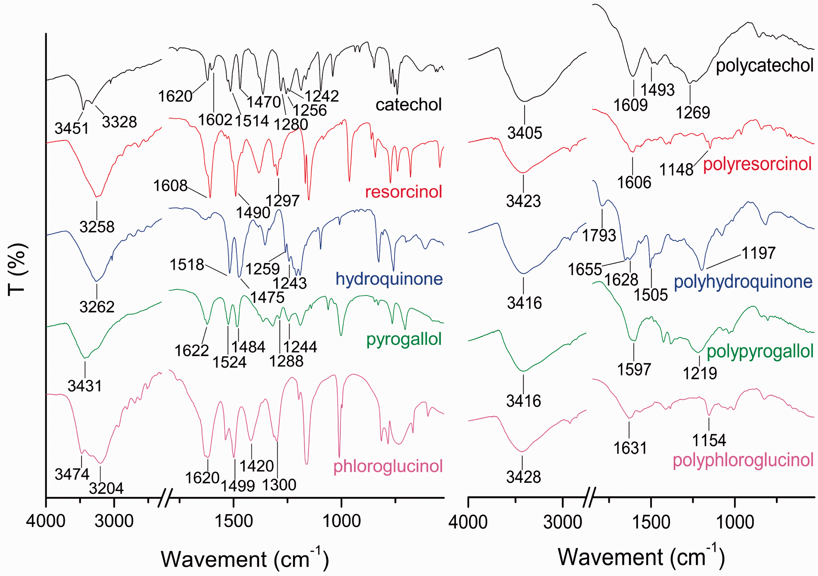
For catechol, the characteristic doublet peaks at 3451 cm−1 and 3328 cm−1 represented the vibrations of its phenolic ortho-hydroxyl structure. On the contrary, the phenolic O-H adsorption bands of polycatechol appeared as one broad peak at 3405 cm−1. This variation can be mainly explained that the hydroxyl groups in catechol monomers were involved into the laccase-mediated oxidative polymerization. Four peaks of catechol at 1620, 1602, 1514 and 1470 cm−1 belonged to the aromatic C = C vibration, while the peaks at 1609 cm−1 and 1493 cm−1 of polycatechol were due to the ortho-disubstitute benzene ring vibrations. In addition, the C-O stretching vibrations for catechol were observed at 1280, 1256 and 1242 cm−1, while the characteristic C-O-C adsorption peak of phenyl ether in polycatechol structure appeared at 1269 cm−1. The intense C-O-C bond absorption for phenyl ether indicated that catechol units in polycatechol structure jointed with each other by ether linkages [35]. Meanwhile, the broad phenolic O-H vibration suggested that there were still a large amount of phenolic O-H groups in polycatechol structure. The chemical structure of the enzymatically produced polycatechol was speculated and is presented in Figure 3(a).
The proposed chemical construction of the enzymatically produced phenolic polymers with catechol (a), resorcinol (b), hydroquinone (c), pyrogallol (d) and phloroglucinol (e), respectively.
For resorcinol, the adsorption bands at 3258 cm−1, 1608 and 1490 cm−1, 1297 cm−1 belonged to the phenolic O-H vibration, the aromatic C = C vibrations and the phenolic C-O vibration, respectively. In comparison, the above IR vibrations of polyresorcinol appeared at 3423, 1606 and 1148 cm−1, respectively. No remarkable vibration peaks of phenyl ether bond were observed in the IR spectrum of polyresorcinol. Moreover, the second and fourth aromatic carbons of resorcinol possess high reactivity and always act as initiating sites of corresponding chemical reactions. As supported above, C-C linkage was assumed the binding bond of resorcinol units in polyresorcinol macromolecules (shown in Figure 3b).
For hydroquinone, the peaks at 3262 cm−1, 1518 and 1475 cm−1, 1259 and 1243 cm−1 represented the phenolic O-H vibration, the C = C vibrations of aromatic rings and the phenolic C-O vibrations, respectively. By contrast, the above vibrations of polyhydroquinone emerged at 3416 cm−1, 1628 and 1505 cm−1, 1197 cm−1, respectively. The new bands at 1793 and 1655 cm−1 was corresponding to the C = O vibrations of quinones [31]. However, the C-O-C vibration bands of phenyl ether between 1300 and 1200 cm−1 were inconspicuous. As given above, it can be supposed that hydroquinone units in polyhydroquinone structure linked with each other by C-C bonds as shown in Figure 3(c).
For pyrogallol, the bands at 3431 cm−1, 1622, 1524 and 1484 cm−1, 1288 and 1244 cm−1 were ascribed to the phenolic O-H vibration, the aromatic skeletal vibrations and the phenolic C-O vibrations, respectively. In comparison, the phenolic O-H vibration and the aromatic skeletal vibration of polypyrogallol were observed at 3416 and 1597 cm−1, respectively. The characteristic C-O-C stretching vibration for phenyl ether was also seen at 1219 cm−1. The adsorption bands of phenolic hydroxyl group and the phenyl ether bond in polypyrogallol indicated that pyrogallol units in the produced polypyrogallol connected to each other by ether linkages [37] as shown in Figure 3(d).
For phloroglucinol, its characteristic phenolic O-H vibrations were at 3474 and 3204 cm−1. The peaks at 1620, 1499 and 1420 cm−1 were attributed to the aromatic skeletal vibrations and the peak at 1300 cm−1 was due to the phenolic C-O vibration. By contrast, the above three adsorptions in polyphloroglucinol appeared at 3428, 1631 and 1154 cm−1, respectively. There was no distinct vibration peak from 1300 to 1200 cm−1 representing the C-O-C stretching vibration of phenyl ether. As suggested above, it can be hypothesized that phloroglucinol monomers concatenated to each other by C-C linkages after the laccase-mediated oxidative polymerization as shown in Figure 3(e).
XPS analysis of jute fabrics
Surface chemical compositions of various jute fabrics obtained by X-ray photoelectron spectroscopy XPS) analysis.

The increase in the nitrogen content of the laccase-treated jute fabric could be attributed to some adsorbed laccase proteins. The nitrogen content of the coated jute fabrics could be the combined contribution of surface coating with nitrogen-free polyphenols and enzyme adsorption. The more the coated polymers on the jute surface, the lower the nitrogen content of jute fabrics is. And the more the adsorbed laccase proteins, the higher the nitrogen content is. The adsorption of laccase proteins on the coated jute surface partly depended on the hydroxyl content of the new surface which is in accordance with the amount and structural property of each coated polyphenol. For catechol, pyrogallol, resorcinol and hydroquinone, the nitrogen contents of jute fabrics coated with their corresponding polymers were lower than that of the control sample, which indicated their surface coating is dominating in the contribution of the nitrogen content. For phloroglucinol, the nitrogen content of the coated jute fabric was higher than that of the laccase-treated sample since the enzyme adsorption took place of the surface coating to be dominant.
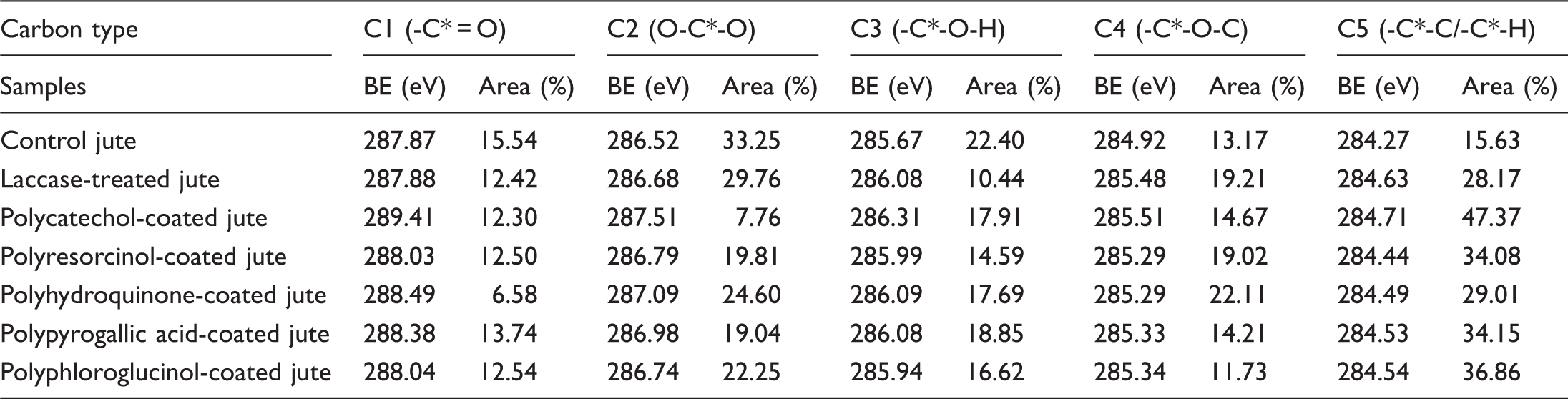
In order to obtain the chemical bonding state of carbon atom on the jute fabric surface, the deconvolution of C1s high-resolution spectra was carried out by curve fitting. As shown in Figure 4, the C1s spectra were deconvoluted into five peaks of C1, C2, C3, C4 and C5. According to the literature [38], the C5 peak with the lowest binding energy originates from a carbon atom bound only to carbon or hydrogen atoms (-C-C/-C-H). The C4 peak represents a carbon atom linked to an ether oxygen atom (-C-O-C). The C3 peak corresponds to a carbon atom bound to a hydroxyl oxygen atom (-C-O-H). The C2 peak represents a carbon atom linked to two non-carbonyl oxygen atoms (O-C-O) which only existed in the cellulose of jute fiber. The C1 peak with the highest binding energy represents a carbon atom bound to one carbonyl oxygen atom (-C = O) in lignin or pectin. The proportions and the peak assignments of these chemical bonds were presented in Table 2. The laccase-treated jute fabric showed decreasing of C3 content and increasing of C4 and C5 contents, which suggested the transformation of phenolic -OH group to phenyl ether and the formation of C-C bond between two aromatic rings after the laccase-mediated oxidation of lignin on the jute fabric surface. After the coating treatments, the C2 contents of the jute fabrics were decreased for the covering of cellulose component on the surface with polyphenols. The more the coated polymers on the jute surface, the lower is the C2 content of C1s spectra. Consequently, the catechol monomer obtained the most coating effect on jute fabrics via the laccase-mediated in-situ oxidative polymerization. The next is pyrogallol, resorcinol, phloroglucinol and hydroquinone in sequence. The C3 and C5 contents of the coated jute fabrics were increased when compared with the laccase-treated jute fabrics as a result of the introduction of polyphenols on surfaces. The C4 proportions of the coated jute fabrics were various. The variations of the C3, C4 and C5 contents on the jute surface all depended on the coated amount and the structural properties of the adsorbed polyphenols and the grafted ones by condensation with lignin.
Deconvoluted C1s spectra of control jute (a), laccase-treated jute (b), polycatechol-coated jute (c), polyresorcinol-coated jute (d), polyhydroquinone-coated jute (e), polypyrogallol-coated jute (f) and polyphloroglucinol-coated jute (g). Proportions and assignments of deconvoluted C1s peaks on the surface of various jute samples.
SEM analysis of jute fabric surfaces
The surface morphology of control, laccase-treated and polyphenol-coated jute fabrics was investigated by SEM and is shown in Figure 5. The surface of the control jute was neatly arranged with some natural impurities covered (Figure 5a). After the laccase treatment the jute surface became smooth and glossy (Figure 5b), which is probably owing to the enzymatic dislodgement and redistribution of bulgy lignins on the surface as a result of the laccase-mediated simultaneous degradation and polymerization of lignin [39]. The jute surfaces coated with different polyphenols were rough and irregular with some particulates or flakes contained as shown in Figure 5(c) to (g), respectively. These materials on the enzymatically coated jute surface were considered the homopolymers of the five polyhydric phenols and attached by way of covalent grafting and non-specific hydrogen-bonding absorption. Besides, the degree of the enzymatic coating was associated with the monomers used. Jute fabrics coated by the laccase-mediated in-situ polymerization of catechol and resorcinol showed more amount of surface coating than those of polypyrogallol, phloroglucinol and hydroquinone, which is approximately in accordance with the conclusion supported by the C2 data of XPS in Table 2.
SEM images of control jute fabric (a), laccase-treated jute fabric (b), polycatechol-coated jute (c), polyresorcinol-coated jute (d), polyhydroquinone-coated jute (e), polypyrogallol-coated jute (f) and polyphloroglucinol-coated jute (g) amplified at 1.00 k.
UV-protective properties of jute fabrics
The effects of the coating treatments with different polyhydric phenols and the incubation time on the UPF of jute fabrics were investigated as shown in Figure 6. After various coating treatments, the UPF values of jute fabrics were increased with different degrees. The increasing extent in UPF of coated jute fabrics possibly depended not only on the coating amount but also on the chemical structure of the coated polymers. The polymeric structure was bound up with the polymerization characteristics of the phenolic monomers. The coating amount of phenolic polymers on jute fabric surface was affected by the incubation time and the selected phenol. The five polyhydric phenols tested for the enzymatic coating showed different trends in UPF of jute fabrics with the increasing of incubation time. In the enzymatic coating reaction, polymerization and depolymerization of polyhydric phenols occurred simultaneously. At the beginning, the phenolic monomers were polymerized dramatically under the catalysis of laccase. The coating resulted from the covalent coupling/grafting and the non-covalent deposition was also enhanced. In the latter stage of the reaction, the laccase-mediated depolymerization became predominant. The coated polymers started to degrade into lower-molecular-weight pieces and fall back to the aqueous medium [39]. The incubation time for different polyhydric phenols to reach their maximum polymerization degree was various. The coating with polycatechol obtained the best UV-protective performance and got its maximum UPF at the incubation time of 48 h. The others were polypyrogallol, polyresorcinol, polyhydroquinone and polyphloroglucinol in order from massive to poor. And their incubation periods for the optimal UPF of jute fabrics were 48 h, 36 h, 24 h and 36 h, respectively.
The UPF values of control, laccase-treated and enzymatically coated jute fabrics with various polyhydric phenols in different incubation periods.
UV resistance of jute fabrics
As shown in Figure 7, the UV resistance of jute fabrics after various coating treatments was studied as compared with the laccase-treated and control samples. The jute fabrics coated with polycatechol and polyresorcinol exhibited strong UV resistance with no UPF dropped after radiation. However, the UPF values of jute fabrics coated with polyhydroquinone, polypyrogallol and polyphloroglucinol after radiation were declined by 22.35%, 24.18% and 33.40%, respectively. These results indicated that the laccase-induced polymeric structures of catechol and resorcinol in jute fabric coatings were more tolerant for suffering the radiation of UV rays.
The UPF comparison of various jute fabric samples before and after radiation.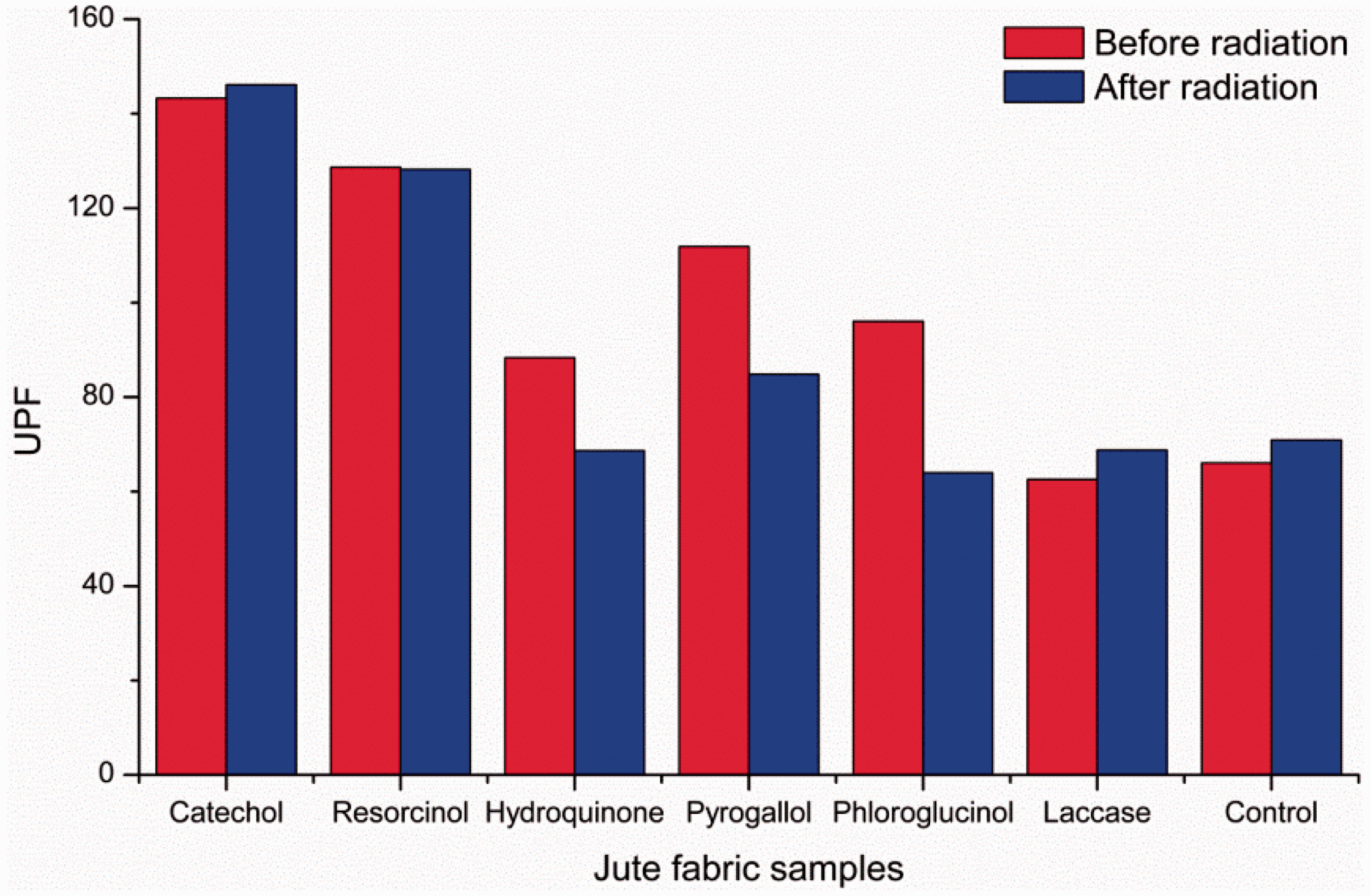
Conclusions
The present work demonstrated that the enzymatic coating via the in-situ polymerization of polyhydric phenols was achieved for enhancing the UV protective properties of technical jute fabrics. The units in the polymer of catechol or pyrogallol were connected to each other by ether bonds. However, monomers of resorcinol, hydroquinone and phloroglucinol were polymerized, respectively, by laccase with C-C bonds jointed together. The enzymatic coatings of jute fabrics with these polyhydric phenols were confirmed by XPS and SEM. The grafting reaction of polyphenols to lignins on the jute surface was proved to be actually existed and the sequence of the coating amounts varied from different polyhydric phenols. The UV protective performance of jute fabrics after the coating of polyphenols was increased in variety, which depended on the coating amount and the chemical structure of the coated phenolic polymers. The enzymatic coating polymerized with catechol obtained the best anti-UV properties. Besides, different polyhydric phenols used in the enzymatic coating showed various trends in UPF of jute fabrics with the increasing of incubation time. The jute fabrics coated with in-situ generated polycatechols or polyresorcinols presented almost no decrease in UPF after radiation, showing excellent UV aging resistance. The enzymatically coated jute fabrics which were UV-resistant could be utilized in technical textiles outdoors, such as packing bags, ropes and textile coverings to enhance their working life and maintain the mechanical properties for a longer time under sunlight.
Footnotes
Declaration of Conflicting Interests
The author(s) declared no potential conflicts of interest with respect to the research, authorship, and/or publication of this article.
Funding
The author(s) disclosed receipt of the following financial support for the research, authorship, and/or publication of this article: This work was financially supported by National Natural Science Foundation of China (51173071), Program for New Century Excellent Talents in University (NCET-12-0883), Program for Changjiang Scholars and Innovative Research Team in University (IRT1135), Fundamental Research Funds for the Central Universities (JUSRP51312B), the Graduate Student Innovation Plan of Jiangsu Province of China (CXZZ13_0752) and the Doctor Candidate Foundation of Jiangnan University of China (JUDCF13023).