
Abstract
HSV-1 is associated with oral lesions. Recently, anti-herpetic activity of different plant species has been investigated. In this study, the effects of Artemisia aucheri aqueous extract on the HSV-1 virus-infected Vero cells were assessed. The highest cell viability occurred in plant aqueous extracts was with a concentration of 75 μg/mL, 1–2 h before viral infection. The IC50 of the aqueous extract of 24.7 μg/ml was calculated. Most percentage of infected cell inhibition (89.6%) was with the chloroform fraction in concentration of 75 μg/ml, and the least percentage of infected cell inhibition (21.7%) was in concentration of 12.5 μg/ml with the ethyl acetate fraction in comparison with untreated control. Moreover, Q-PCR results revealed that the expression of genes UL46 and US6 were significantly reduced in the presence of different treatments utilized in the experiment. In conclusion, the present study proposes that aqueous extracts of medicinal plant Artemisia aucheri have anti-viral property and may be considered as a remedy for HSV-1 treatment.
Introduction
Human alphaherpesviruses are among a group of viruses that produce viral infections in the majority of humans. Human alphaherpesviruses comprise three members, herpes simplex virus (HSV) 1 and 2 and varicella zoster virus (VZV). HSV-1 is transmitted by contact with an infected person who has reactivations of the virus. HSV-1 is typically associated with oral lesions. [1]. HSV-1 contain at least 74 genes (or open reading frames, ORFs) within their genomes, although speculation over gene crowding allows as many as 84 unique protein coding genes by 94 putative ORFs. These genes encode a variety of proteins involved in forming the capsid, tegument, and envelope of the virus, as well as controlling the replication and infectivity of the virus [1]. The complete 152-kbp genomic DNA sequence of HSV-1 was published in 1988 [2]. In the whole genome of HSV-1, they recognized 72 genes which encode 70 distinct proteins. This genome contains two extended regions of unique sequence (UL contains 56 viral genes and US contains 12 viral genes), each of which is bounded by a pair of inverted repeat elements (TRL-IRL and IRS-TRS).
Treatment against these viruses usually involves general-purpose antiviral drugs that interfere with viral replication, reduce the physical severity of outbreak-associated lesions, and lower the chance of transmission to others [3]. It has been reported that the daily use of antivirals such as acyclovir and valacyclovir can reduce reactivation rates. However, the extensive use of anti-herpetic drugs has led to the development of drug resistance, which in turn leads to treatment failure [4]. For this reason, anti-herpetic activity of different plant species has been investigated [5–7].
Artemisia aucheri Boiss is a medicinal and aromatic plant that belongs to Asteraceae family. There are around 500 species of Artemisia in Asia, Europe, and North America and 34 species of this family are found all over Iran [8]. Artemisia aucheri is limited mostly to mountainous landscapes with high slope and sandy soils. This species is native in Iran and its distribution extends to West Tibet and West Himalaya. This plant has many medicinal properties and is useful in traditional medicines for the treatment of some diseases. Artemisia aucheri extraction has cytotoxic effects in cancer treatment; it has anti leishmanial effects, and essential oil of A. aucheri seed and aerial parts acts against Escherichia coli, Staphylococcus aureus, and Listeria monocytogenes [9, 10]. Similarly, A. aucheri is well known for its medicinal uses and food value, and also used for ornamental purpose and as a soil stabilizer in disturbed habitats. The chemical constituents of this plant species has been reported as anti-viral compounds against herpes simplex viruses [5].
Having said about different medicinal uses of this plant species, we carried out the present study with the aim to investigate the effect of A. aucheri aqueous extract on the HSV-1 virus-infected Vero cells derived from the kidney of an African green monkey.
Results
Counting Vero cells in infection with HSV-1 virus and aqueous extract of plant
Trypan blue exclusion test of cell counts revealed about 3–4 million cells in the 25 cm2 flask, that after viral infection, the cytopathic effect (CPE) was completed after 4 days. Counting the cells in time intervals of 24, 48, and 72 h, revealed that the cell viability is different by the viral infection time and plant aqueous concentrations (25, 50, 75, and 100 μg/mL). The lowest the cell viability occurred in 25 μg/mL concentration of plant aqueous extract in 1–2 h after viral infection, while the highest the cell viability occurred in plant aqueous extracts concentration of 75 μg/mL, in 1–2 h before viral infection.
IC50 result
Measurements of the aqueous extract’s and aqueous fraction’s effect
For obtaining the IC50 of aqueous extract and aqueous fractions (ethyl acetate, chloroform, and petroleum ether) by MTT Assay, 3 × 10⁵ Vero cell/ml with aqueous extract different concentrations (10, 25, 50, and 100 μg/ml) has been cultured in a 96-well plate culture (each concentration has been repeated 3 times). At the end, the IC50s were calculated by using GraphPad Prism8 (San Diego, USA).
The IC50 of the aqueous extract (Aq-E = 24.715 ± 1.162 μg/ml) and fractions including: the IC50 of the petroleum ether fraction (Ether-F = 18.047 ± 0.794 μg/ml), the chloroform fraction (Chloro-F=16.638 ± 0.110 μg/ml), the ethyl acetate fraction (Ethyl-F = 45.512 ± 4.068 μg/ml), and the IC50 of the acyclovir as controls (ACV=15.071 ± 0.078 μg/ml) was calculated.
Most percentage of infected cell inhibition (89.6%) was with the chloroform fraction in concentration of 75 μg/ml and the least percentage of infected cell inhibition (21.7%) was in concentration of 12.5 μg/ml with the ethyl acetate fraction in compared with untreated control (virus) (Figure 1). Aqueous extract’s and aqueous fraction’s preventing percentage on HSV-1 virus was significant (p<0.01). The pairwise comparison between the groups was also significant (p <0.01) and the chloroform fraction with the lowest concentration of IC50 = 16.63 μg/ml had the greatest effect on virus titer that was comparable to acyclovir in virus inhibition (Figure 2). Percentage of infected cells inhibition after infection with HSV-1 virus and in the presence of different concentrations of aqueous extract (Aq-E) ethyl acetate fraction (Ethyl-F), chloroform fraction (Chloro-F) and petroleum ether fraction (Ether-F) in comparison to control group (virus). The most percentage of infected cells inhibition (89.6%) was with the chloroform fraction and the least percentage of infected cells inhibition (21.7%) was with the ethyl acetate fraction in comparison with untreated control (virus). The values of IC50 of aqueous extract, petroleum ether fraction, chloroform fraction, and the ethyl acetate fraction on HSV-1 virus using MTT Assay. Values had difference significantly from each other according to GraphPad Prism8. The effect of aqueous extracts and aqueous fractions in inhibition of HSV-1 virus titer was significant (p<0.01). The chloroform fraction with the lowest concentration of IC50 = 16.63 μg/ml had the greatest effect on virus titer that was comparable to acyclovir in virus inhibition.

Q-PCR results
Reduction of gene expression of UL46 of HSV-1 virus in different treatments used.

Treatments that were significant in the table with asterisk have been shown. The highest rate of reduction of gene expression of UL46 of HSV-1 virus was observed at the same time with chloroform fraction.
Ethyl acetate 4 h after infection 3.668, 0.003*.
Reduction of gene expression of US6 of HSV-1 virus in different treatments used.

In all treatments, reduction of gene expression of US6 of HSV-1 with aqueous extract of A. aucheri, petroleum ether, and chloroform and ethyl acetate fractions was significant. The highest of the fold changes in 4 h after infection with total extract and chloroform fraction were observed.
In general, the present study revealed that aqueous extract of A. aucheri significantly reduce the titer of virus and the expression level of the genes UL46 and US6, in vero cells inoculated with HSV-1 virus at MOI = 0.1. Moreover, some of the treatments used like, total plant aqueous extract, as well as chloroform fractions, have a higher level of effect on reducing virus expression (p = 0.01).
Discussion
The present study revealed that aqueous extract of A. aucheri can reduce significantly the HSV-1 virus titration and the virus genes expression. This clearly adds up to the literature, that shows medicinal plants’ extracts have anti-viral properties. For example, El-Toumy evaluated the anti-herpetic activity against herpes simplex virus type 1 (HSV-1) in 25 Egyptian plants extracts [7]. They carried out experiment in vitro and on Vero cell lines by cell viability and reported that extracts of two plant species, namely, Euphorbia cooperi N.E.Br. Ex A. Berger (Euphorbiaceae) and Morus alba L. (Moraceae) showed potential anti-herpetic activity. These extracts contained pure flavonoid compounds; namely, 7-galloyl catechin, gallic acid, kaempferol 3-O-β-(6″-O-galloyl)-glucopyranoside, quercetin 3-O-β-(6″-O-galloyl)-glucopyranoside, curcumin, quercetin, and kaempferol which exhibited significant inhibition. Similarly, Parsania reported that the plant extracts of Mentha pulegium L. (the mint family) has antiviral activity, and that direct treatment of HSV-1 with the plant extract resulted in 1.7 log10 TCID50 reduction in virus titers after 1 hour compared to the control [6].
Khan et al. [5] reviewed anti-HSV substances from natural sources, including both extracts and pure compounds from herbal medicines, and reported that traditional medicines, like Ayurvedic, traditional Chinese (TCM), Chakma medicines, are good and potential sources for promising anti-HSV drugs. They also found that phenolics, polyphenols, terpenes (e.g., mono-, di-, tri-), flavonoids, sugar-containing compounds, are promising anti-herpetic agents, and concluded that natural products from medicinal plant extracts are very important source of anti-HSV agents. Moreover, Yang [11] investigated geraniin and 1, 3, 4, 6-tetra-O-galloyl-beta-D-glucose (1346TOGDG), isolated from the acetone extract of Phyllanthus urinaria L., were examined for their activity against HSV-1 and HSV-2 in vitro. They reported that geraniin actively suppressed HSV-2 infection, whereas 1346TOGDG effectively inhibited HSV-1 infection.
Experimental section
Plant material
We used A. aucheri Boiss of the genus Artemisia L. that contains many species with medicinal properties [12]. Fifty grams of aerial parts of A. aucheri were collected from Semnan to Shahroud regions in Semnan province and dried in the shade. The species of these collected plants were approved by Forests, Range and Watershed Management Organization, the Ministry of Jihad-Agriculture of Iran.
Aqueous extract and fractions
The plant was soaked in 500 mL of distilled water for 2 days, then gently heated for half an hour to reach the boiling point (but did not bubble). Then, the solution was filtered with a filter paper and placed under a hood to dry. It was collected by scratching in a pre-weighed container and packed with paraffin and stored at −20°C.
We used aqueous soluble extracts of A. aucheri. The plant extract was dissolved in distilled water. This solution was passed through a decanting funnel, after 1/2 h, to which polar as well as non-polar solvents were added. The most non-polar fraction of plant extracts is Ether de pétrole (Petroleum ether), (Merck, Germany), which was removed from the extract after slight agitation. We repeated this procedure three times and finally the fraction isolated was dried under a chemical hood and measured accordingly. The rest of the extract was collected in the next stage of chloroform (Merck,Germany) solvent extraction similar to the above method, but this time, the chloroform solvent was placed in the lower part (lower phase). Finally, the ethyl acetate extract (polar solvents) (Merck, Germany) was collected with all the mentioned steps in three stages. The final step was freeze drying at −20°C [13].
Treatments
We used acyclovir 50 μg/mL (ACV), (Exir Pharmaceutical, Iran.) as drug control as well as A. aucheri aqueous extract, along with fractions of 25, 50, 75, and 100 μg/mL, at the time of virus infection and 4 h after virus infection.
Cell culture
Vero cells derived from the kidney of an African green monkey (University Jihad, Iran) were cultured in DMEM (Dulbecco’s Modified Eagle Medium) with high glucose (GIBCO Germany), 1% penicillin/streptomycin (GIBCO Germany), and kept in CO2 incubator at 37°C. When the cell density reached over 90%, they were treated with trypsin–EDTA (0.25%) solution. These cells were stained with trypan blue dye (10%), and the number and percentage of vital cells were determined. Then, about 105 cells/ml cells were transferred to new culture flask.
Virus multiplication
Vero cells were infected with HSV-1 virus at MOI 1:10 and kept in CO2 incubator for 24 h and after the appearance of cytopathogenic effect (CPE), they were stored at freezer at −80°C.
Determination of titer of virus
The cells were cultured in a 96-well cell culture plate. We prepared 10 logarithmic dilution of the virus stock. Each viral dilution, Vero cells as cell control, as well as virus control, in four replications was used. A 100 μL of viral dilutions was added to each replicate and after 1 h, 200 mL of medium containing 5% FBS was added. The CPE was daily observed by inverted microscope and finally the fifty-percent tissue culture infective dose (TCID50) of virus was determined by the Reed Muench method [14].
Aqueous extracts effect on virus
In order to determine the effect of the IC50 of aqueous extract and aqueous fractions with concentrations (25, 50, 75, and 100 μg/mL) on virus, the Vero cells were cultured on 96-well cell culture plates. About 200 μL of Vero cells (1 × 10⁵ cell/ml) were placed in the wells and were incubated at 37°C for 24 h. Then, the cell culture was infected with virus at (MOI = 0.1), and incubated for 24 h, to which 10 μL of MTT was added and incubated at 37°C in 5% CO2 for 3–4 h. Then, we added 100 μl of the MTT solvent to each well and covered the wells with aluminum foil. Next, it was placed on a shaker for 15 min to dissolve the sediment particles. Absorption was then measured at 570 nm [15] and the IC50s were calculated by using GraphPad Prism8.
Q-PCR of the genes UL46 and US6 (gD)
We used the FavorPrep™ Tissue Total RNA Mini Kit for RNA extraction. The Vero cells were inoculated with HSV-1 and plant aqueous extract with fraction concentration of 75 μg/mL. The RNA concentration was determined by NanoDrop.
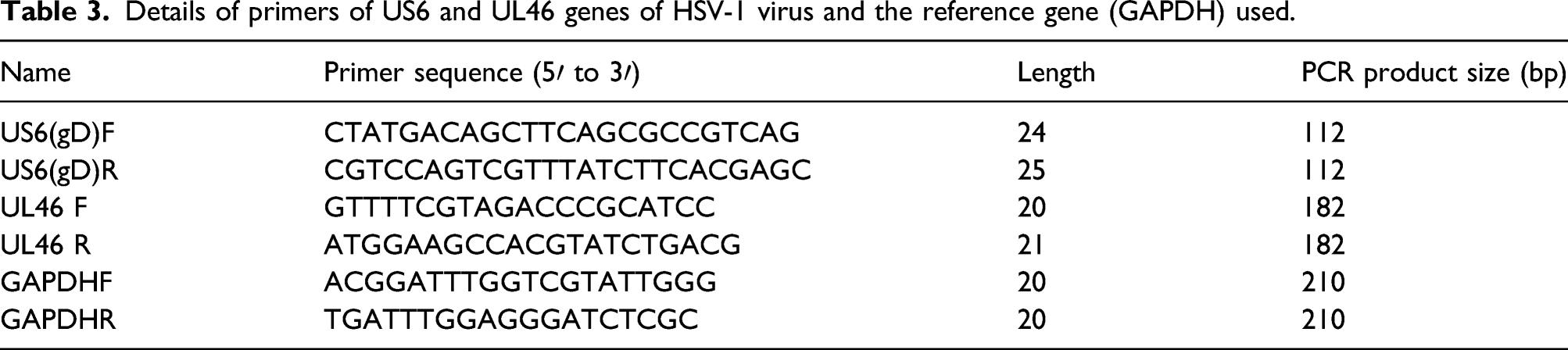
Details of primers of US6 and UL46 genes of HSV-1 virus and the reference gene (GAPDH) used.
Conclusion
In conclusion, the present study proposes that aqueous extracts of medicinal plant A. aucheri have anti-viral property and may be considered as a remedy for HSV-1 treatment.
Footnotes
Acknowledgements
We would like to thank the financial support of the Islamic Azad University and Blood Transfusion Research Center, High Institute for Research and Education in Transfusion Medicine.
Declaration of conflicting interests
The author(s) declared no potential conflicts of interest with respect to the research, authorship, and/or publication of this article.
Funding
The author(s) received no financial support for the research, authorship, and/or publication of this article.