
Abstract
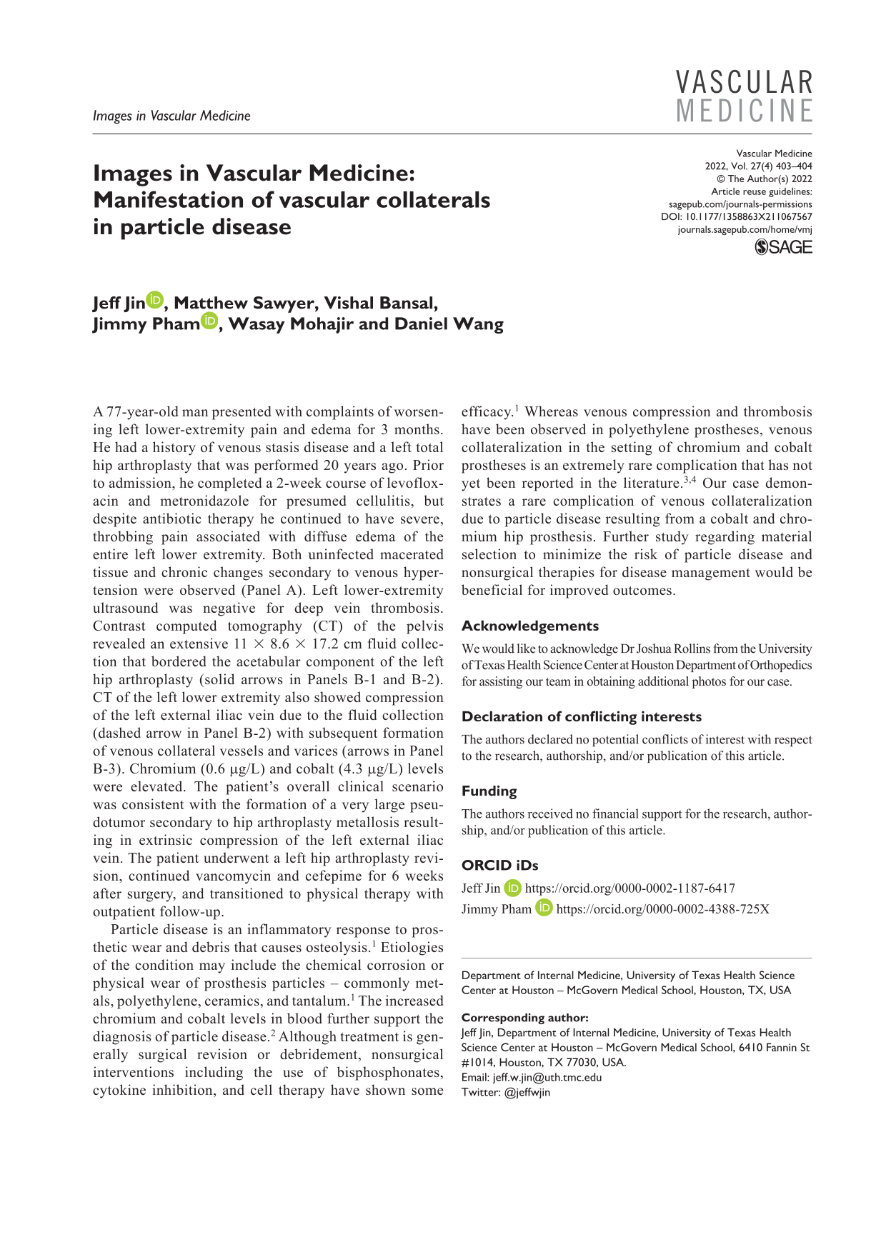
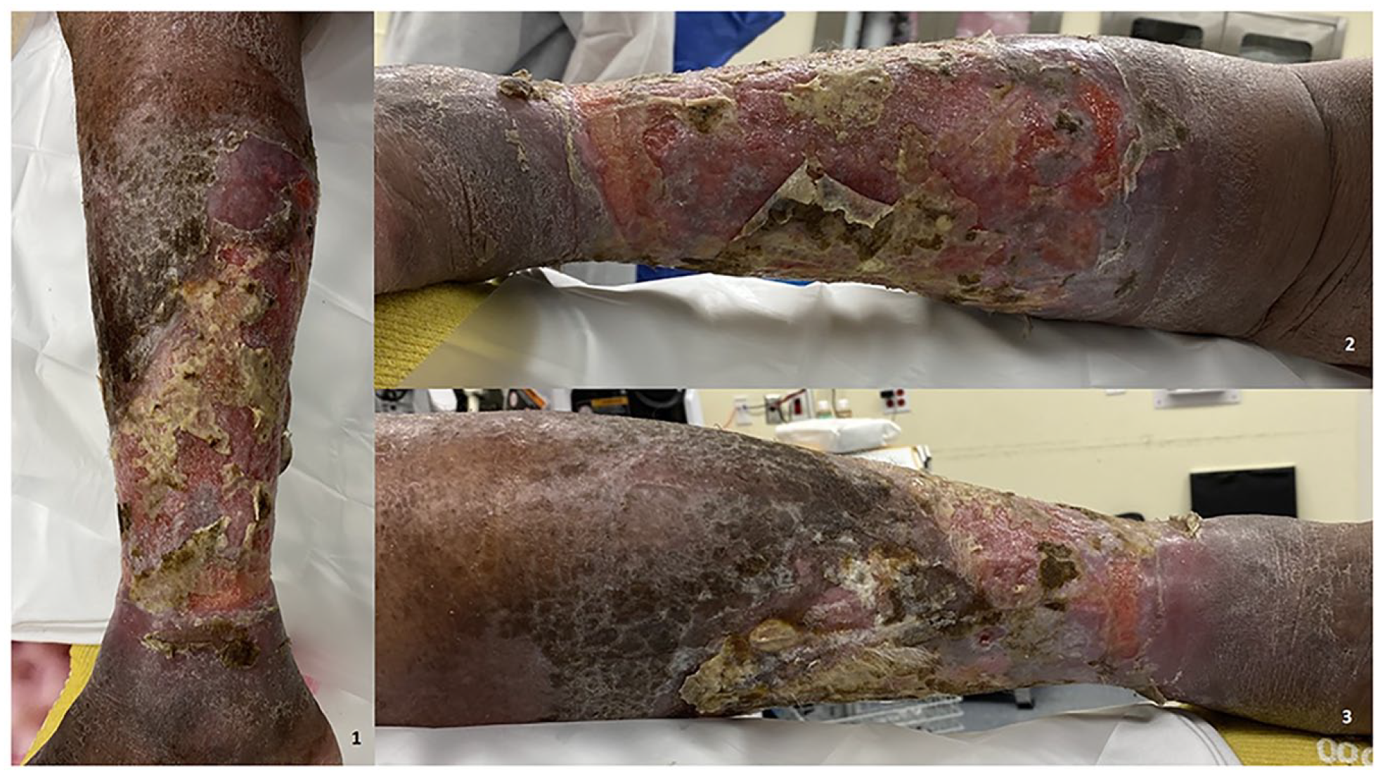
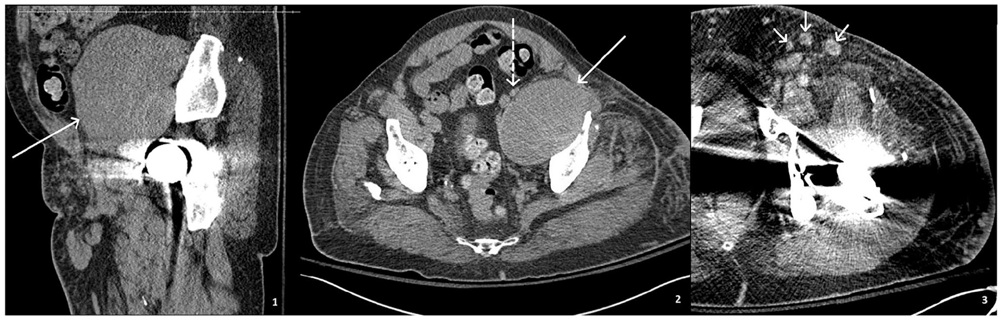
A 77-year-old man presented with complaints of worsening left lower-extremity pain and edema for 3 months. He had a history of venous stasis disease and a left total hip arthroplasty that was performed 20 years ago. Prior to admission, he completed a 2-week course of levofloxacin and metronidazole for presumed cellulitis, but despite antibiotic therapy he continued to have severe, throbbing pain associated with diffuse edema of the entire left lower extremity. Both uninfected macerated tissue and chronic changes secondary to venous hypertension were observed (Panel A). Left lower-extremity ultrasound was negative for deep vein thrombosis. Contrast computed tomography (CT) of the pelvis revealed an extensive 11 × 8.6 × 17.2 cm fluid collection that bordered the acetabular component of the left hip arthroplasty (solid arrows in Panels B-1 and B-2). CT of the left lower extremity also showed compression of the left external iliac vein due to the fluid collection (dashed arrow in Panel B-2) with subsequent formation of venous collateral vessels and varices (arrows in Panel B-3). Chromium (0.6 μg/L) and cobalt (4.3 μg/L) levels were elevated. The patient’s overall clinical scenario was consistent with the formation of a very large pseudotumor secondary to hip arthroplasty metallosis resulting in extrinsic compression of the left external iliac vein. The patient underwent a left hip arthroplasty revision, continued vancomycin and cefepime for 6 weeks after surgery, and transitioned to physical therapy with outpatient follow-up.
Particle disease is an inflammatory response to prosthetic wear and debris that causes osteolysis. 1 Etiologies of the condition may include the chemical corrosion or physical wear of prosthesis particles – commonly metals, polyethylene, ceramics, and tantalum. 1 The increased chromium and cobalt levels in blood further support the diagnosis of particle disease. 2 Although treatment is generally surgical revision or debridement, nonsurgical interventions including the use of bisphosphonates, cytokine inhibition, and cell therapy have shown some efficacy. 1 Whereas venous compression and thrombosis have been observed in polyethylene prostheses, venous collateralization in the setting of chromium and cobalt prostheses is an extremely rare complication that has not yet been reported in the literature.3,4 Our case demonstrates a rare complication of venous collateralization due to particle disease resulting from a cobalt and chromium hip prosthesis. Further study regarding material selection to minimize the risk of particle disease and nonsurgical therapies for disease management would be beneficial for improved outcomes.
‘Images in vascular medicine’ is a regular feature of Vascular Medicine. Readers may submit original, unpublished images related to clinical vascular medicine. Submissions may be sent to: Heather Gornik, Editor in Chief, Vascular Medicine, via the web-based submission system at http://mc.manuscriptcentral.com/vascular-medicine
Footnotes
Acknowledgements
We would like to acknowledge Dr Joshua Rollins from the University of Texas Health Science Center at Houston Department of Orthopedics for assisting our team in obtaining additional photos for our case.
Declaration of conflicting interests
The authors declared no potential conflicts of interest with respect to the research, authorship, and/or publication of this article.
Funding
The authors received no financial support for the research, authorship, and/or publication of this article.