
Abstract

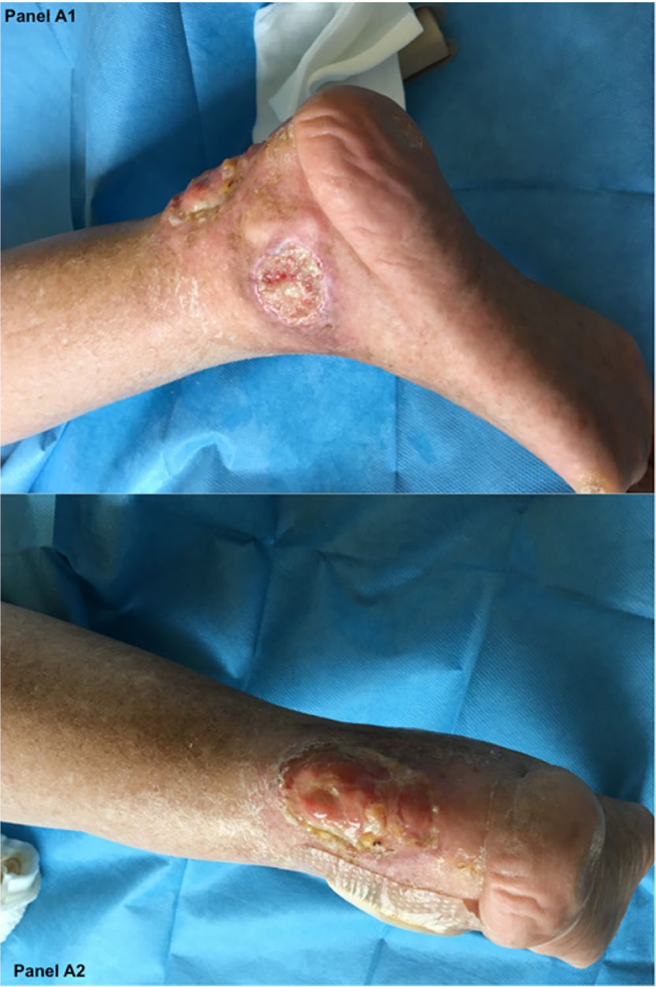
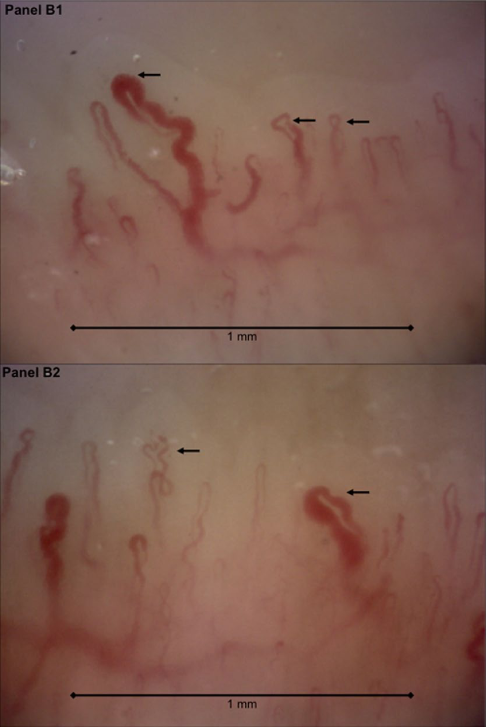
A 49-year-old male was referred to our rheumatology division for suspicion of a scleroderma spectrum disorder. He spontaneously suffered from large, deep ulcers on both legs, around the left Achilles tendon, lateral malleolus and elbows (Panels A1 and A2). Lower extremity pulse examination and vascular Doppler ultrasound were normal. A complete examination showed scleroderma-like skin involvement of the abdomen and lower and upper limbs, except for the hands. Furthermore, flexion contractures of the wrists, ankles and feet, and lower limb paresthesia were detected. He also reported xerostomia and xerophthalmia. His medical history was notable for long-standing progressive dysphonia that had evolved into a high-pitched hoarse voice. Moreover, he had bilateral cataract surgery at 18 years of age, osteopenia, premature hair loss and graying, and short stature. Raynaud’s phenomenon and telangiectasias were not present. Further investigations, including autoantibodies, were all negative except for mild hypercholesterolemia, and no organ involvement was identified. Nailfold capillary abnormalities were consistent with a scleroderma-like pattern characterized by disorganization of capillary architecture: giant, tortuous and crossing capillaries (black arrows); and a mild decrease in the capillary number (Panels B1 and B2, magnification 200×: arrows) and a mild decrease in the capillary number.
Panels A1 and A2.
Panels B1 and B2.
As the patient did not fulfill the criteria for systemic sclerosis, other rare diseases were taken into account. Genetic screening revealed heterozygosity of the WRN gene (exon 24) and a heterozygous splicing mutation in c.1577-3C>A (exon 13). Based on these findings, he was diagnosed as having Werner syndrome (adult progeria). This is a rare autosomal recessive disorder characterized by an array of features consistent with accelerated aging. The impaired DNA damage repair and telomere end maintenance might explain the premature cell senescence. In particular, mesenchymal stem cells are severely affected in their role of maintenance of vascular homeostasis, thus supporting premature vascular involvement. To the best of our knowledge, this is the first report describing the nailfold capillaroscopy abnormalities in this rare condition.
‘Images in vascular medicine’ is a regular feature of Vascular Medicine. Readers may submit original, unpublished images related to clinical vascular medicine. Submissions may be sent to: Heather Gornik, Editor in Chief, Vascular Medicine, via the web-based submission system at http://mc.manuscriptcentral.com/vascular-medicine
Footnotes
Declaration of conflicting interests
The authors declared no potential conflicts of interest with respect to the research, authorship, and/or publication of this article.
Funding
The authors received no financial support for the research, authorship, and/or publication of this article.