
Abstract

Keywords
Introduction
Patients with suspected vascular disease may undergo further evaluation of the blood vessels using one of several advanced vascular imaging techniques. Two types of imaging—computed tomography angiography (CTA) and magnetic resonance angiography (MRA)—are non-invasive tests that can provide detailed anatomical pictures of the arteries. Occasionally, some patients are asked to undergo a minimally invasive imaging test called catheter-based angiography, which directly examines the structure of the blood vessels. Depending on the clinical scenario, arterial blockages can be opened or repaired at the time of this procedure.
All three types of these vascular imaging studies help evaluate for arterial diseases such as narrowings (blockages) of the arteries due to plaque (atherosclerosis). Blockages in the leg arteries, peripheral artery disease (PAD), can lead to pain in the extremities, especially with exertion. Narrowing of the abdominal arteries, such as renal and mesenteric arteries, can occur for a variety of reasons and cause abdominal pain, high blood pressure, or kidney disease. Carotid artery plaque and narrowing can limit blood flow to the brain, causing strokes, mini-strokes, or transient blindness. Imaging studies can help evaluate for other vascular diseases, including dilatation of vessels (aneurysms), tearing of the vessels (dissection), blood clots (thrombosis), and abnormal connections between blood vessels (arteriovenous fistulas). Your doctor can help you identify which test is appropriate for evaluation of your symptoms.
For either CTA or MRA, patients may drink clear liquids and take medications prior to the imaging procedure. Depending on the imaging center, patients will be asked to refrain from eating for approximately 2 to 4 hours prior to the appointment. Patients undergoing catheter-based angiography should confirm dietary and medication restrictions with their physician beforehand, as each test may vary depending on the clinical situation. Patients should wear loose, comfortable clothing on the day of the procedure and be prepared to change into a patient gown. The physician will determine which study is most appropriate, as each can provide slightly different information. More detailed descriptions of each test are provided below.
Your physician has ordered the following test (see checked box □):
□ Computed tomography angiography (CTA)
Computed tomography angiography (CTA) is a specific type of CT scan that requires the placement of an intravenous line (IV) in the arm that allows contrast to be injected into the blood vessels while the patient is being scanned (Figure 1). This contrast is similar to a dye and it highlights the blood vessels and shows them in greater detail. This allows vascular diseases such as aneurysms, dissections, blood clots, or arterial blockages to be seen more readily, as shown in Figure 2. Some patients may undergo a CTA to provide a ‘roadmap’ prior to certain surgical procedures. Prior to the test, patients may be asked to take a blood test to ensure that they have normal kidney function because there is a small risk that the contrast may affect kidney function. Patients with previously known kidney trouble or allergies to intravenous contrast agents should inform the physician before undergoing this study, as pre-treatment with medications may be needed prior to study.

CTA (computed tomography angiography) of the legs showing normal arteries without any blockages.
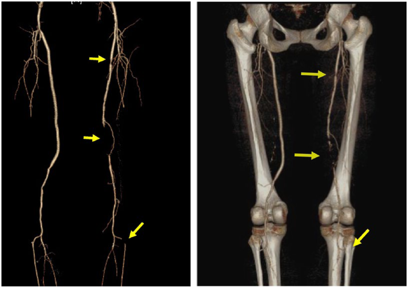
Left: Abnormal CTA (computed tomography angiography) of legs showing narrowing and blockages of arteries (arrows). Right: CTA of lower extremities from the same patient with reconstructed bones overlay. Arrows indicating sites of arterial blockages.
During the test, the patient lies on a movable exam table that slides back and forth into a donut-shaped scanner. The machine is often open on both sides of this cylinder, and patients do not usually experience feelings of claustrophobia. As contrast is injected into the blood vessels through the intravenous line, some patients experience a warm sensation throughout the body, which is normal and should pass quickly. Other possible effects include a metallic or salty taste in the mouth, headache, nausea, and/or vomiting. If these effects occur, they usually last only for a short period. During this process, the CT scanner emits X-rays in a circular fashion around the patient’s body to help form detailed images of the blood vessels.
The test takes a few minutes and allows the physician to view a higher level of anatomical detail than other types of imaging such as vascular ultrasound. This test uses a small dose of radiation, but the risks related to this exposure are minimal. There is a theoretical risk of cancer related to excessive cumulative exposure to radiation over a person’s lifetime. Patients should always review with their physician all potential risks and benefits of the test. Patients who are pregnant, planning to become pregnant, or very young may want to consider an alternative form of imaging to avoid exposure to any radiation.
Once the procedure is complete, the computer will take about 15 to 20 minutes to process the data to form the final detailed images. These images are then reviewed and interpreted by a reading physician. While CTA provides high-resolution imaging of the blood vessels, sometimes additional testing (such as catheter-based angiography) may be required depending on the clinical scenario.
□ Magnetic resonance angiography (MRA)
Magnetic resonance angiography (MRA) is a technique similar to magnetic resonance imaging (MRI) in that a large magnetic field and radio waves are used to create pictures of the blood vessels and other body parts (Figures 3 and 4A). As with the other vascular imaging studies, patients may have this test if there is concern for vascular diseases that include aneurysms, dissections (as shown in Figure 4B), clots, or arterial blockages. As with CTA, the physician may request an MRA to provide a ‘roadmap’ of the arteries in order to plan for certain vascular procedures.
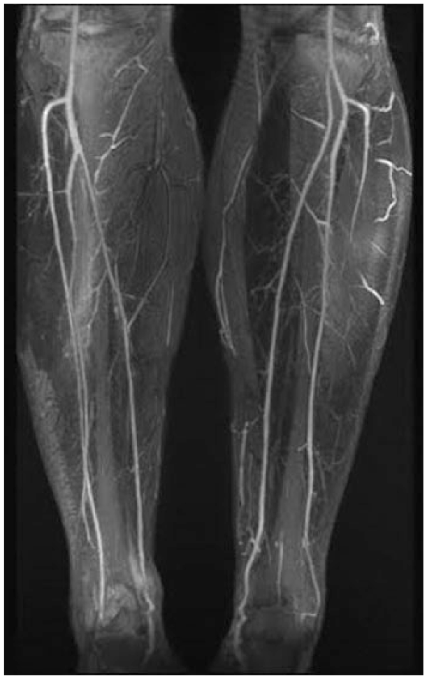
MRA (magnetic resonance angiography) of the legs showing normal calf arteries without any blockages.
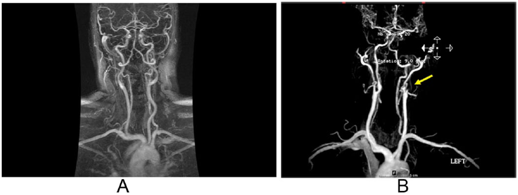
(A) MRA (magnetic resonance angiography) of the neck showing normal arteries without any blockages. (B) MRA of the neck showing an abnormal left internal carotid artery. An abrupt blockage of flow (arrow) is caused by a dissection, or a tear, in the lining of the artery.
Total imaging time may vary depending on the specific body part being evaluated, but the test should require approximately 30 to 45 minutes. Patients should not wear any jewelry or metallic objects to the test as they will be placed in a magnetic field. Metal that is brought into the magnetic field can become dangerous to the patient and other health professionals nearby. Similarly, anyone with prosthetic implants that contain metal (i.e. older hip replacements), pacemakers/defibrillators, or other metallic objects embedded within their body should review the risks of this test with their physician. It is important to note that some of the newer medical devices such as pacemakers are now safe to undergo MRI/MRA imaging, but patients should confirm this with the physician who implanted their device.
While MRA does not expose patients to radiation, a gadolinium-based contrast may be injected through an intravenous line during the exam. Patients usually are asked to take a blood test prior to the procedure to make sure that they have normal kidney function prior to the contrast injection because abnormal kidney function can increase the risk of a serious skin condition called nephrogenic systemic fibrosis (NSF). Patients who have ever had an allergic reaction to gadolinium or a similar product should inform their doctor prior to the procedure. All pregnant women should also inform their doctor prior to the test.
During the test, the patient is placed on a movable examination table that goes into a large cylindrical magnet. The magnet scans the body and may make loud noises as it is collecting information. The machine emits radio waves to alter the orientation of hydrogen atoms in the body. As these hydrogen ions move back to their regular position, the magnet detects their energy changes and creates an image based on this information. For this reason, it is important that patients lie completely still throughout the length of the test. Some MR machines are enclosed, so patients with claustrophobia may want to discuss the need for medications to reduce anxiety with their physician prior to the test.
Once the procedure is complete, the computer will take about 15 to 20 minutes to process the data to form the final detailed images, which will be reviewed by a physician. If the results show any abnormalities, patients may be referred for further testing or procedures.
□ Catheter-directed angiogram
A catheter-directed angiogram is a minimally invasive procedure to evaluate the blood vessels for narrowing or blockages. It is performed by a vascular interventionalist, who may be a cardiologist, vascular surgeon, or interventional radiologist. Prior to the angiogram, patients are asked to refrain from eating or drinking for a number of hours, but may take all their medications as prescribed unless informed otherwise. During the test, the patient lies flat on a table in an environment similar to an operating room, and the skin is prepared in sterile fashion to minimize the risks of infection. A small tube (called a catheter) is first inserted into a leg or arm artery. Then, contrast is injected through the catheter while X-ray images are taken (Figure 5). Patients with allergies to contrast or iodine, or with pre-existing kidney disease and/or diabetes, should alert their physician prior to the test. Most procedures do not require general anesthesia that requires a breathing tube, but will use local anesthetic to numb the area of the body where the catheter will be inserted. Intravenous medication may also be given to make the patient drowsy or sleepy during the procedure.
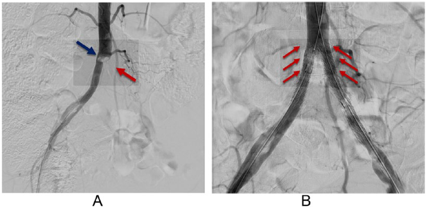
Catheter-directed angiogram showing (A) a complete blockage in the left common iliac artery (red arrow) and a narrowing in the right common iliac artery (blue arrow), and (B) resolution of the blockage after successful angioplasty and stenting of both vessels (multiple red arrows).
The site at which the catheter is placed may vary, depending on which blood vessels need to be examined. Quite often, the catheter is placed in the artery near the groin crease (common femoral artery) or at the wrist (radial artery). After the catheter has been placed, the vascular interventionalist uses X-rays to follow the catheter as it is advanced within the blood vessels to the area of interest. The test allows the interventionalist to see specific areas within the blood vessels and provide a definitive diagnosis. If the injected contrast can travel through the visualized blood vessel without difficulty, the physician will see that no significant blockages are present.
If blockages are found in the arteries, a physician will recommend lifestyle modifications that include dietary changes, quitting smoking, and potentially starting new medications. At times, the vascular interventionalist may proceed immediately following the diagnostic angiogram (Figure 5A) to perform an angioplasty procedure to open the blockages. To achieve this, special balloons are inflated in the artery and small metal scaffolds called stents may be used (Figure 5B). In more advanced disease, surgical procedures to bypass blocked arteries may be required.
Most angiograms take about 1 hour to complete, but more complicated procedures will need additional time. Once the procedure is complete, most patients are monitored for a few hours. Depending on the complexity of the procedure, patients may be asked to stay in the hospital overnight. As with any test or procedure, patients should review the risks and benefits with their physician. Risks from an angiogram may include kidney injury or failure, allergic reaction, bleeding or hematoma formation, blood vessel tear (dissection), stroke, or long-term risks of radiation.
Summary
The three aforementioned advanced vascular imaging tests—CTA, MRA, and catheter-directed angiogram—are commonly performed to help physicians diagnose and potentially treat a variety of vascular conditions. These tests can provide additional information after a thorough vascular physical examination and ultrasound imaging. Patients’ physicians will determine which study is most appropriate, as each can provide slightly different information.
Footnotes
Declaration of conflicting interests
The authors declared no potential conflicts of interest with respect to the research, authorship, and/or publication of this article.
Funding
The authors received no financial support for the research, authorship, and/or publication of this article.