
Abstract

What is an IVC filter?
An inferior vena cava filter, or IVC filter, is a small medical device that is used to prevent blood clots from traveling from the legs to the circulation of the lungs.
The veins are the tubes that return blood from the body tissues to the heart and lungs. A blood clot that forms in the deep veins of the legs is called deep vein thrombosis (DVT). These blood clots may break off and travel through the large vein of the abdomen called the inferior vena cava (IVC) to the heart and the circulation of the lungs. This condition, called pulmonary embolism (PE), may cause shortness of breath, chest pain, and a racing heartbeat, and if severe can lead to loss of consciousness, heart failure, and even death.
When is an IVC filter used?
The treatment for blood clots in the legs or lungs is blood-thinning medication, also known as anticoagulants. However, some patients with blood clots may not be able to take blood thinners safely, such as in cases of bleeding or high risk for bleeding, or during and immediately after surgery. In these cases, an IVC filter may be inserted.
Generally, IVC filters are recommended only when patients have recent (fresh) blood clots and cannot take blood-thinning medications. Occasionally, though, they may be used in addition to blood thinners in patients who have had multiple episodes of PE, or in patients with large blood clots in the legs or lungs. Some patients who have had repeated blood clots in the lungs (PEs) that have caused increased lung blood pressure (a condition known as pulmonary hypertension) may have an IVC filter inserted. IVC filters are generally not recommended for the routine treatment of blood clots except in these special situations.
There are a number of different types of IVC filters available (Figure 1). One of the earliest IVC filters was called a Greenfield filter after its inventor, Dr Lazar Greenfield, and is still in use today. Occasionally the term Greenfield filter is used generically to refer to any type of IVC filter, although this is technically incorrect.
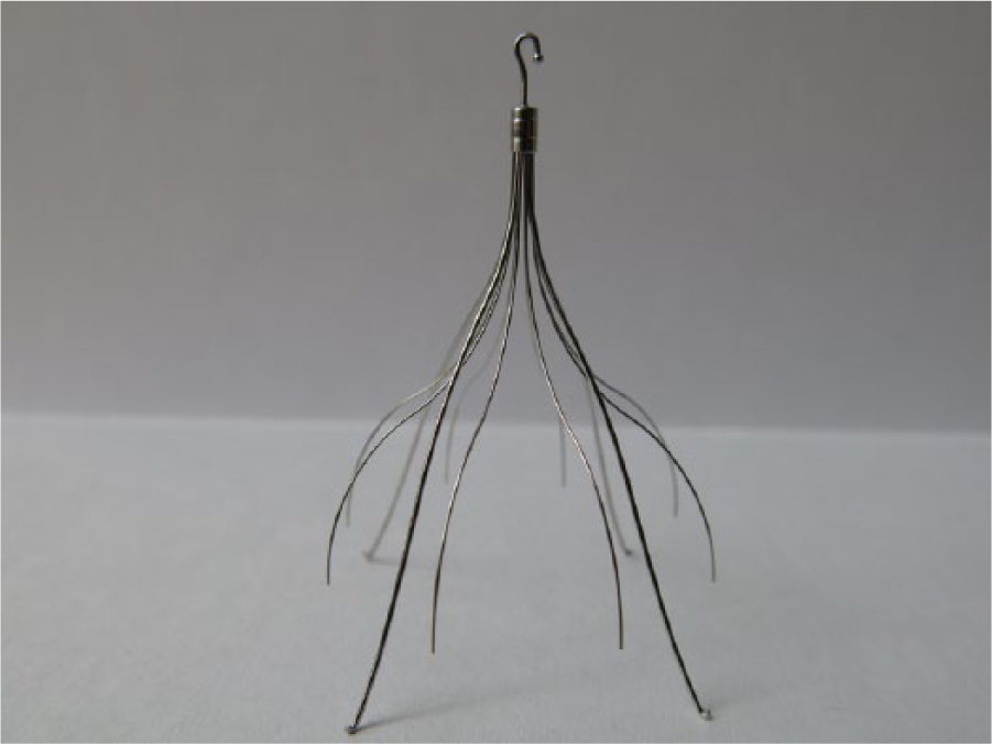
An example of a retrievable IVC filter. The hook at the top of the device allows for its removal. The filter is approximately 2 inches long. (Photo courtesy of Dr Ying Wei Lum, Johns Hopkins University School of Medicine.)
IVC filters are made from titanium, stainless steel, or other non-reactive metals. They fit into the inside of the IVC and allow blood to pass through freely to return to the heart and lungs, but trap larger blood clots – much like a strainer or a sieve. There are many different shapes and types of IVC filters available for use in patients. Most IVC filters that are used today are designed to be removed at a later time when no longer necessary. These are called ‘retrievable’ IVC filters.
How is an IVC filter put in?
The procedure to insert an IVC filter takes place in a specialized room called an angiography suite or catheterization lab, with the patient lying on a table under an X-ray camera. Patients are usually given medication to reduce anxiety or make them drowsy. To help prevent infection, the skin at the insertion site is cleansed and the patient is covered with sterile drapes, with just the cleaned insertion site showing. The person performing the procedure may be a surgeon, interventional radiologist, or cardiologist, assisted by nurses or other technicians. The team performing the procedure will wear sterile gowns, gloves, caps, and masks to keep the procedure as clean as possible.
IVC filters are most commonly inserted (put into the body) through a large vein in the groin, or occasionally the neck. The skin at the insertion site is numbed with a local anesthetic, and then a needle is inserted into the vein. A small wire is guided through the needle into the vein, and the needle is removed. Then a small tube is guided over the wire and into the vein, and the wire is removed. Contrast material, or dye that lights up with real-time X-ray, is injected into the tube to make a picture of the large veins of the leg and the IVC.
If the IVC is suitable in size and anatomy, the IVC filter is inserted through the tube and into the IVC, where it is expanded to attach to the vein walls. Another picture is taken to confirm that the IVC filter is positioned properly, and then the tube is removed from the vein. The person performing the procedure holds pressure over the vein to prevent excess bleeding, and the puncture site is covered with a clean bandage. Usually the procedure requires no stitches.
IVC filters can be inserted while a patient is hospitalized or as an outpatient; the patient may return home after a short period of observation in the recovery room. Patients may return to their normal activities shortly after IVC filter placement.
The procedure for IVC filter removal is similar to that for inserting it; however, the removal site is usually through the large jugular vein in the neck. As with the insertion procedure, a catheter is placed in the vein, through which the person performing the procedure uses a snaring device to grasp the filter, collapse it into the tube, and remove it from the body.
Insertion and removal procedures usually are quite short, requiring less than 20 minutes.
What are the complications?
Complications of IVC filter insertion are uncommon. They are divided into complications related to insertion and complications related to the filter itself.
Insertion complications
All procedures that puncture the skin carry a small risk of bleeding, bruising, pain, and infection. Precautions are taken to minimize these risks, including the cleaning procedures noted above, holding pressure at the insertion site after catheters are removed, and using local anesthetic medications during the procedure.
IVC filter complications
Rarely, IVC filters may become dislodged from their position in the IVC and tilt or move slightly within the vein. When this occurs, the filter may need to be retrieved and if still required – for instance, if the patient is still at risk for clotting and cannot take a blood thinner – replaced with a new, properly positioned filter. IVC filters may rarely become damaged, often when one of the struts or tines of the filter breaks loose. The strut may travel in the bloodstream and become lodged in another vein or organ. Occasionally the IVC filter may puncture the wall of the vein, which may or may not cause pain, depending on the structures involved.
IVC filters occasionally can fill with blood clots, leading to severe swelling of the legs (Figure 2). When this occurs, the patient may require a procedure to remove clots from the filter in addition to blood-thinning medications. Fortunately, complete clotting of the filter is quite rare.

An IVC filter (white arrow) in position in the inferior vena cava, the large vein of the abdomen. The dark area below and extending above the filter is clotted blood that has developed around the filter (black arrowheads).
Sometimes, if a removable IVC filter stays in the body too long, the body forms a coating around it that makes removal difficult or impossible. In such cases, if the filter is not causing a problem, it is left in place. In rare cases it may be removed surgically, with an incision made in the abdomen to reach the IVC.
Summary
IVC filters are medical devices that are placed in the large vein of the abdomen to prevent blood clots from traveling from the veins of the legs to the lungs. They are most commonly used when a patient cannot receive blood thinners to treat leg blood clots (DVT). IVC filter insertion can be done as an outpatient under local anesthesia, with only a puncture to a vein in the leg or neck. Most IVC filters are designed to be removed when no longer necessary. IVC filter complications are uncommon.
Footnotes
Declaration of conflicting interest
The authors declare that there is no conflict of interest.
Funding
This research received no specific grant from any funding agency in the public, commercial, or not-for-profit sectors.