
Abstract
Venous thoracic outlet syndrome is a complex but rare disease that often can have excellent outcomes if quickly recognized and treated. The syndrome results from compression of the subclavian vein along its exit from the thoracic cavity and frequently affects young otherwise healthy patients. Modern diagnosis is made with a combination of clinical exam, appropriate non-invasive imaging, and, finally, contrast venography, which can be both diagnostic and therapeutic. Treatments have evolved over time to the point where patients can undergo less extensive procedures than previously performed and still maintain excellent outcomes. One of the most important predictors of outcome is the initiation of treatment within 14 days of symptoms. Hence, the importance of the accurate and prompt diagnosis of this syndrome in patients with an upper-extremity deep vein thrombotic episode cannot be further underscored. This review is a concise summary of the background and treatment algorithm for this patient population.
Keywords
Introduction
Thoracic outlet syndrome (TOS) is a term that describes the potential compression of three neurovascular structures: in order of frequency, specifically the brachial plexus, subclavian vein or subclavian artery. Most commonly, patients will present with isolated signs and symptoms specific to the distinct anatomic structure that is being compressed. As such, neurogenic thoracic outlet syndrome (90–95%) is most frequently encountered, followed by venous (5%) and, least commonly, arterial (1%). In this article the venous component of this syndrome will be discussed.
Some confusion exists over the terminology describing this condition and will be worth discussion. Venous thoracic outlet syndrome (VTOS) refers to compression and thrombosis of the axillosubclavian vein and is quite frequently referred to as effort thrombosis or Paget–Schroetter syndrome. The term ‘effort thrombosis’ is a slightly older term, which was coined before the etiology of vein compression was understood, leading physicians of the time to attribute it purely to exertion. There is a subset of patients who have symptoms but no thrombosis, which is termed intermittent obstruction or McCleery syndrome. Secondary thrombosis of the vein may occur possibly due to injury or iatrogenic causes such as central line or pacemaker lead placement, but is not related specifically to compression. 1 However, it is important to distinguish primary thrombosis from secondary thrombosis. Primary effort thrombosis (now known as Paget–Schroetter syndrome) was first described in 1875 by Paget 2 and again in 1884 by Von Schroetter. 3 In 1934, Matas reviewed the condition as an expert witness in the case of a young man who developed the problem in the course of his job and sought compensation. This was in the first mention of the condition in American medical literature. 4 In 1948, the term Paget–Schroetter syndrome was finally coined by Hughes. 5
Paget–Schroetter syndrome describes compression and eventual thrombosis of the subclavian vein, which occurs inside of a triangle bordered by the first rib, clavicle and the subclavius muscle/costoclavicular ligament. A second triangle also exists formed by the anterior scalene, middle scalene and first rib, through which the subclavian artery and nerve trunks of the brachial plexus pass. Compression of this second triangle can result in either arterial or neurogenic TOS or both. Knowledge of these anatomic relationships is important to the understanding of both the etiology and management of these conditions.
There has been much historical debate and controversy regarding management of VTOS. Initial management consisted of anticoagulation alone with relatively poor results. 6 This approach was abandoned after thrombolysis became available in the 1970s. Another important development, transaxillary thoracic outlet decompression by means of first rib resection, was described in 1965 by Roos 7 and this was later modified to include scalenectomy as a way to reduce recurrence. 8 The modern form of this operation is frequently termed first rib resection with scalenectomy (FRRS). Other approaches to the thoracic outlet, including the more traditional supraclavicular and infraclavicular approaches, were preferred by others contributing to the controversy over the optimal approach. Central to the modern discussion of management is the timing and sequence of thrombolysis, FRRS through various approaches, and treatment of residual defects in the damaged vein. Review of the data supporting each of these interventions will be provided, as well as a summary of the current recommendations and algorithm in use at our institution.
Epidemiology
Venous TOS is a rare problem, thought to occur with an incidence of 1/100,000 people per year.1,9 The population most likely to be affected is young active males in their 30s. Males are affected with a 2:1 predominance over females. 10 Most often the right arm is affected, which is thought to be due to a higher proportion of right-handed dominance in society.1,10,11 Athletes are another population likely to be affected. A recent study from a single institution identified 41 athletes treated for TOS over 13 years. 12 A total of 14 of these patients were diagnosed with VTOS, resulting in about one case per year. Athletes are at particular risk due to the repetitive upper extremity motions involved in many sports, including baseball, swimming, weight lifting and others. Occupations which require repetitive overhead arm movement can also put patients at risk. Additionally, thrombophilia has been identified as a risk factor predisposing to thrombosis in VTOS by several authors.11,13,14
It is important to mention that, in our experience, VTOS patients are frequently underdiagnosed or misdiagnosed in the community. This may be due to the relatively uncommon nature of the disease and unfamiliarity many practitioners have. Many times patients have seen several providers before a referral to a vascular specialist is made and a diagnosis reached. This can have implications on the success of therapy, specifically thrombolytic therapy – as described later.
Anatomy/pathophysiology
Paget–Schroetter syndrome or effort thrombosis results from repeated compression injury to the subclavian vein. The axillary vein is a continuation of the brachial vein, and ascends towards the thorax. The basilic vein inserts into the axillary vein at variable locations, but usually near the axilla. The axillary vein becomes the subclavian vein as it passes over the first rib underneath the clavicle. It then joins the internal jugular vein to become the brachio-cephalic vein. The term axillosubclavian is used to encompass both the axillary and subclavian portions of the vein. This term is often used to describe the location of the injury/deep vein thrombosis due to backward propagation of thrombus from the subclavian vein to the axillary veins and occasionally to involve the basilic and brachial veins as well.
To reach the internal jugular vein, the subclavian vein must traverse the thoracic outlet. The roof of the tunnel is the clavicle, the floor is the first rib. The sides are formed by the subclavius/costoclavicular ligament medially and the anterior scalene muscle laterally. 15 The clavicle and first rib join together at a fulcrum and allow extreme force to be applied between them, in the region the vein resides (Figure 1). This problem is further exacerbated by activities that require repetitive overhead movement of the upper extremity, such as pitchers, mechanics, weightlifters and others. This is thought to result in multiple chronic insults to the vein over time, leading to inflammation, scaring and eventual thrombosis. 16 After thrombosis occurs, collateral vessels will develop frequently. This can help alleviate symptoms and delay presentation if venous drainage is adequate. Distal propagation of the thrombus can subsequently obstruct the collateral veins, resulting in the classic acute presentation. 17 The subclavius muscle is another important structure in the thoracic outlet. This muscle, as its name implies, runs underneath the clavicle and can compress the subclavian vein.
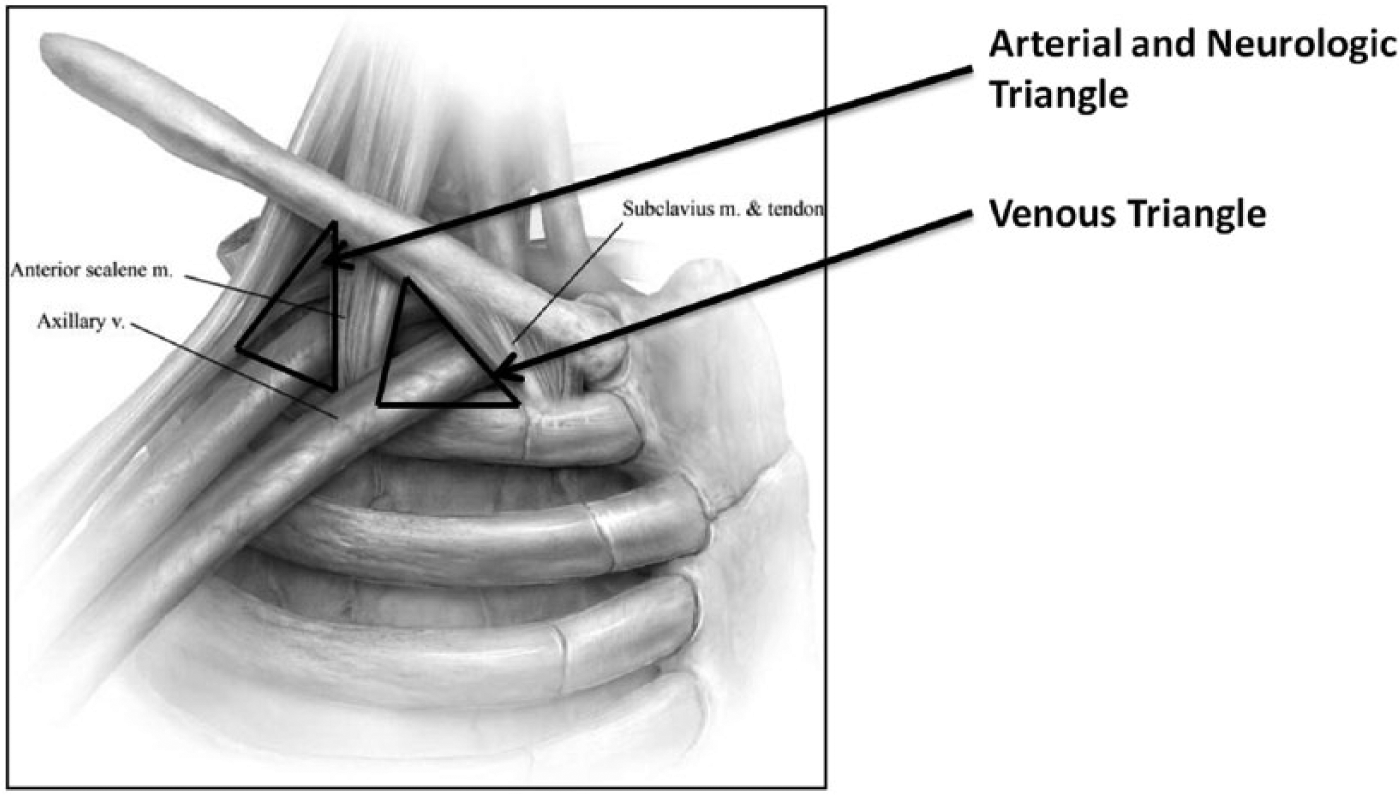
A graphic representation of the thoracic outlet. Note the venous exit is medial and the arterial and neurologic exits are lateral. Note also the lever formed by the first rib and clavicle. (Reproduced from ref. 1, with permission from Elsevier.).
It has been shown in normal, asymptomatic individuals that the subclavian vein can be compressed and flow interrupted through provocative tests,18,19 implying these tests have a high sensitivity but low specificity. Given that normal individuals can experience vein compression without symptoms, it is not clear exactly what abnormality predisposes those who are affected to develop disease. Anatomic variations in the position or size of the costoclavicular ligament and hypertrophy of the anterior scalene or subclavius muscle in conjunction with multiple repetitive compression cycles have been postulated. It has also been reported by Sheng et al. that patients presenting with VTOS have a high concurrent prevalence of occult first rib fractures and osteophytic degeneration. 20 In their series, 43% of the patients treated over a 12-month period had evidence of occult first rib fracture. This was most prominently seen in patients over 30 years of age. These fractures were detected at the time of operation by close inspection of the resected specimen. It is unclear how the presence of these fractures impacts the development of VTOS, if at all. More data will be needed to determine to what extent this finding exists in age-matched controls. Finally, it is important to note that up to 29% of patients with TOS have been reported to have skeletal anomalies, including cervical ribs, clavicular anomalies, and isolated first rib aberrations. 21 In the majority of these cases the anomalies were congenital. It is likely a combination of many factors that explain why some patients will go on to develop disease while others will not, even with demonstrated vein compression.
Some individuals develop symptoms, without evidence of thrombosis, termed intermittent obstruction or McCleery syndrome. 22 This was described in 1951 by McCleery et al. as an intermittent obstruction of the subclavian vein resulting from compression by the subclavius and anterior scalene muscles. These patients are thought to be at risk for developing thrombosis and should undergo definitive treatment with first rib resection and scalenectomy. It is important to distinguish patients presenting with McCleery syndrome from those with neurogenic TOS. Frequently a component of arm swelling will be present in McCleery syndrome and absent from neurogenic TOS. Pain, numbness and tingling are common symptoms to both syndromes.
Clinical presentation
Diagnosis
Diagnosis of VTOS is accomplished in most patients through an initial history and physical exam followed by a confirmatory imaging study, most often venous duplex ultrasound. In many patients the diagnosis will be fairly certain with history and physical exam alone. As previously described, a sudden swollen, blue, painful upper extremity in a young active male is the hallmark presentation. In older patients, alternative causes for sudden arm swelling/venous thrombus should be considered, such as pancoast tumor or malignancy. Lymphedema can also present with swelling, but is much more likely to be chronic in nature. Of historical interest, provocative physical maneuvers were previously used to guide diagnosis such as Adson’s test, but have no bearing on modern diagnosis. Adson’s test involves rotation of the head toward the affected arm and feeling for loss of a radial pulse during hyperabduction maneuvers of the arm. A positive test can be suggestive of a narrowed thoracic outlet space. However, this test is not very reliable and has been shown to be highly inaccurate in the diagnosis of TOS in general because up to 40% of the normal population can have a positive test. 24 The presence or absence of this sign should not impact the diagnosis of VTOS.
Duplex ultrasound has become the recommended imaging study for initial diagnostic purposes. It has both a high sensitivity (78–100%) and specificity (82–100%) for diagnosis of thrombus. 25 Findings that are suggestive for Paget–Schroetter syndrome are evidence of thrombosis and/or complete occlusion of the subclavian vein with a loss of flow. The entire portion of the vein must be examined to avoid missing a potential thrombus and to assess the extensiveness of the thrombus if present. In the absence of any findings of thrombosis or in situations where McCleery’s syndrome is suspected, the arm should be placed in abduction maneuvers and flow should be assessed in the subclavian and axillary veins at the different arm positions. Flow characteristics of the vein are measured with color Doppler (Figure 2): significantly increased velocities can suggest a narrowing at the thoracic outlet and absence of flow suggests total occlusion. It is important to note that it can be challenging to visualize the subclavian vein as it traverses the costoclavicular junction and this may obscure a potential thrombus.
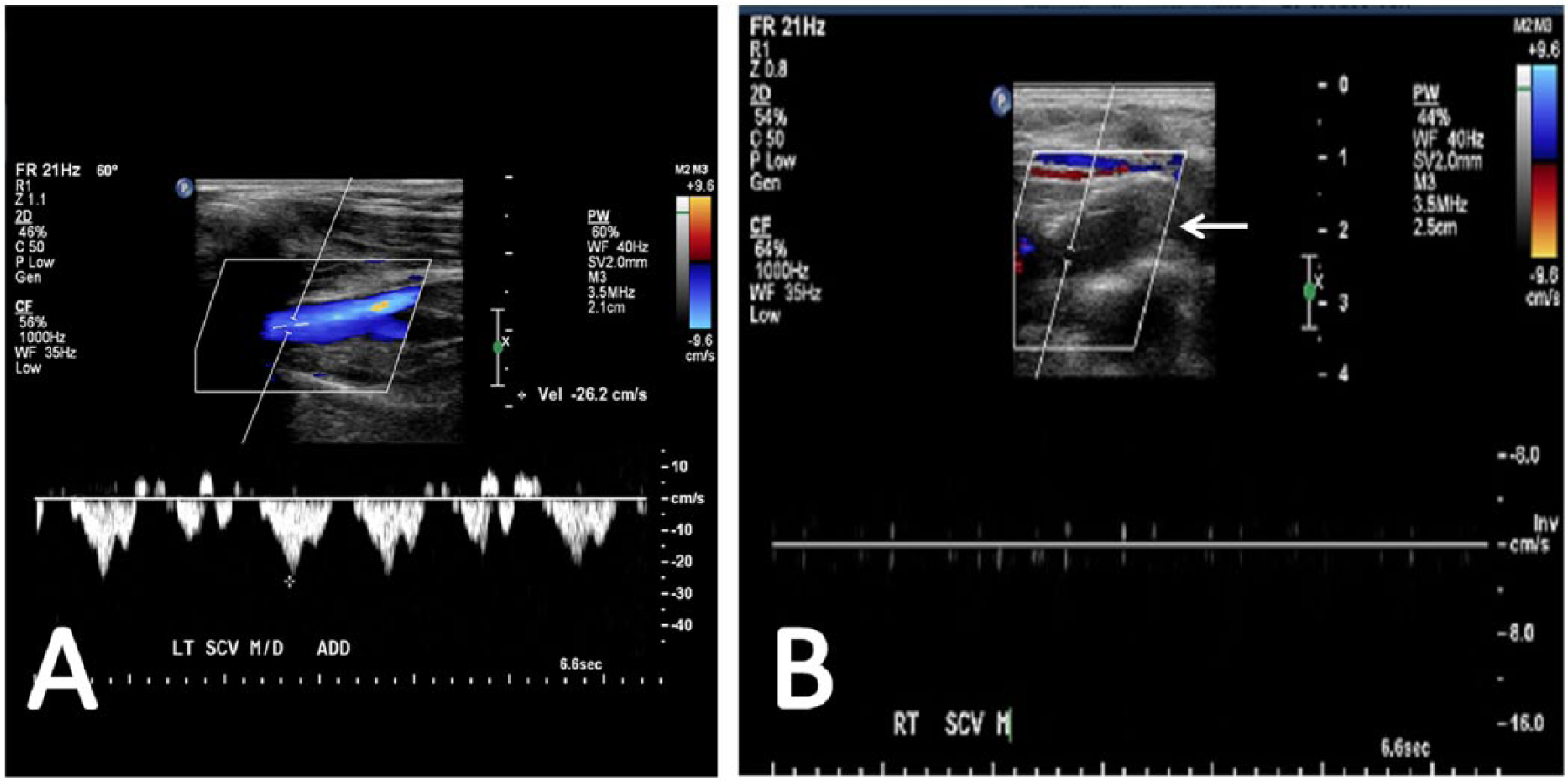
(A) Duplex ultrasound image demonstrating the normal axillosubclavian vein. (B) Thrombosed axillosubclavian vein, as demonstrated by lack of both color flow and venous waveform, with the presence of an acute thrombus (see arrow).
Contrast venography was previously the recommended initial test for diagnosis. Now it is indicated in patients with symptoms who have demonstrated pathology on duplex ultrasound and in whom an initial endovascular intervention is planned. In addition, it may be indicated in patients where a high clinical suspicion exists but a negative duplex ultrasound has been obtained due to the limitations of ultrasound previously described. The venous system is accessed with ultrasound guidance through either the brachial or basilic veins instead of the cephalic vein, as the cephalic vein inserts more centrally on the subclavian vein and can lead to a missed thrombus. 24 A positive test will demonstrate occlusion of the subclavian vein at the costoclavicular junction (Figure 3). Flow from collateral veins bypassing the thrombus may be observed, depending on the duration of the problem. In patients without thrombus, the arm should be imaged both in adduction and abduction before obstruction is ruled out. Venography is also useful to demonstrate venous stenosis. A greater than 50% stenosis is considered significant and is an indication for intervention.
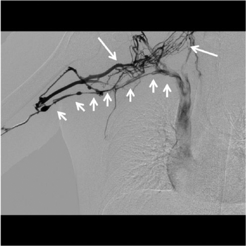
A contrast venogram demonstrating near-complete occlusion of the subclavian vein due to thrombus, prior to intervention. Large white arrows indicate collateral venous drainage around thrombus. Small white arrows indicate the native axillary and subclavian vein.
Several studies have reported on the incidence of thrombophilia in patients with Paget–Schroetter syndrome. The studies quote a range of 8% to 67% of patients presenting with primary thrombosis that will test positive for thrombophilia.11,13,14,26 Recommendations on this topic remain controversial. In a protocol devised by one author, the presence of a prothrombotic state found at the time of primary thrombosis was an indication for lifelong anticoagulation and was also associated with a significantly higher complication rate during therapy. 13 Indeed, while there might be a slightly increased incidence of thrombophilia in patients with VTOS, empirically good results have been reported by many without the institution of lifelong anticoagulation. There is currently no consensus at this time on the need for testing. At our institution it is not routine practice to screen for hypercoagulable disorders; this is reserved for those who report a family history or previous unprovoked thrombotic events. Further study will be needed before routine screening can be recommended.
Treatment
Treatment for this condition historically centered on anticoagulation and symptomatic treatment. The problem with anticoagulation alone is a significant rate of disability, recurrent thrombosis and persistent symptoms, which have been demonstrated in several studies.6,26 Prolonged anticoagulation is also lifestyle-limiting for the younger patient population who are most often affected, due to both the anticoagulation and the restrictions on arm use, which were previously suggested. Symptomatic treatment includes pain control and compression. It has been previously suggested that compression sleeves may provide some benefit in reduction of post-thrombotic syndrome; 27 however, this is based on extrapolation of treatment recommendations for lower-extremity DVT. The incidence of upper-extremity post-thrombotic syndrome is certainly rarer and less frequent than the lower extremity. At this point, we recommend upper-extremity compression for symptomatic patients with persistent swelling prior to thoracic outlet decompression.
Thrombolysis is accomplished by accessing the vein percutaneously and inserting a catheter in an antegrade fashion into the clot. An attempt is made to cross the thrombus. If successful, a catheter with side ports is placed across the lesion. A continuous infusion of recombinant-tissue plasminogen activator (r-tPA) is used most frequently to lyse the clot over a period of 6–48 hours. After a successful thrombolysis, a repeat venogram should be performed to demonstrate vein patency and hyperabduction compression maneuvers performed to demonstrate the location of venous compression under venography (Figure 4). If the lesion cannot be crossed, a catheter with an endport can be used to attempt mechanical aspiration of the thrombus, but this is typically less successful. An alternative to catheter-directed thrombolysis is pharmacomechanical thrombolysis (PMT). PMT involves use of a catheter coupled with a mechanical device which assists with clot fragmentation and, in conjunction, the dispersion of a thrombolytic agent. There is evidence to suggest both methods are equivalent with respect to the success of thrombolysis.33,34 However, PMT requires less time to clot dissolution than thrombolysis with r-tPA alone and is the method of choice for many.
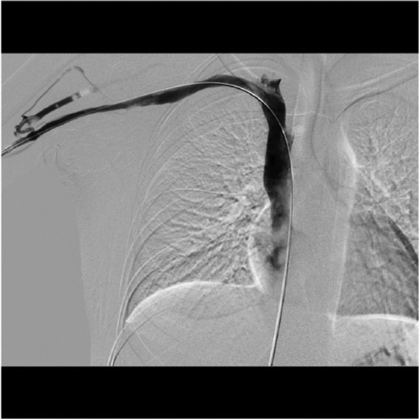
A completion venogram following successful intervention on the thrombus seen in Figure 3 (note the significant loss of collateral flow).
While the short-term benefits of thrombolysis, including decreased swelling, improved pain and re-establishment of venous patency, are apparent, it is unfortunately not the definitive treatment in patients with VTOS. One series reported a 23% rate of recurrent thrombosis within 13 months after thrombolysis. 35 This is representative of many studies which suggest up to one-third of patients will go on to rethrombose without additional treatment. It was realized over time that treatment of the thrombus alone was not sufficient. The extrinsic compression on the vein must be addressed in order to prevent recurrence.
FRRS has been advocated over a number of years to allow complete decompression of the thoracic outlet. Many approaches have been described. In 1962, Falconer and Li reported 11 cases of thoracic outlet syndrome treated with FRRS using a supraclavicular approach. 36 In 1966, Roos developed the method for first rib resection via a transaxillary approach. 7 In 1968, an infraclavicular approach was described by Gol. 37 Some basic principles apply regardless of which approach is chosen. First, removal of the first rib close to the sternomanubrial junction medially and within a few millimeters of the transverse spinal process posteriorly (Figure 5). Incomplete resection is a risk for recurrence of the problem. If a cervical rib is present, it should be removed along with the first rib. One report on the incidence of bony abnormalities in TOS suggests that these are much more common in arterial TOS rather than VTOS. 21 Second, debulking the subclavius and anterior scalene muscle is important to ensure the superior and lateral compression on the vein is released. Finally, conducting a complete circumferential venolysis of the subclavian vein is important to ensure there is no residual compression from fibrotic tissues which can develop after repeated insults to the area.
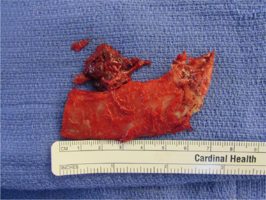
A specimen after first rib resection with attached anterior scalene muscle.
Each approach to thoracic outlet decompression has benefits and limitations. At our institution the transaxillary approach is preferred. The transaxillary approach is advantageous because it allows for excellent exposure of the anterior portion of the first rib, where the compression occurs. The entire rib can be resected and the most medial portion of the rib can also be completely disarticulated and removed from the manubrium. The entire subclavian vein is visible and can be accessed all the way to the jugular vein for a complete venolysis. Finally, the incision is the most cosmetically pleasing of the approaches available. Some of the down sides to this approach compared to the others include more difficulty with positioning and difficulty in conducting open venous reconstruction if needed; fortunately, in our opinion, this is rarely necessary. The other approaches also provide excellent exposure, but at the cost of a more noticeable scar which may or may not be of concern to the patient.
Deciding the order and timing of the interventions described above has been a source for much debate. In 1989, Kunkel and Machleder described a seminal protocol that integrated the treatments described previously, in an orderly approach. 38 Patients in this protocol were initially treated with thrombolysis, followed by 3 months of anticoagulation. After 3 months the patients were brought back and a venogram was repeated; if residual compression on the vein was seen or the patient remained symptomatic then a first rib resection was recommended. After surgery, balloon angioplasty was used to correct any remaining intrinsic venous defects. This was later further validated in a follow-up study of 50 patients, with a reported 93% of patients who remained asymptomatic if venous patency was established. 39 While this protocol showed good results, one critique was on the waiting period between initial thrombolysis and surgery. Up to 10% of patients can have a repeat episode of thrombosis after the initial thrombolysis during the 3-month waiting period, with also a small risk of pulmonary embolism. 39 In addition, this waiting period is a time of disability for the patient, which can be particularly distressing in young active patients. Early first rib resection has been suggested as a modification to the Kunkel and Machleder protocol by several authors.32,40,41 This approach has the benefit of minimizing the risk of reocclusion after thrombolysis, while maintaining a low rate of complications related to surgery.
At our institution, approximately 50% of the patients we treat for TOS are for VTOS. Our current practice is tailored depending on the presentation. Owing to our referring practice, we treat patients who present acutely within days or up to several months (and occasionally years) after the initial thrombotic episode. We recommend establishment of a diagnosis with a careful history and physical exam followed by duplex ultrasound. Our preference is to follow an early resection strategy for patients who present acutely. In our experience, not all patients will require venography or thrombolysis. 28 Our decision to use venography or thrombolysis is dependent on the severity of patients’ symptoms. If symptoms are not severe, usually anticoagulation alone followed by surgical decompression 2–4 weeks later will suffice. However, if thrombolysis is performed, our preference is to complete a transaxillary FRRS soon after thrombolysis has been completed. We recommend surgical decompression even if the vein remains occluded after thrombolysis, as over 90% of these patients will recanalize the vein over time.28,42
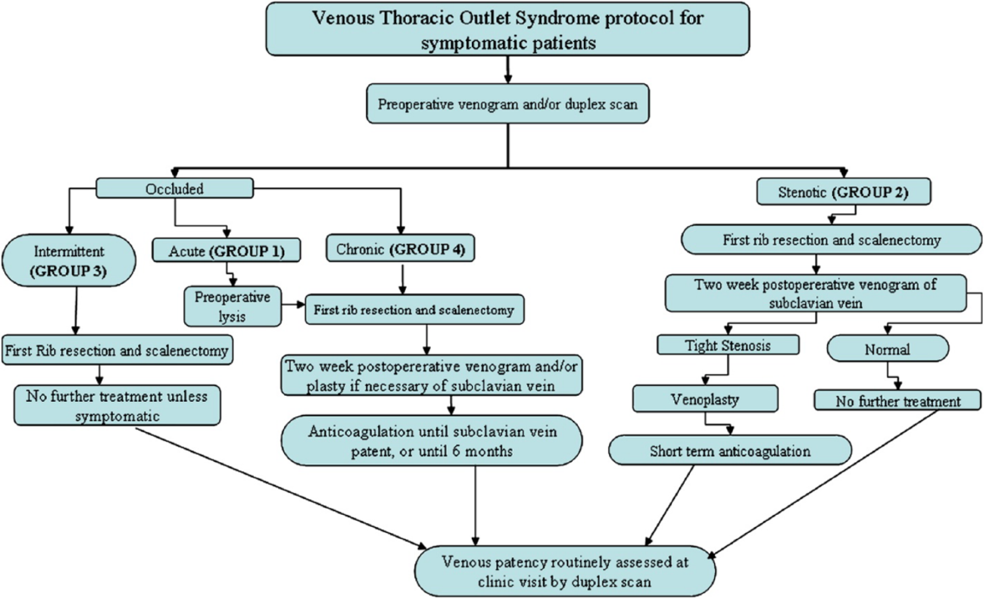
This flow chart is representative of the approach used at our institution. The ‘occluded’ (Groups 1, 3, 4) represents those with Pagett–Schroetter syndrome. The ‘stenotic’ (Group 2) represents those with McCleery syndrome. *Not reflected in this algorithm is the use of immediate venous reconstruction at the time of first rib resection, which is preferred by some. (Reproduced from de León RA. Mutiple treatment algorithms for successful outcomes in venous thoracic outlet syndrome. Surgery 2009; 145: 500–507, with permission from Elsevier.)
Outcomes
A good outcome is defined as patency of the vein and resolution of the presenting symptoms in the absence of anticoagulation or pain medications. In addition, return of function of the arm and return to occupation or sports are important criterion for a good outcome. Good outcomes can be expected if Paget–Schroetter syndrome is quickly recognized and treated appropriately. In patients who undergo prompt thrombolysis followed by successful first rib resection, surgical result rates as high as 95–98% are reported.1,28 In high-performance athletes, it has been reported that about 85–100% will return to competitive sports.12,17 In non-athletes, a 77% return to work rate has been reported. 43 Overall, most authors in high-volume institutions report excellent outcomes regardless of the surgical approach undertaken.
Conclusion
The treatment for thoracic outlet syndrome has continued to evolve over the past century as our knowledge of the disease has improved. Understanding has grown to describe the mechanism of injury involving venous compression, scaring, and eventual thrombosis from what was previously thought to be related to overexertion only. What was previously a permanently debilitating condition for many patients in the prime of their working years has been rendered much less so. Early recognition and treatment is essential to a successful outcome. A patient with VTOS can now expect excellent outcomes if treatment is rendered promptly. Much of this success is owed to the development and evolution of thrombolysis and safe surgical decompression techniques.
Footnotes
Declaration of conflicting interest
The authors declare that there is no conflict of interest.
Funding
This research received no specific grant from any funding agency in the public, commercial, or not-for-profit sectors.