
Abstract
Molluscum contagiosum (MC) is a viral cutaneous infection common in children. It is characterized by umbilicated, skin-colored papules that typically resolve without treatment over several months to years. Immune response to the virus may cause inflammatory reactions, including molluscum dermatitis, inflamed molluscum, Gianotti-Crosti syndrome-like reaction, erythema annulare centrifugum, or even a generalized id reaction (a reactive inflammatory process driven by a separate condition that stimulates the immune system). We report a unique case of a granuloma annulare-like id reaction secondary to immune recognition of MC in a pediatric patient followed by rapid resolution of their MC.
Case Report
A 6-year old girl with history of molluscum contagiosum (MC) for 12 months presented to the pediatric dermatology clinic with new-onset lesions. Physical examination revealed umbilicated, skin-colored papules on the chin, axilla, and thigh, some of which were inflamed and erythematous (Figure 1). In addition, there were new symmetric, erythematous, annular plaques without scale on her elbows and knees (Figures 2 and 3). She had no known history of atopic dermatitis (AD) and prior treatment of the MC included ZymaDerm, an over-the-counter topical medication with active ingredients including silver nitrate, echinacea angustifolia, fucus vesiculosus, and thuja occidentalis. Based on clinical presentation, she was diagnosed with inflamed molluscum and a granuloma annulare (GA)-like id reaction (a reactive inflammatory process driven by a separate condition that stimulates the immune system). The non-inflamed MC lesions were treated with cantharidin 0.7% solution and the GA-like plaques were treated with triamcinolone 0.1% ointment. At her follow up in 11 weeks, all the MC lesions had resolved and her GA-like id reaction had cleared within several days of using triamcinolone.

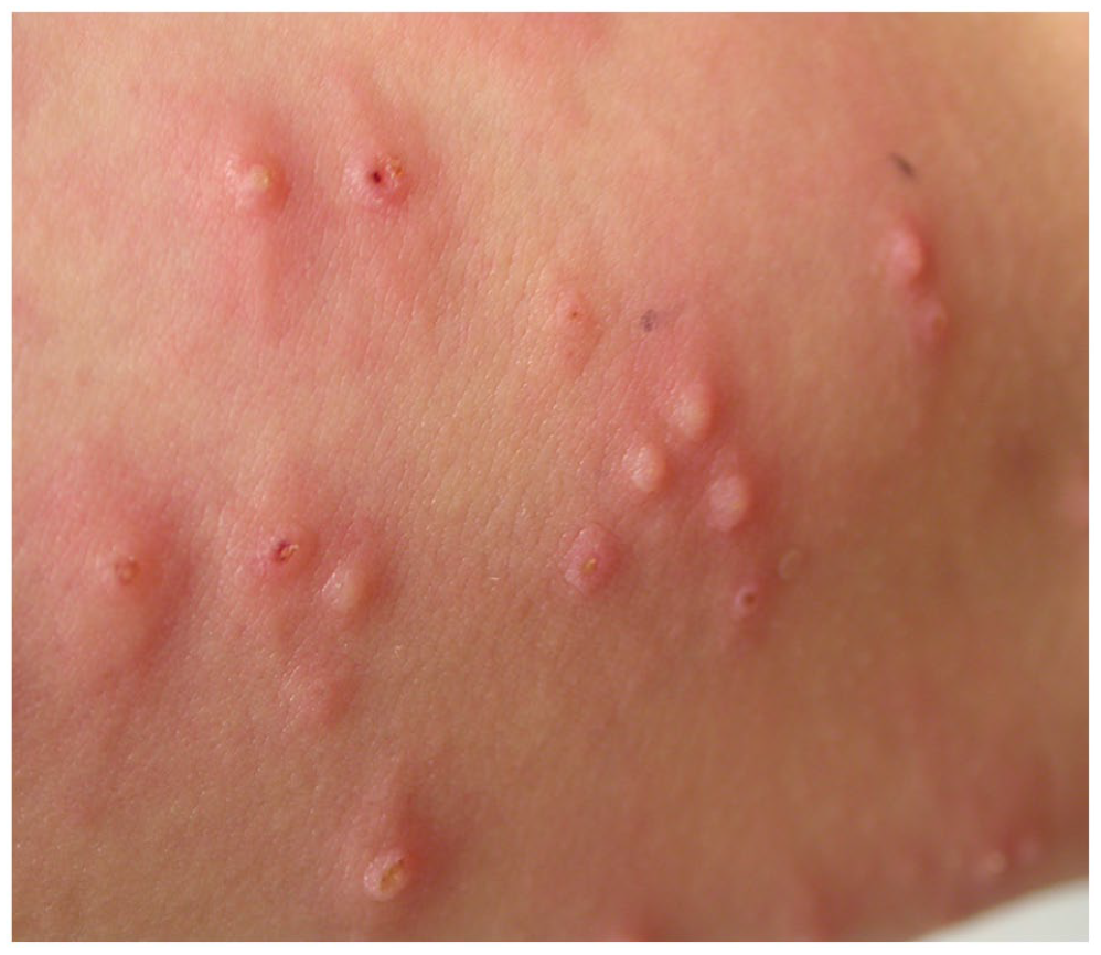
Inflamed, erythematous papules and umbilicated, flesh-colored papules distributed around the left axilla.

Erythematous, annular plaque without scale on both elbows.

Erythematous, annular plaque without scale on the right knee.
Discussion
Children can develop inflammatory reactions due to immune recognition of MC, all of which typically resolve following clearance of MC lesions. 1 Molluscum dermatitis occurs in up to 39% of patients with MC and is characterized by eczematous patches or plaques surrounding MC lesions. 1 (Figure 4) Children with a history of AD were found to be significantly more likely to develop molluscum dermatitis compared to children without a history of AD (50.6% vs 31.8%; P < .001). 1 Inflamed MC lesions have been reported in 23% of children. 1 Characterized by erythema, edema, and painful pustular or fluctuant lesions, this reaction can often be mistaken with a bacterial infection, leading to unnecessary incision and drainage, antibiotics, financial burden, and familial distress. 2 Paradoxically, it is postulated that these lesions translate a mounting immune response to the MC virus prior to its clearance and have become known as the “beginning of the end (BOTE)” sign 2 (Figures 5-7). Gianotti-Crosti syndrome-like reactions (GCLRs) are also associated with immune recognition and imminent resolution of MC.1,3 Occurring in only 5% of children, GCLRs are predominantly characterized by pruritic, new-onset erythematous papules, plaques, and papulovesicles distributed symmetrically on the extensor surfaces of extremities.1,3 In addition, there are several reports of children with irregular, annular scaling plaques surrounding inflamed MC lesions who were diagnosed with erythema annular centrifugum (EAC).4,5 There is also a case report of a child presenting with a generalized id reaction in response to MC and associated molluscum dermatitis. 6

Molluscum dermatitis encircling a molluscum lesion on the back of the right thigh.
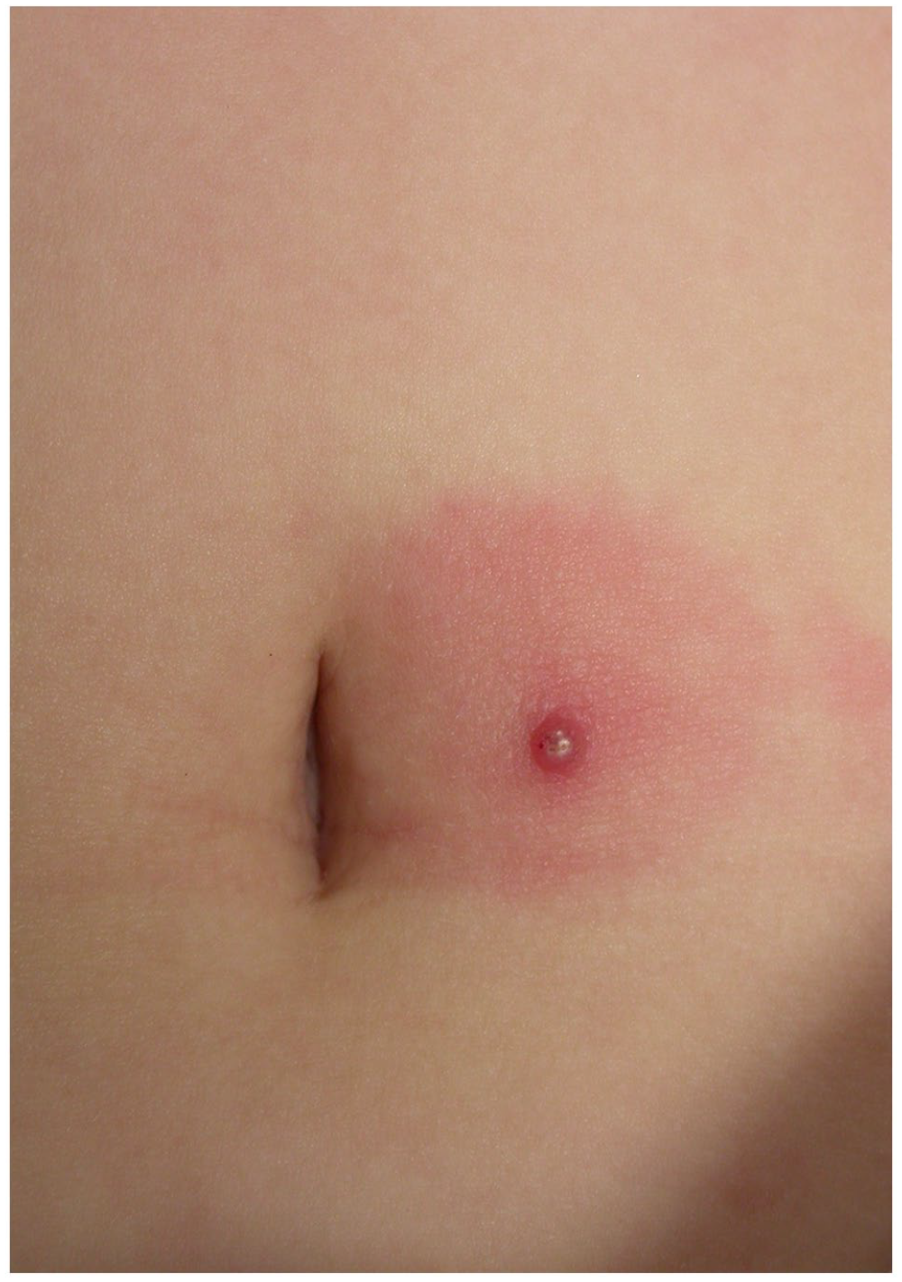
Inflamed molluscum (BOTE sign) near the umbilicus.

Inflamed molluscum with lymphangitis.
Inflamed molluscum post cantharidin treatment.
Here, we present a case of a GA-like id reaction in a pediatric patient with MC and inflamed molluscum. GA is a self-limited, cutaneous reaction of unknown etiology that may be triggered by infections, vaccinations, or local minor trauma in children. 7 Diagnosis is typically clinical but can be confirmed by histology, which is characterized by a mucinous degenerating core of collagen and a surrounding infiltrate of lymphocytes and histiocytes. 7 To our knowledge, GA-like id reaction to MC has not been previously reported. Given the lymphocytic component, we hypothesize that this reaction signals activation of the immune response and forthcoming clearance of MC, especially in the context of inflamed MC lesions. This is consistent with our patient’s rapid clearance of both her MC and GA-like id reaction shortly after her initial visit.
Conclusion
MC is a common viral cutaneous infection that can be associated with numerous inflammatory reactions in children including molluscum dermatitis, inflamed MC lesions, GCLRs, EAC, and even a generalized id reaction.1 -6 In some cases, these reactions are associated with immune recognition of the virus and impending clearance. We submit a unique case of a GA-like id reaction in a pediatric patient with MC and inflamed molluscum. Given the lymphocytic component of GA, we hypothesize that this reaction signifies activation of the immune response and forthcoming clearance of MC, which is consistent with our patient’s rapid clearance of both her MC and GA-like id reaction shortly after her initial visit.