
Abstract
Lung cancer, colon cancer, breast cancer, and prostate cancer are the leading causes of death in developed countries. Many cancers display non-specific signs in the early stage of the disease, thus making early diagnosis often difficult. We focused on nestin as a new biomarker of possible clinical importance in the early diagnosis and monitoring of cancer. The expression of nestin takes place at an early stage of neural differentiation, but no expression of the nestin gene can be revealed in normal, mature adult tissues. Nestin plays an important role in the development of the central nervous system and contributes to the organization and maintenance of cell shape. Nestin was found to be a marker of microvessel density, which in turn has proven to be a reliable prognostic factor for neoplastic malignancies in patients. Nestin expression correlates with an increased aggressiveness of tumor cells. The role of nestin in cancers of the colon and rectum, liver, central nervous system, lung cancer, breast cancer, melanoma, and other cancers has been reviewed in the literature. Associations between nestin expression and prognosis or drug-resistance may help in disease management. More research is needed to understand the molecular mechanisms of nestin expression and its role in possible targeted therapy.
Introduction
Cancer contributes significantly to the reduction of average lifespan. Lung cancer, colon cancer, breast cancer, and prostate cancer are the leading causes of death in developed countries. 1 According to the Global Burden of Disease data, the total number of deaths due to neoplasms increased from nearly 5.7 million in 1990 to 8.2 million in 2013, which is an increase by 45.6% globally. During the same period of time, the number of deaths due to colon and rectum cancer increased by 57.3% (from 490 200 to 771 100), breast cancer by 44.2% (from 327 000 to 471 000), prostate cancer by 82.8% (from 157 100 to 292 700), and tracheal, bronchus, and lung cancers by 56.5% (from 1 050 000 to 1 639 600). 2 A low survival rate can be noticed in elderly patients and in those from Eastern Europe. 3
Many cancers display non-specific signs in the early stage of the disease, thus making early diagnosis often difficult. Maringe et al 4 report that only about 10-20% patients being diagnosed with colon or rectal cancer are in the Duke’s A stage. The VITAL study indicates that only 22.6% of respondents were diagnosed with local or in situ lung cancer. 5 The early stage of cancer is associated with better prognosis and longer survival rates, which draws attention to the importance of reliable screening tools and biomarkers which would help to diagnose cancer at an early stage and to properly monitor the progress or regress of the disease. The stage of the disease at diagnosis is an important predictor of mortality for cancer and therefore, new biomarkers of the neoplastic process are being sought to facilitate the early detection of neoplastic diseases.6 -8 We focused on nestin as a new biomarker of possible clinical importance in the diagnosis and monitoring of cancer in the light of currently approved biomarkers for clinical use.
Characteristics of the Protein Nestin
Nestin was first described in animal studies on the neural development in mammals. Rodent studies on the monoclonal antibody Rat-401 allowed for the identification of cells at an early stage of their development which displayed some morphological features similar to radial glia. 9 This was then identified as nestin—a class VI intermediate filament (IF) protein, which was considered as a marker for identifying the state of a stem cell. The expression of nestin takes place at an early stage of neural differentiation, but no expression of the nestin gene can be revealed in normal and mature adult tissues. In a murine model, it was found in muscle precursors, but not in mature muscle cells. 10 Nestin plays an important role in the development of the central nervous system (CNS) and, as with other IFs, participates in the organization and maintenance of cell shape; at this stage, it is distributed across the intracellular space in a wave-like pattern with a pronounced perinuclear accumulation.11,12 The presence of nestin was also detected in the cell nuclei of glioblastoma, but its role there remained unclear. 13 The expression of nestin becomes downregulated immediately at the point of transition from a proliferating stem cell to a postmitotic neuron. 10 The expression of nestin may be induced by various pathologic conditions such as injury, neurodegenerative diseases of the brain, or the neoplastic process.14,15 However, it is also present in a broad variety of cells, including proliferating vascular endothelial cells, basal cells of the mammary glands, and liver stem cells.16 -18 Following the hypothesis that CNS tumor cells might display a gene expression pattern similar to that of the CNS stem cell from which they arise, the research evaluated nestin in neoplastic lesions of the brain and spine. 12 In addition, nestin as a biomarker of a stem cell was found in malignant tumors originating in other organs.
Nestin was found to be a valuable marker of microvessel density (MVD), which in turn has proven to be a prognostic factor for patients with neoplastic malignancies. A significant association between MVD and liver metastasis proves that a tumor’s potential to grow, spread, and metastasize is connected with tumor-associated angiogenesis and the fast growth of new vessels around the tumor as well as in the cancer tissue itself.19,20 MVD is thus the essential prognostic factor, but the most important information is delivered by the number of new blood vessels because they have leaky and fragile basement membranes which allow tumor cells to enter into circulation and spread over the body, whereas a mature vessel wall is far harder for cancer cells to penetrate. 21
Nestin expression correlates with an increased aggressiveness of tumor cells. The anti-apoptotic function of nestin through inhibiting caspase activation exerts a pro-survival effect on cancer cells. 22 Recent observations have revealed an association between the overexpression of nestin and neoplastic characteristics with a poor prognosis in different cancers. The correlations between nestin protein expression and tumor aggressiveness (the ability to metastasize and spread; how advanced the disease is), clinicopathological features, and poor survival rates has been confirmed for many tumors.23 -25
There are some latest studies on molecular mechanisms of nestin action regarding tumor progression. One of them suggests that type VI intermediate filaments (IF) play an important role in mediating cell stiffness, that is, nestin increases cancer cell metastasis by reducing cell stiffness. Yamagishi et al proved that metastatic ability of cells was inhibited by nestin knockout as the knockout cells were stiffer. Nestin enhances cell body softness and flexibility of the cytoskeleton. They indicated that knockout of nestin in tumor cells may be a new therapeutic strategy for the inhibition of cancer metastasis. 26 Wang et al suggested that nestin may determine survival by means of oxidative stress. They revealed that Nestin and Nrf2 (NFE2-related factor 2) were involved in a positive-feedback loop that mediate the antioxidant responses and cellular redox homeostasis. They concluded that the Nestin–Nrf2 signaling pathway could be targeted as novel therapeutic approaches for cancer treatment. 27 Li et al revealed that the tumor-promoting effects of nestin were mediated by binding to Gli3, a zinc finger transcription factor that negatively regulated hedgehog signaling. Their findings uncovered in Gli3 a therapeutic target to treat the malignancies. 28
Colorectal Cancer
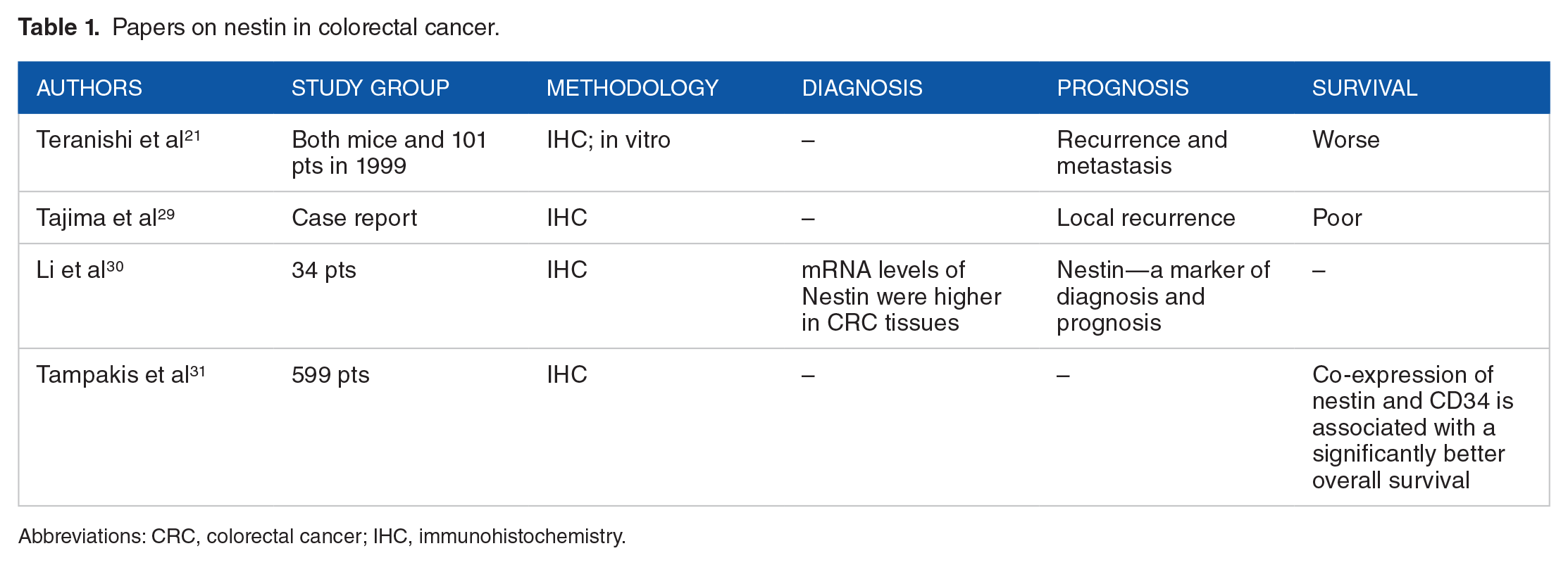
A lot of endothelial markers stain and identify the newly formed small blood vessels and the existing large blood vessels. Teranishi et al 21 on an animal model showed that the CD34-stain pattern of colon and rectal cancer blood vessels differs from those stained by nestin. CD34 was revealed in endothelial cells of large blood vessels with an average diameter of 9.67 μm, whereas nestin was found in vessels with an average diameter of 9.06 μm. Significant differences between median diameters of blood vessels were also observed (8.82 vs 6.30 μm) in human colorectal cancer tissue. In the infiltrating border of the tumor the stronger expression of nestin than of CD34 was found. In addition, MVD detected by nestin appeared to be a better prognostic factor for survival than MVD discovered by CD34.
Research on the correlation between increased nestin expression and the clinical characteristics of colon and rectal cancer are limited. Tajima et al 29 described a case of a patient with a cancer of the descending colon with overexpression of several proteins, including nestin which indicated a poor prognosis. The patient had a recurrence approximately 1 month after surgery and 2 months later, died. Li et al showed that nestin is connected with tumorigenesis in colon and rectal cancer. 30 Expression of nestin was higher in cancer tissue in comparison with normal tissues. They observed nestin staining both in the endothelium of small-sized tumor vessels and stromal cancer cells. What is more, in vitro tests showed that a knockdown of nestin arrested the cell cycle at S-phase, and inhibited the cellular proliferation and migration. These findings prove that nestin can be used as a prognostic marker and it can also be a new therapy for cancer patients.
Tampakis et al in their large retrospective cohort study analyzed 599 patients with resectable colorectal cancer. They concluded that combined expression of nestin and CD34 is associated with a significantly better survival irrespectively of age, gender, T and N stage, grade, type of invasive margins, and microsatellite instability. 31
The compilation of papers on nestin in colorectal cancer is presented in Table 1.
Papers on nestin in colorectal cancer.
Abbreviations: CRC, colorectal cancer; IHC, immunohistochemistry.
Liver
In the normal adult human liver, the presence of stem cells with multiple differentiating capabilities has been described. Those human liver stem cells are positive for stem cell markers such as nestin among others. After differentiating in mature hepatocytes, markers for immature cells become downregulated. 18 One of the hypotheses behind the carcinogenesis is p53 mutation, which is common in liver cancers. Loss of p53 functions facilitates the dedifferentiation of mature hepatocytes into nestin-positive progenitor-like cells, which then may be further differentiated in liver carcinomas. 32
In liver cancers, increased expression of nestin is associated with increased levels of other biomarkers and a poor prognosis. A significant correlation between microvessel density and liver metastasis proves that a tumor’s potential to grow, spread, and metastasize is related to tumor-associated angiogenesis and the fast growth of new vessels around the tumor as well as in the cancer tissue itself.19,20 Yang et al 33 assessed a set of biomarkers related hepatic stem/progenitor cell or tumor angiogenesis in specimens collected from patients with hepatocellular carcinomas. Nestin overexpression was found in patients who experienced a recurrence after the complete resection of the tumor. Nestin staining was localized in small tumor cells. Nestin appeared to be a prognostic factor for overall survival and recurrence free survival as well as an independent factor for tumor recurrence. A significantly elevated expression of nestin was detected in drug-resistant cell lines of hepatocellular carcinoma, but nestin knockdown in drug-resistant cell lines increases chemosensitivity. The role of nestin in chemoresistance makes it a good candidate for targeted therapy. 34
Malvi et al carried out a retrospective study on 28 patients with resected mixed primary liver cancers. The authors concluded that nestin could be considered as the tool in differential diagnosis of intermediate-cell carcinomas. 35 Vasuri et al prospectively examined 39 liver HCC nodules from 22 consecutive patients and identified four different morphovascular HCC patterns using CD34 and nestin. They concluded that a subclassification of HCC is possible preoperatively by means of MRI. 36
The compilation of papers on nestin in liver cancer is presented in Table 2.
Papers on nestin in liver cancer.

Abbreviations: HCC, hepatocellular carcinoma; IHC, immunohistochemistry; OS, overall survival; RFS, relapse-free survival.
CNS Cancers
Primary CNS cancers are rare, with the incidence accounting for 1.4% of new cancer diagnoses in the United States, but they contribute to greater morbidity and mortality than other malignancies.1,2 For this reason, efforts are being made to discover more about markers which help to diagnose and monitor the disease. Among the CNS tumors, the astrocytic tumor group is the most common histologic type of cancer with an overall incidence of 2.98/100 000. 37 Although evidence of the role of nestin is strongest in gliomas, the assessment of nestin expression has been carried out performed for other CNS tumors as well.
Nestin expression was confirmed both in low- and high-grade tumors; however, most of the gliomas that expressed high levels of nestin were high-grade gliomas such as anaplastic astrocytomas, anaplastic oligodendrogliomas, anaplastic oligoastrocytomas, and glioblastomas. 38 Expression of nestin was detected in neoplastic astrocytes and endothelial cells and correlated with tumor grade. 39 Many other researchers have confirmed the occurrence of a positive correlation between the level of nestin expression and the degree of malignancy in CNS tumors.24,40
The level of nestin expression translates into clinical outcomes. The level of nestin was found to be a strong prognostic factors for total survival in astrocytoma WHO grades II and III. 38 Higher expression of nestin, either alone or with a combination with other markers, is predictive of a significantly lower 5 year survival rate, overall survival, or progression-free survival. 38 ,40 -42
In meningiomas, which are mostly benign, higher expression of nestin was found in anaplastic and atypical lesions compared to benign ones, but the difference was not statistically significant. 43 In benign meningothelial meningiomas WHO grade I, nestin was located predominantly in the walls of blood vessels. 44 Nestin positive staining was found in gliomatosis cerebri cells; it was stronger in more undifferentiated cells. 45 High grade intracranial schwannomas also express nestin as do those located in other organs of the human body.38,46 Nestin-positive neuronal cells were found in gangliogliomas and malignant peripheral nerve sheath tumors.47,48
In ependymoma cells, nestin expression was detected in 55% of cases. The level of nestin expression was higher in grade III than in grade II tumors. In addition, nestin expression was significantly more often detected in tumors which showed an endothelial proliferation, increased microvascular density, and necrosis. 49 The occurrence of nestin positivity was an independent marker for poor progression-free survival and overall survival. 50
The assessment of other biomarkers helps to establish both clinical features and prognosis. Common expression of nestin and vimentin indicates an enhanced motility and invasive potential of astrocytoma cells. 51 High nestin expression and no IDH1/2 mutations are associated with shorter survival in patients with invasive astrocytomas. 42 Increased co-expression of VEGF in the high grade astrocytoma and ependymoma, the co-occurrence of CD133 positivity in gliomas, and the co-expression of microtubule-associated protein 2 (MAP2) in gangliogliomas are associated with the nature of the tumor being more invasive and with worse clinical outcomes.39,41,47,50
A simplified predictive model based on the status of expression of biomarkers such as CD133, CD44, nestin, and MVD appeared to be a good predictor of recurrence-free survival and overall survival in patients with a hepatocellular carcinoma. The model had better sensitivity than other indices (tumor size, number, differentiation, encapsulation, vascular invasion, TNM stage, CLIP score, γ-glutamyl transferase, and α-fetoprotein). 33
Behling et al 52 retrospectively analyzed 113 patients with glioblastoma (GBM) and concluded that nestin had no independent prognostic impact and showed no differences regarding age and clinical status. Matsumoto et al retrospectively examined 95 cases of primary surgically resected GBM. They found out that nestin was significantly overexpressed in non pseudopalisading perinecrotic lesions (non-Ps), but it was not—in lesions with pseudopalisading features (Ps) in GBM tissues. 53
The compilation of papers on nestin in CNS cancer is presented in Table 3.
Papers on nestin in CNS cancer.

Abbreviations: CNS, central nervous system; GC, gliomatosis cerebri; IHC, immunohistochemistry; MPNST, malignant peripheral nerve sheath tumor; OS, overall survival; PFS, progression-free survival.
Lung Cancer
Lung cancers are another group of tumors with poor prognosis.1,2 Nestin, like other cancer stem cell markers, is considered a good predictor for the poor prognosis and clinicopathological characteristics; however, some controversies regarding this remain. About 80% of lung cancer cases constitute non-small cell lung cancer (NSCLC), and thus the majority of studies include this histological type of lung cancer. Reports from the literature show that nestin can be detected in almost 90% of NSCLC cases, while no expression is observed in alveolar and bronchial epithelial cells retrieved from adjacent tissues. Overexpression of nestin was found to be associated with poorly differentiated phenotypes, TNM stage, tumor size, and proliferative index.54 -57 Overexpression of nestin also correlates with shorter survival in patients receiving adjuvant chemotherapy.54,55 A similar effect of nestin on tumor characteristics was also observed in other lung cancers such as adenocarcinoma. 57 Only about 40% of malignant pleural mesothelioma (MPM) cases express nestin, but in those nestin positive tumors, high expression of nestin is associated with decreased overall survival. 58
In in vitro models, knockdown of nestin expression resulted in the inhibition of tumor cell proliferation and a decreased ability of NSCLC cells to form colonies. 54 The possibility of modifying nestin expression makes it a promising candidate for targeted therapies. 57
The compilation of papers on nestin in lung cancer is presented in Table 4.
Papers on nestin in lung cancer.

Abbreviations: IHC, immunohistochemistry; MPM, malignant pleural mesothelioma; NSCLC, non-small cell lung cancer.
Breast Cancer
In normal mammary glands, nestin is expressed in cell within the basal/myoepithelial layer. It may play a role in the maintenance of successive regenerative cycles characterized by cellular proliferation and terminal differentiation of the epithelial portion of the gland. It was revealed that nestin is expressed together with other markers of the regenerative compartment (deltaN-p63, and CK14), which are characteristic for stem cells, and is also expressed in highly aggressive and poorly differentiated basal epithelial phenotypes of breast cancer. 17 Elevated expression of nestin commonly occurs within the basal breast cancer subtype with mutations in the BRCA1 gene.17,59 Nestin is usually expressed in triple-negative and in inflammatory breast cancers subgroups.60,61 On the other hand, expression of other markers correlates with the present of nestin. Co-expression of CK14 and p63 was confirmed in the epithelial, columnar basal and filamentous cells of the mammary gland. 17 Co-occurrence of melatonin receptor 1 and nestin was found in higher tumor stages (TII/III) with a higher risk of relapse, while a lack of co-staining for those markers correlated with lower tumor stages. 62
Nestin expression is associated with shorter survival rates. In patients with locally advanced (T4) breast cancer, the presence of nestin predicted poor 5 year survival. 63 Patients with nestin expression have higher rates of disease recurrence with a distant metastasis than nestin negative patients. 60 The presence of nestin is independently associated with reduced breast cancer specific survival.59,64 Clinically, expression of nestin indicates increased invasiveness of breast cancer and can serve as a prognostic factor for worse outcomes.
The compilation of papers on nestin in breast cancer is presented in Table 5.
Papers on breast cancer.

Abbreviations: BRCA, breast cancer; IHC, immunohistochemistry.
Melanoma
Melanoma is the most invasive cancer of skin malignancies. Despite the fact that only 4% of all skin cancers are various types of melanoma, they are responsible for 74% of all skin cancer related deaths. 2 The expression of nestin was found in a wide range of skin lesions, but a higher percentage of nestin-positive cells can be found in nodular melanomas than in dysplastic nevi or superficial spreading melanoma. 65 A significant positive correlation has been identified between melanoma T stage and nestin expression. Nestin expression is higher in tumors over 2 mm thick than those below that thickness. It is also higher in compound than in junctional nevi. In advanced stage T4 tumors, elevated nestin expression is often located in the peripheral area or the invading front of the tumor, which may suggest its role in cell migration or invasion. 66 Nestin can also be useful in staging as it helps to differentiate nodal melanocytic nevi from metastatic melanomas during sentinel lymph node evaluation. 67
There are many melanocytic markers which are sensitive to melanoma. Expression of SOX2 is useful in differential diagnosis in cases where metastatic disease is suspected. 67 BRN2 and SOX10 together with nestin were detected in most primary and metastatic melanoma cell lines. 60 It was found that only some transcription factors can regulate nestin expression. Silencing SOX10 and SOX9 results in a decrease in nestin protein production, while the downregulation of BRN2 does not affect nestin expression. 68 Currently, there is ongoing research on the mutual associations between biomarkers, as patterns of expression of nestin together with other melanoma biomarkers can help predict melanoma invasiveness. 69
The compilation of papers on nestin in melanoma is presented in Table 6.
Papers on nestin in melanoma.

Abbreviations: IHC, immunohistochemistry.
Other Cancers
The ongoing research on nestin expression confirms its importance in many cancers. The presence of nestin protein was found in cytoplasm of the pancreatic ductal adenocarcinoma cell, but only about 30% of those cancers express nestin. Nestin expression varied across the different pancreatic cancer cell lines, but it had no effect on survival; no difference in the overall 2 year survival rate was observed between nestin-positive and nestin-negative patients. The presence of nestin expression correlated with nerve invasion and cancer-positive surgical margins, which may translate into greater motility and aggressiveness of nestin-positive tumor cells.70,71 Nestin expression was also found in several other types of pancreatic tumors such as pancreatoblastoma, solid pseudopapillary neoplasm, undifferentiated carcinoma, serous cystic adenoma, and mucinous cystic neoplasm predominantly located in the sarcoma part of the tumors. 72
Similarly to pancreatic cancers, nestin positivity was found in 34% of esophageal squamous cancer. Expression of nestin was significantly associated with a shorter median survival time and a shorter progression-free survival time. Expression of nestin was positively correlated with expression of other proliferation and apoptosis markers, suggesting the role of nesting invasive characteristics of tumor cells.73,74 An experimental model shows that nestin knockdown enhances chemotherapeutic sensitivity of paclitaxel to esophageal carcinoma, which indicates the possibility of a new therapeutic approach in cancer patients. 74
In serous ovarian cancer, high expression of nestin is associated with an aggressive malignant phenotype and a poor prognosis. 75 In patients with a clear cell renal cell carcinoma, the presence of nestin-positive endothelial cells helps to predict an early relapse, especially in the pT1 subgroup of patients, who have a good prognosis. 76
Conclusions
The assessment of nestin expression, either alone or in combination with other biomarkers, may improve the diagnostic process in many cancers and may help to identify certain types of cancer in clinically problematic cases. The pattern of nestin expression gives information about the status of angiogenesis, tumor cell differentiation, and invasiveness. Associations between nestin expression and prognosis or drug-resistance may help in disease management. More research is needed to understand the molecular mechanisms of nestin expression and its role in possible targeted therapy.
Footnotes
Funding:
The author(s) received no financial support for the research, authorship, and/or publication of this article.
Declaration of conflicting interests:
The author(s) declared no potential conflicts of interest with respect to the research, authorship, and/or publication of this article.
Author Contributions
All the authors contributed equally to this work.