
Abstract
Dentigerous cysts are the most common type of developmental odontogenic cysts. Multiple devices has been described for decompression. The current case report describes the use of a custom-made decompression appliance, designed through a digital workflow, in managing dentigerous cysts. A 7-year-old male patient with no prior medical history was referred to our oral surgery department due to swelling on the left side of the lower jaw. Upon intraoral examination and cone-beam computed tomography (CBCT), a provisional diagnosis of an inflammatory dentigerous cyst related to the impacted premolar was made. A digital decompression appliance was planned using EXOCAD (Exocad Gmbh, Darmstadt, Germany), and produced using a stereolithography (SLA) 3D printer. The appliance were delivered on the day of the cystostomy after extraction of the deciduous molar (tooth 85). In this report, the advancements in digital design technologies were explored enabling the creation of customized cyst decompression devices. Various stages of the design process were discussed, including 3D modeling, material selection, and the integration of digital workflows in the fabrication process. Additionally, the benefits of using such devices were addressed, including improved patient outcomes, enhanced precision in treatment, and the reduction of surgical complications.
Introduction
Dentigerous cysts are the most common type of developmental odontogenic cysts, typically associated with unerupted teeth. Standard treatment involves either enucleation, marsupialization or decompression, with the treatment strategy depending on the cyst’s size and its proximity to vital anatomical structures.1,2 Unlike decompression, which employs devices to keep the cyst cavity open, marsupialization does not require such appliances. Both methods, are considered as definitive treatment options for dentigerous cyst management.3,4
Decompression is used to decrease the volume of the cystic cavity by reducing internal pressure with the aid of a device placed into the cystic lumen in the treatment of odontogenic cysts.1,2
Multiple devices have been described such as the use of nasogastric tubes, urethral catheters or luer syringes.4,5 However, the advent of digital technologies has introduced innovative tools and techniques, enhancing both precision and effectiveness of the surgical intervention.
The reduction in the cystic cavity can be followed by panoramic radiography or volumetric tomography.1-3
This report aimes to describe the efficacy of a custom-made decompression appliance designed using a digital workflow in managing dentigerous cysts, focusing on fabrication steps and clinical outcomes compared to conventional methods.
Case Presentation
A 7-year-old male patient, with no prior medical history, was referred to our oral surgery department due to swelling on the left side of his mandibule. Upon intra oral examination a slight mobility of the temporary second mandibular molar was noted (tooth 85), using the FDI tooth numbering system.
A panoramic radiograph (Figure 1) revealed a well-defined radiolucent lesion associated with an impacted second premolar germ (tooth 45). Further evaluation with cone-beam computed tomography (CBCT) assessed the extent of the lesion and proximity to the inferior alveolar nerve and mental foramen.
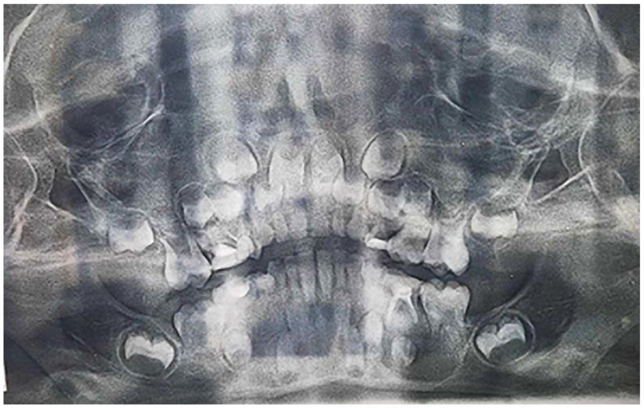
Panoramic radiograph showing a radiolucent image associated with an impacted second mandibular premolar germ.
Based on these findings, a provisional diagnosis of an inflammatory dentigerous cyst related to the impacted premolar was made. A preventive approach was chosen to facilitate proper eruption of the second premolar. Cyst decompression was planned via the extraction socket of the deciduous molar (tooth 85). A biopsy was also performed to confirm the diagnosis.
The patient’s legal guardian understood the procedure as part of the treatment and provided informed consent. (Attached file + last page)
*Device Design and Fabrication
The digital workflow began with the acquisition of a Standard Tessellation Language (STL) file representing the maxillary and mandibular arches, along with the patient’s occlusal relationship, using an intra oral scan Aoralscan 3 (Shining 3d, Hangzhou, China). Through of a specialized CAD software (EXOCAD Dental CAD 3.1 rijeka (Exocad Gmbh, Darmstadt, Germany)), the digital model was processed to design a custom removable decompression appliance.
The Computer-Aided Design process included the virtual extraction of the retained deciduous tooth, followed by the placement of a prosthetic tooth precisely adapted to the mesiodistal space, ensuring no contact with the opposing tooth. (Figure 2). The same module used for designing removable prostheses was utilized to construct the base and clasps, ensuring precise adaptation to adjacent teeth. A cylindrical drainage tube was integrated by perforating both the prosthetic tooth and the device’s base. Its length and diameter were customized to the patient’s cyst anatomy, as determined by Cone Beam Computed Tomography (CBCT).

Digital work flow for the device’s design: (a) virtual removal of the decidious tooth, (b) design of the device’s base, clasps and replacement tooth, (c) device adjustment, (d and e) incorporation of the cylindar drainage tube, and (f) final design of the device’s base and replacement tooth.
A bonding agent was applied to securely attach the replacement tooth to the 3D-printed resin base.

Delivery of the 3D decompression appliance during the same procedure.

Clinical presentation at 2-week postoperative of follow-up.
A panoramic radiograph obtained 6 months after the decompression procedure showed a significant reduction in the size of the odontogenic cyst and progressive bone regeneration around the affected area, with the permanent premolar nearly erupting in the correct axis and position (Figure 5). Given the favorable resolution of the lesion and the natural eruption of the tooth, no further orthodontic tooth traction is necessary at this stage.

Panoramic radiograph 6 months after decompression.
Discussion
Various decompression devices have been described in the literature,6 -9 designed to drain fluids secreted by the cyst epithelium through a tube extending into the cyst cavity. Some appliances were made from polyethylene tubing 10 and sutured to the soft tissues to prevent instability or loosening of the tube within the cyst cavity. 11 Other devices can be anchored to the bone using fixation screws 12 or attached to neighboring teeth with wire ligatures or orthodontic brackets. 13 Additionally, nasogastric tubes, urethral catheters, luer syringes, intravenous administration sets, dual nasal trumpet stents, and saline cuffs have also been used for cyst decompression.14-18
The integration of digital workflows in the design and application of decompression devices for dentigerous cysts offers several advantages over traditional methods, including increased accuracy, reduced procedural time, and improved patient outcomes. 19 Removable appliances used for decompression allowed our patient to thoroughly clean the device and rinse the cystic cavity while maintaining space and esthetic function by replacing the missing teeth during the healing period. In fact, the presence of adjacent teeth and the clasp design are key factors in ensuring the device’s stability throughout the treatment period.
While conventional decompression methods involved manual adjustments and trial-and-error approaches, often extending the duration of the surgical procedure, digital workflows provided significant time savings and reduced the risk of complications.20-23 A recent study reporting a case series of 6 odontogenic cysts treated with decompression devices fabricated using digital workflows showed sufficient bone regeneration without complications during enucleation and follow-up. 5
Although the initial cost of digital equipment and software may exceed that of traditional methods, the long-term benefits—such as enhanced precision, fewer complications, and faster recovery times—can offset these expenses. With ongoing technological advancements and increased accessibility, the cost of digital processes is expected to decrease, making them a more viable option for various clinical settings in the future. 24 Research should prioritize improving digital workflows to maximize the advantages of digital technologies in decompression procedures.21,24
Conclusion
Incorporating digital processes into the development of decompression tools for cysts represents a significant advancement in dental practice. The use of imaging, computer-aided design, and 3D printing has resulted in a tailored, precise solution. This approach not only improves outcomes but also paves the way for personalized treatments in oral and maxillofacial surgery. Further research and extended monitoring are recommended to confirm the effectiveness and durability of the device across various clinical situations.
Footnotes
Acknowledgements
The authors would like to thank the oral surgery department in Farhad Hached university hospital and all the staff for allowing the procedure to be performed.
Ethical Considerations
Ethical approval was not required.
Author Contributions
Ghada Bouslama and Aya Mtiri contributed in performing the experiment and drafting the manuscript. Ghada Bouslama and Hajer Zidani performed the analysis and partly the manuscript preparation. Lamia Oualha and Souha Ben Youssef participated in revising the manuscript and providing important intellectual content. All authors have provided approval of the final manuscript.
Funding
The author(s) received no financial support for the research, authorship, and/or publication of this article.
Declaration of Conflicting Interests
The author(s) declared no potential conflicts of interest with respect to the research, authorship, and/or publication of this article.