
Abstract
Purpose:
To report a rare incidence of corneal endothelial cell loss following airbag injury.
Observation:
A 27-year-old female who sustained a motor vehicle accident with airbag deployment presented with hand motion vision in the left eye. Ocular examination demonstrated corneal abrasion, significant corneal stromal edema with descemet folds, and a central pachymetry of 999 µm. The patient was managed with topical steroids and antibiotic drops. One week later, specular microscopy revealed a central endothelial cell count of 2200/mm2 with pleomorphism, polymegathism, and a decreased central corneal thickness of 569 µm. A repeat of specular microscopy 6 months later showed a decreased central endothelial cell count of 1611/mm2 with recovered visual acuity of 20/30.
Conclusion:
Corneal endothelial loss is a severe complication of ocular airbag injury. Serial ophthalmic assessment is recommended because endothelial cell loss may continue for some time after the initial impact.
Introduction
Direct ocular trauma secondary to airbag injury is diverse and can be sight-threatening. Pearlman et al 1 reported that the cornea is the most common anatomical part involved. Corneal abrasion followed by hyphema is the most common ocular insult. 1 Corneal endothelial cell loss, however, is less common. Herein, we report a case of corneal endothelial cell loss following airbag injury.
Case Report
A 27-year-old female, who is medically free with no past ocular history, presented with vision loss in the left eye after being involved in a motor vehicle accident. The patient reported wearing a seat belt and driving at a speed of 80/km/hour when she hit another vehicle. The airbag system immediately deployed, and she had no extraocular trauma. Initial examination revealed visual acuity of 20/30 in the right eye and hand motion in the left eye with no relative afferent pupillary defect. Periocular examination revealed mild periorbital swelling in the left eye. The anterior segment examination demonstrated a central corneal epithelial defect with marked corneal stromal edema and descemet folds (Figure 1A). The remaining ocular examination was unremarkable, including the right eye. Anterior segment optical coherence tomography (AS-OCT) demonstrated profound corneal stromal edema with descemet folds and a thickness of 999 µm (Figure 1B). The patient was started on topical prednisolone acetate 1% Q6H, moxifloxacin 0.5% Q6H, and cyclopentolate 1% Q8H. One week later, specular microscopy revealed a central endothelial cell count of 2200/mm2 with pleomorphism, polymegathism, and a decreased central corneal thickness of 569 µm (Table 1). Topical prednisolone acetate 1% was tapered over 3 weeks, and the other treatments were discontinued. Six months later, the previously noted corneal edema has resolved (Figure 2A and B), repeated specular microscopy revealed a decreased central endothelial cell count of 1611/mm2 (Table 1). and the visual acuity has recovered to 20/30. Specular microscopy of the un-injured right eye demonstrated a normal endothelial cell count (Figure 3).

(A) Slit lamp photo of anterior segment showing central diffuse corneal edema with descemet folds and (B) AS-OCT at the time of presentation showing a significant corneal edema with descemet folds.
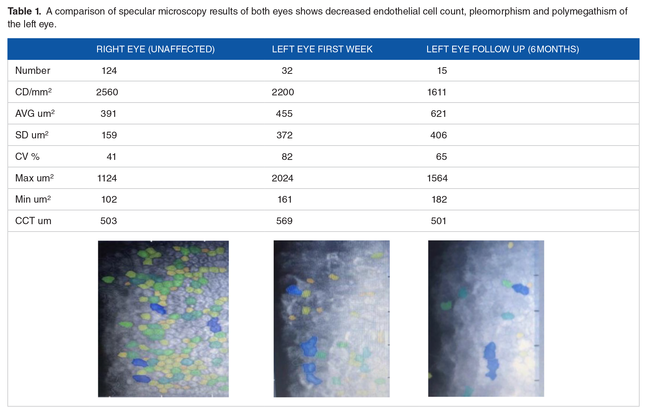
A comparison of specular microscopy results of both eyes shows decreased endothelial cell count, pleomorphism and polymegathism of the left eye.

(A) Slit lamp photo of anterior segment at 6 months showing clear cornea and (B) AS-OCT at 6 months demonstrates decreased corneal thickness.

Specular microscopy of the right eye demonstrates normal corneal endothelial cell count.
Discussion
The corneal endothelium plays a significant role in maintaining corneal clarity and hydration. It is generally believed that the corneal endothelium has a limited ability to replace lost cells; thus, damage involving this structure can have a deleterious impact on vision. Kazumi Fukagawa and his colleagues probably mentioned the first report of endothelial cell loss following airbag injury. 2 They demonstrated a 51% reduction in the endothelial count by specular microscopy in a young lady who incurred airbag ocular trauma. Years later, Geggel et al reported a similar case of a 38-year-old female who sustained multiple ocular injuries secondary to airbag trauma. The patient developed persistent corneal edema that failed to resolve despite aggressive treatment with steroids and eventually required corneal transplantation. 3 In a small series of pediatric airbag-associated ocular trauma cases, Walker et al. obtained an endothelial cell count of 9 eyes (6 subjects, median age of 11.5 years) that sustained airbag injury after a median of 3.8 years and compared them with age-matched controls. They found a statistically significant difference of 547 cells/mm2 between the mean endothelial cell count of both groups. One patient developed bullous keratopathy and required corneal transplantation. 4 A recent report described a 50-year-old female who developed paracentral focal corneal edema with a decreased endothelial cell count of 1430/mm2 following an airbag injury. Improvement in the endothelial count to 1850/mm2 was observed 1 month later, along with the full resolution of edema and recovery of vision. 5 Unfortunately, our patient had a significantly reduced endothelial count at follow-up after 6 months, although the patient’s corneal edema has resolved. However, it is unclear whether this endothelial cell loss will continue over time and potentially lead to bullous keratopathy or not. It has been reported that the normal mean endothelial cell count for healthy individuals aging 20 to 30 years is 2933.75 cells/mm2 ± 345.92 (mean ± SD). 6 Although the underlying mechanism of endothelial cell loss is unclear, a possible explanation is that it is due to sudden cellular expansion after the contusion injury. A driver’s position near the airbag and inflator power were associated with more significant endothelial cell loss. 2 In addition, it has been shown experimentally that during severe ocular trauma, the anterior-posterior diameter of the globe can decrease by up to 41%. This deformation may allow the corneal endothelium to come into contact with the crystalline lens and iris, leading to damage and subsequent loss. 7
Conclusion
Corneal endothelial loss following airbag injury is an exceedingly rare and critical complication. Prognosis is variable and potentially related to the magnitude of the initial impact. Regular ophthalmic assessment is recommended because endothelial cell loss may continue with time.
Footnotes
Funding:
The author(s) received no financial support for the research, authorship, and/or publication of this article.
Declaration of conflicting interests:
The author(s) declared no potential conflicts of interest with respect to the research, authorship, and/or publication of this article.
Author Contributions
All authors contributed to manuscript revision and approved the final version of the manuscript.
Patient Consent
The patient consent to publish the case report was obtained. This report does not contain any personal information that could lead to the identification of the patient.
Data Availability Statement
Data sharing is not applicable to this article as no datasets were generated or analyzed during the current study.