
Abstract
The study investigated the antidiabetic potentials of the fruit extract of Parquetina nigrescens with the aim of justifying its folkloric antidiabetic usage in some part of Nigeria. Acute toxicity test of the plant extract was assessed using Lorke’s method. Its antidiabetic activities were assayed in α-amylase, α-glucosidase, glucose, and streptozotocin-induced diabetic rats’ models at various doses with acarbose and glibenclamide (5 mg/kg) as positive controls. Molecular docking studies were performed to identify the antidiabetic constituent of the extract and elucidate its possible mechanism of action. The estimated median lethal dose
Introduction
A medicinal plant has been described as ‘natural plant material utilized in the absence of industrial processing for the treatment of diseases on a local or regional scale’.1,2 It is also any plant containing substances that are effective therapeutically or are precursors for drug manufacture in one or more of its organs, such as seeds, root, bark, leaf, fruit, stem, skin, flowers, or even the entire plant. They produce and store a variety of metabolites that have physiological impacts on living organisms.3,4 The universal role of plants in the treatment of diseases is exemplified by their employment in all the major systems of medicine, irrespective of the underlying philosophical premise. 5 Many drugs of importance of today such as digoxin, tubocurarine, reserpine, vincristine, vinblastine, and artemisinin were obtained from plants.1,5 Hence, continued investigation of ethnomedicinal plants for newer drugs or templates for semisynthesis is imperative.
Diabetes mellitus is a syndrome characterized by inappropriate fasting or postprandial hyperglycaemia and its metabolic consequences which include disturbed metabolism of carbohydrate, protein, and fat which results from a deficiency of insulin secretion or its action. 6 It is a major cause of morbidity and mortality both in developing and developed countries, and the incidence is rising rapidly with sub-Saharan Africa experiencing the largest percentage increase between 2013 and 2035. 7 Diabetes has become more common over the world, with estimates of 151 million adults in 2000, 194 million in 2003, 246 million in 2006, 285 million in 2009, 366 million in 2011, 382 million in 2013, 415 million in 2015, and 425.0 million in 2017. According to a recent report, 463 million persons worldwide have diabetes in 2019, representing 9.3% adult populace (20–79 years). This number is expected to surge to 578 million (10.2%) in 2030 and 700 million (10.9%) in 2045. 8
Enzymes are directly involved in the pathophysiology of diabetes and plays significant roles in its management through their inhibition or activation by standard drugs or antidiabetic plants. 9 Alpha amylase, alpha glucosidase, sodium-glucose cotransporter-2 (SGLT2), dipeptidyl peptidase IV (DPP IV), peroxisome proliferator activated receptor gamma (PPAR-γ) and 11β-Hydroxysteroid dehydrogenase (11β-HSD) are among the important enzymes that can provide mechanistic insight to the method through which phytochemicals manage diabetes. Alpha amylase and alpha glucosidase are enzymes responsible for the breakdown of complex carbohydrates to simple sugar molecules in the mouth and small intestine, respectively. 10 Sodium-glucose cotransporter-2 increases the blood glucose level of diabetic subjects by facilitating the reabsorption of glucose from the urine into the kidney, while DPP-IV increases the incretin levels thereby improving glucose tolerance and enhancing insulin secretion.11,12 Peroxisome proliferator activated receptor gamma function by increasing insulin sensitivity of body cells such that they absorb more glucose resident in the blood stream while 11β-HSD facilitates insulin resistance by body cells through the conversion of cortisone to cortisol.13,14 Therefore, the inhibition of alpha amylase, alpha glucosidase, SGLT-2, DPP-IV, and 11β-HSD, and the activation of PPAR-γ is a major strategy to manage diabetes in human subject.
Parquetina nigrescens (Afzel.) Bullock (Asclepiadaceae), commonly known as “Ogbo” among Yoruba speaking people in Nigeria, is a perennial shrub found in equatorial West Africa.15,16 It is commonly used in different parts of West and East Africa for the treatment of several ailments which include diarrhoea, gonorrhoea, menstrual disorders, insanity, intestinal worm infections, skin lesions, and erectile dysfunction.17,18 The root bark is used in the management of diabetes mellitus in Akure, Ondo State of Nigeria. 19 It has been reported for its antimicrobial, gastrointestinal protective,20,21 antioxidant, 22 analgesic, anti-inflammatory, 16 and antidiabetic activities.19,23 Nigrescigenin, periplogenin, strophantidin, strophantidol, 16-acetyl strophantidin, 16-hydrostrophantidin, noradrenaline, cymarin, and isorhoifolin have been isolated from the plant.6,24 Convallatoxin was isolated from ethylacetate fraction of the methanolic root bart extract as one of its antidiabetic constituents. 19
Drug candidate identification through computational methods has gained wide acceptability because it offers a cheaper, faster, efficient, and reliable route compared to the existing methods. 25 Molecular docking is a widely explored method of identifying hit molecules through their binding energy and orientation in the binding pocket of enzymes. 26 It also offers mechanistic insight into the mode of action of phytochemicals in the treatment of diseases. 27 This work was designed to investigate the antihyperglycaemic effects of the methanol fruit extract of P nigrescens in in vitro and in vivo antidiabetic models. It also uses molecular docking method to identify the chemical compounds that may be responsible for its in vitro and in vivo antidiabetic activities as well as unveil the mechanism through which the hit molecules manage diabetes through multitarget strategy.
Methodology
Materials and equipment
Rotary evaporator (RE301/601/801 model, Yamato Scientific America, Inc., U.S.A), chiller (Churchill, Instrument Co. Ltd, U.K), vacuum pump (MB 338618 model, Edwards High Vacuum Int., England), oven (Hearson & Co. Ltd, London), Mettler electronic weighing balance (AB 54 model, Mettler Toledo, U.S.A), ultraviolet (UV) lamp (254 and 366 nm) (Grant Instrument, U.K), Oral cannula, ACCU-CHEK Glucometer (model GB 11558973, Roche, Germany) with ACCU-CHECK test strips (Roche, Germany), UV spectrophotometer, Dutrao (Model SM 600, Shang Yhai Yong Chuang Medical Instrument Co. Ltd) spetrophotometric microplate reader, sodium citrate, citric acid, streptozotocin, Glibenclamide® (Sigma-Aldrich Co. LLC, U.S.A), and potassium hydroxide.
Plant materials and extraction
Parquetina nigrescens leaves were collected at Ede Road, Ile-Ife. It was authenticated at the herbarium of the Department of Botany, Obafemi Awolowo University, Ile-Ife and Herbarium specimen with voucher number, IFE 17513 was deposited. The leaves were air-dried, powdered, and macerated in methanol inside a volumetric flask for 72 hours and mechanically agitated at intervals. The extract was filtered, and the marc was re-extracted 3 times and concentrated in-vacuo to obtain a yield of 14.52% w/w.
Solvent partitioning of the extract
The extract of P nigrescens
Animals
Healthy Wistar albino rats weighing between 120 and 180 g of either sex bred under standard conditions (temp. 27 ± 3°C, relative humidity 65%) at the animal house, Department of Pharmacology, Faculty of Pharmacy, Obafemi Awolowo University, Ile-Ife, Nigeria were used for the experiment. They were fed on a standard pellet diet (Bendel Feeds, Nigeria), and water was given ad libitum.
Acute toxicity test
The experiment was carried out in 2 phases by orally administering methanol extract
Antidiabetic studies
In-vitro α-amylase inhibitory activity of the extract
The assay was evaluated using modified procedure of McCue and Shetty. 30 A volume of 100 µL of extract or acarbose (positive control) and 100 µL of 0.02 M phosphate buffer (pH 6.9 with 0.006 M sodium chloride) containing α-amylase from Aspergillus oryzae (0.5 mg/mL) were added to each tube and incubated at 25°C for 10 minutes. After preincubation, 100 µL of 1% starch solution in 0.02 M sodium phosphate buffer (pH 6.9 with 0.006 M sodium chloride) was added to each tube. The reaction was stopped with 200 µL of dinitrosalicylic acid colour reagent. The test tubes were incubated in a boiling water bath for 5 min, then cooled to room temperature. The reaction mixture was then diluted by adding 1.5 mL distilled water, and absorbance was measured at 540 nm using a microplate reader (SpectraMax, USA) by adding 200 µL in 96-well plates. The α-amylase inhibitory activity was expressed as % inhibition and also the concentrations of extract/Acarbose resulting in 50% inhibition of enzyme activity (IC50) were determined.31,32

In-vitro α-glucosidase inhibitory activity of the extract
The assay was evaluated using modified procedure of McCue and Shetty. 30 Alpha glucosidase from Saccharomyces cerevisae was purchased from Sigma. A volume of 50 µL of extract or standard drug (acarbose) and 100 µL of 0.1 M phosphate buffer (pH 6.9) containing α-glucosidase solution (1.0 U/mL) were incubated in 96-well plates at 25°C for 10 min. After preincubation, 50 µL of 1 mM p-nitrophenyl-glucopyranoside solution in 0.1 M phosphate buffer (pH 6.9) was added to each well. The reaction mixtures were incubated at 25°C for 20 minutes and stopped by adding 200 µL of 1M Na2CO3. The absorbance readings were recorded by micro-plate reader 405 nm and compared to a control which had 50 µL of buffer solution in place of the extract/acarbose. The α-glucosidase inhibitory activity was expressed as % inhibition and also the concentrations of extract/acarbose resulting in 50 % inhibition of enzyme activity (IC50) were determined.30,32

Antihyperglycaemic effect of extract on glucose-induced hyperglycaemic rats
This experiment was carried out by using groups of 6 rats each fasted for 18 hours that were orally given 10 g/kg of glucose. After 0.5 hour (time point 0), rats having blood glucose levels ⩾ 7.0 mmol/L (126 mg/dL) were considered hyperglycaemic and given (p.o.) vehicle (Tween 80 [1 %] in distilled water) (negative control) extract (100, 200. and 400 mg/kg) separately and 5.0 mg/kg glibenclamide (positive control). At 0.00, 0.50, 1.00, 2.00, and 4.00 hours, blood drop from each rat’s caudal vein was placed on to a glucometer strip inserted into the glucometer. The percentage decrease in blood glucose level at these time points was calculated and compared to the negative and positive controls.33-35
Antihyperglycaemic effect of extract on streptozotocin-induced diabetic rats
Diabetes was induced in 18 h fasted rats by intraperitoneally injecting the rats with freshly prepared solution of streptozotocin (65 mg/kg) in 0.1 M sodium citrate buffer (pH 4.5). The blood glucose levels of the rats were observed after 72 hours of induction and they were left for 5 days afterwards. Rats with fasting blood sugar (FBS ⩾ 11.0 mmol/l) were considered diabetic and separately divided into 4 groups of 8 rats viz; negative control that were orally given 1% Tween 80 in distilled water, test groups that received, 100 and 200 mg/kg (doses with the highest activity from glucose-induced hyperglycaemic experiment); the positive control group that were administered with glibenclamide (5 mg/kg). Each group was treated daily accordingly for 14 days while blood glucose levels were monitored on days 1, 4, 7, 10, and 14, and the percentage blood glucose reduction was determined and compared with that of the control.36-38
Protein and ligand preparation
The three-dimensional (3D) structures of α-amylase, α-glucosidase, SGLT-2, PPAR-γ, 11-betahydroxylsteroiddehydrogenase, and dipeptidylpeptidase-4 enzymes were obtained from the Protein Data Bank (www.rcsb.org) with PDB IDs 4GQR, 3L4Y, 3DH4, 5LSG, 1BHS and 3D4L, respectively. The amino acid residues in the binding sites of these enzymes were identified and visualized using PyMOL by considering residues with 5 Å, while those that were without co-crystalized ligand was viewed with Computed Atlas of Surface Topography of Proteins (CASTp) server (see supplementary material, Table S1). Then, the co-crystallized ligand, water molecules, co-factors and ions were removed and the pure proteins were saved in pdb format for molecular docking studies.
Ligand construction
The 3D chemical structures of standard drugs Acarbose (for α-amylase and α-glucosidase) and 27 phytoconstituentS15,24 previously isolated and identified from P nigrescens were downloaded from the PubChem database in sdf format (see supplementary material, Table S2). The energy minimization of the ligands was executed using the in-built Open Babel GUI plugin of the PyRx 0.8 software. All minimized chemical structures were converted and saved in PDB format.
Molecular docking studies
The molecular docking protocol was first validated before docking studies were performed. The validation was performed by re-docking the co-crystallized ligand into binding pocket of each enzyme. Root mean square deviation (RMSD) values of the resulting re-docked and co-crystallized ligand were estimated using PyMOL software.
To perform the virtual screening, the PDB files of the individual target proteins were loaded on MGL Tool where polar hydrogen atoms were added to each to each protein and nonpolar hydrogen atoms were merged and the resulting proteins were saved in PDBQT format. Each protein was thereafter loaded in PyRx 0.8 software and the grid box centre and the dimension of the enzyme binding site was set. Docking runs for each ligand was carried out using the built-in Autodock Vina tool with an exhaustiveness value of 50. 39 Result output with the best docking pose, lowest RMSD value, and binding energy was selected. The hydrogen bonding, hydrophobic, electrostatic, and pi-interactions of each protein-ligand complex was analysed using discovery studio software 2020.
Results and Discussion
Safety profile of P nigrescens
In the acute toxicity test of the fruit extract of P nigrescens using Lorke’s method in this study, no death or any changes in the breathing pattern of the rats were recorded during the period of the experiment. In addition, the extract did not cause any adverse effects on the skin, gastrointestinal, sensory and nervous systems of the rats when its geometric graded doses up to 5000 mg/kg were given. Hence, the extract had
Antidiabetic studies of P nigrescens fruit extract
In the α-amylase inhibitory study, the methanolic fruit extract of P nigrescens demonstrated significant inhibition of the breaking down of complex sugars into simple sugars with increase in concentration from 62.5 to 1000 (µg/mL) similar to the positive control, acarbose. Parquetina nigrescens extract with IC50 value of 0.05 µg/mL that was significantly lower than 0.81 µg/mL of acarbose indicated a significantly better antihyperglycaemic effect of the extract than acarbose (Table 1).
Alpha amylase inhibitory activity of P nigrescens fruit extract.

Data show the mean ± SEM (n = 6). Values with different superscripts within columns are significantly different (P < 0.05, 1-way analysis of variance followed by the Student–Newman–Keuls’ test).
Similar to the α-amylase inhibitory experiment (Table 1), the methanolic fruit extract of P nigrescens also showed a significant increase in activity with increase in concentration of the extract the same way as acarbose. This indicated that the extract could prevent elevation of blood glucose levels in diabetic individuals. Acarbose however, with IC50 value 0.06 µg/mL was significantly more active than the extract with the IC50 of 0.08 µg/mL (Table 2). The combination of the good α-amylase and α-glucosidase effects of the extract as demonstrated in this study (Tables 1 and 2) showed the extract to be a potential antihyperglycaemc agent with extrapancreatic mechanism of action.
Alpha glucosidase inhibitory activity of fruit extract of P nigrescens.

Data show the mean ± SEM (n = 6). Values with different superscripts within columns are significantly different (P < .05, 1-way analysis of variance followed by the Student–Newman–Keuls’ test).
Results of antihyperglycaemic studies such as Oral Glucose Tolerance Test (OGTT) in animals using insulin-stimulating drugs like glibenclamide as positive controls can be extrapolated to type 2 diabetes state in humans.33,34,35,40 Early extra-pancreatic and late insulin-stimulating mechanisms of action has been reported as the mechanisms of action of glibenclamide and was therefore used in this study to investigate the possible mechanism of action of the plant extract. 41
The results of the study showed that the negative control group of rats that received 10 g/kg glucose solution gave a time-dependent (0.5-4 h) reduction in blood glucose levels. This was caused by homeostasis mechanism of the rats showing the healthy state of its pancreas.35,42,43 The extract of P nigrescens at 100 and 400 mg/kg was devoid of activity at 0.5 to 2 h but gave 8.5% and 2.5% effect at 4 h that was significantly lower than glibenclamide (5 mg/kg). However, the 200 mg/kg of the extract elicited comparable (P > 0.05) activity to glibenclamide at 1 to 4 h indicating highest activity of the extract at this dose. Furthermore, similar profile of activity of the extract at 200 mg/kg with glibenclamide at all time points suggested insulin stimulating as its major mechanism of action at this dose (Table 3). 41
Dose-related glucose-lowering effect of P nigrescens fruit extract.

Abbreviations:
Data show the mean ± SEM blood glucose levels at the different time points expressed as percentages of levels at 0 h (To), n = 6. Values in parentheses represent the percentage reductions in blood glucose levels relative to negative control for each time point. Values with different superscripts within columns are significantly different (P < 0.05, 1-way analysis of variance followed by the Bonferroni t-test).
Generally, partitioning the extract into different organic solvents improved the antihyperglycaemic effect of the extract. The n-hexane fraction
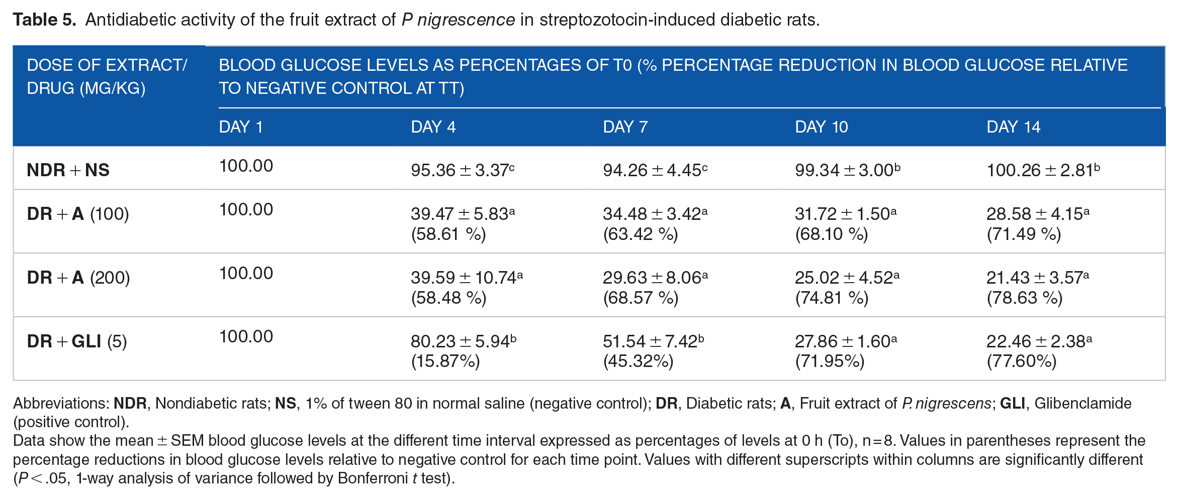
In streptozotocin-induced diabetic rats model, 100 and 200 mg/kg doses that showed effectiveness in the glucose-induced antihyperglycaemic study both in the extract and partition fractions (Tables 3 and 4) were used to further validate the antidiabetic folkloric claim of the plant. The result showed that the nontreated diabetic rats were persistently hyperglycaemic throughout the 14 days of the study which confirmed that the diabetes induced in the rats by the administered streptozotocin was permanent (Table 5). The extract of P nigrescens at 100 mg/kg gave 58%, 64%, 68%, 72% blood glucose levels reduction on days 4, 7, 10, and 14, respectively, while its 200 mg/kg gave a comparable (P > .05) activity of 59%, 69%, 75%, and 79 % on the same days, respectively, which indicated that increasing the dose of the extract did not appreciably increase its activity. The positive control, glibenclamide (5 mg/kg) however, elicited 16%, 45%, 72% and 78% antidiabetic on days, 4, 7, 10, and 14, respectively, that was significantly lower (P < .05) than that of the extract on days 4 to 7 but comparable (P > .05) on days 10 to 14. This showed that the extract at the tested doses was effective in reducing hyperglycaemia with earlier onset of action compared to glibenclamide (Table 5).
Dose-related glucose-lowering effect of the partition fractions P nigrescens fruit extract.

Abbreviations:
Data show the mean ± SEM blood glucose levels at the different time interval expressed as percentages of levels at 0 h (To), n = 6. Values in parentheses represent the percentage reductions in blood glucose levels relative to negative control for each time point. Values with different superscripts within columns are significantly different (P < .05, 1-way analysis of variance followed by Bonferroni t test).
Antidiabetic activity of the fruit extract of P nigrescence in streptozotocin-induced diabetic rats.
Abbreviations:
Data show the mean ± SEM blood glucose levels at the different time interval expressed as percentages of levels at 0 h (To), n = 8. Values in parentheses represent the percentage reductions in blood glucose levels relative to negative control for each time point. Values with different superscripts within columns are significantly different (P < .05, 1-way analysis of variance followed by Bonferroni t test).
Docking protocol validation and molecular docking analysis
The co-crystallized and re-docked ligand showed similar orientations in each case, and the calculated RMSD values between them were ⩽2.0 Å (Supplementary Material [Table S1]), indicating that the docking protocol is reproducible.
Molecular docking is a widely explored method in computer-aided drug design that helps in identifying drug candidates, predicting their extent of binding and orientation in the binding pocket of the receptors implicated in the diabetes pathophysiology. 26 The binding energy of glibenclamide was used as cut-off point against in case of enzymes without a standard drug while the marketed drug identified as the inhibitor of enzymes were used to select hit molecule where applicable and presented in Table 6.
Interaction analysis of hit molecule against the target receptors.

The result of molecular docking on α-amylase showed that isorhoifolin elicited the best binding energy of −9.1 kcal/mol compared to acarbose (−6.7 kcal/mol). Isorhoifolin formed hydrogen bonding interactions with Arg526 at 2.8576 Å, Glu404 at 1.7959 Å, Asp203 at 2.2134 Å, and Tyr299 at 2.3828 Å. Also, 2 amino acid residues (Trp406 and Phe450) formed hydrophobic interactions with isorhoifolin atoms. Furthermore, the ligand formed pi-interactions (alkyl, anion, cation, pi-pi stacked) with amino residues (Trp441, Trp539, Trp539, Phe575, and His600; Figure 1A).

Interaction diagram of isorhoifolin-4GQR (A), isorhoifolin-3L4Y (B), isorhoifolin-3L4Y (C), isorhoifolin-3DH4 (D), isorhoifolin-5LSG (E), and isorhoifolin-1BHS (F).
Isorhoifolin elicited the considerably high binding energy against α-glucosidase (−9.4 kcal/mol) compared to acarbose −7.0 kcal/mol. The phytochemical formed hydrogen bonding interactions with Glu233 (2.9106 Å), Glu233 (2.3171 Å), Asp197 (1.8375 Å), and Asp300 (3.3559 Å). Three amino acid residues (Leu162, Ile235, and His101) participated in hydrophobic interactions with isorhoifolin. Also, isorhoifolin established pi-interactions with Leu162, Ala198, Lys100, and electrostatic interaction with His101 (Figure 1B).
Isohoifolin elicited a good binding energy against SGLT2 enzyme with a binding energy of −9.5 kcal/mol as compared to the standard drugs; dapagliflozin (−8.6 kcal/mol) and glibenclamide (−9.2 kcal/mol). The ligand formed hydrogen bonding interactions with Tyr176 (2.1769 Å), Ser368 (1.8797 Å), Ile270 (2.9714 Å), and Val185 (2.5535 Å). Three amino acid residues (Tyr269, Tyr269, and Tyr138) formed 3 hydrophobic interactions with isorhoifolin, while pi-interactions were established with Ile270, Arg273, and Ile270 (Figure 1C).
Isorhoifolin gave a binding energy of −10.3 kcal/mol against PPAR-γ enzyme compared to glibenclamide (−9.6 kcal/mol). The phytochemical formed hydrogen bonding interactions with Arg288 (2.8163 Å), Glu343 (2.7433 Å), Arg280 (2.6198 Å), Glu295 (2.9249 Å), Ile326 (1.7505 Å), Ser289 (2.0059 Å), Ser342 (3.1474 Å), and Ser342 (2.9001 Å). Five amino acid residues (Ile341, Gly284, Cys185, Gly284, and Cys188) formed 3 hydrophobic interactions with isorhoifolin. Isorhoifolin also formed pi-interactions with Arg288, Ile341, and Arg288 (Figure 1D).
In the molecular docking studies of the phytoconstituents against 11-betahydroxylsteroiddehydrogenase, isorhoifolin elicited the best binding energy of −10.8 kcal/mol. The ligand formed 8 hydrogen bond interactions with Ser11, Arg37, Val66, Asn90, Gly92, Asn152, Thr35, and Gly9 at bond distances 3.0418 Å, 2.9827 Å, 2.4389 Å, 2.8657 Å, 1.9777 Å, 3.0866 Å, 2.7873 Å, and 1.9290 Å, while 6 hydrophobic interactions were formed withTyr155, Arg37, Val66, Val113, Ile14, and Met193 (Figure 1E).
Isorhoifolin gave a high binding energy of −9.4 kcal/mol against DPP-IV enzyme. It formed 7 hydrogen bond interactions with Arg61, Ile107, Thr156, Trp216, Ser106, Trp157, and Trp157 at bond distances 2.0599 Å, 2.1345 Å, 2.5708 Å, 2.5240 Å, 2.8419 Å, 3.4192 Å, and 3.2326 Å. However, no hydrophobic and pi-interactions were formed by the ligand (Figure 1F).
Elucidation of interactions between protein-ligand complexes gives a mechanistic insight into the extent of stability and binding energy elicited during molecular docking. Hydrogen bond and hydrophobic interactions play a huge role in binding affinity and stability of phytochemicals. 44 Hydrophobic interactions are formed between lipophilic groups of phytochemicals and nonpolar side chains of amino acid residues at the active site of receptors. 45 The lipophilic groups of isorhoifolin formed better hydrophobic interactions with the nonpolar chains of amino acid residues of alpha amylase, alpha glucosidase, SGLT2, DPP-IV, and PPAR-γ enzymes, thereby contributing to its stability and its high binding energy.
Pi-interactions like pi-alkyl and pi-sigma are part of noncovalent interactions formed by ligand in protein’s binding pocket, thereby contributing to the stability of drug candidates in the receptor’s binding pocket. 46 These interactions are observed with the electron and aromatic group of phytochemicals and are involved in drug intercalation and charge transfer in receptors binding pocket. 45 Isorhoifolin established good pi-sigma and pi-alkyl interactions with amino acid residues at the active site of alpha amylase, alpha glucosidase, SGLT2, 11β-HSD, and PPAR-γ enzymes.
Conclusion
The results of this study confirmed that the fruit extract of P nigrescens possessed a significant antidiabetic effect by inhibiting incursion of glucose into the blood stream as well as acting through insulin stimulation. The antihyperglycaemia activity was enhanced from further partitioning of the extract in order of increasing polarity. The binding energies obtained for the molecular docking studies identified isorhoifolin as the hit molecule against alpha amylase, alpha glucosidase, SGLT2, DPP IV, PPAR-γ, and 11β-HSD as the phytochemical that may be responsible for the antidiabetic property of the fruit extract. The phytochemical is a polar chemical compound that may be isolated from the ethyl acetate fraction owing to its polarity. Hence, it may also be among the major constituents responsible for the antihyperglycaemic activity of the ethyl acetate fraction. The isolation, characterization, structure elucidation, in vitro and in vivo antidiabetic studies of isorhoifolin is recommended for future studies.
Supplemental Material
sj-docx-1-bbi-10.1177_11779322231223857 – Supplemental material for Evaluation of the Antidiabetic Activities of the Fruit of Parquetina nigrescens (Afzel.) Bullock and In Silico Identification of Its Antidiabetic Agent
Supplemental material, sj-docx-1-bbi-10.1177_11779322231223857 for Evaluation of the Antidiabetic Activities of the Fruit of Parquetina nigrescens (Afzel.) Bullock and In Silico Identification of Its Antidiabetic Agent by Marcus D Ayoola, Yetunde B Ogundeko, Temiloluwa D Obanleowo, Deborah O Omole, Blessing N Chukwu and Kolade O Faloye in Bioinformatics and Biology Insights
Footnotes
Funding:
The author(s) received no financial support for the research, authorship, and/or publication of this article.
Author Contributions
This work was carried out in collaboration among all authors. The author, MDA designed and supervised the work of ODT, ODO, CBN, and OYB, while FKO co-supervised the work. Manuscript was prepared by MDA and FKO, read by all authors and approved for publication, while author MDA processed for publication
Declaration of Conflicting Interests:
The author(s) declared no potential conflicts of interest with respect to the research, authorship, and/or publication of this article.
Supplemental Material
Supplemental material for this article is available online.
References
Supplementary Material
Please find the following supplemental material available below.
For Open Access articles published under a Creative Commons License, all supplemental material carries the same license as the article it is associated with.
For non-Open Access articles published, all supplemental material carries a non-exclusive license, and permission requests for re-use of supplemental material or any part of supplemental material shall be sent directly to the copyright owner as specified in the copyright notice associated with the article.