
Abstract
Transcutaneous electrical nerve stimulation (TENS) is extensively used as pain relief through endorphins release. Moreover, recent findings showed a role in the activation of the autonomic nervous system (ANS); it was evidenced by modification in the heart rate variability and ANS-related marker. The objective of this pilot study is to evaluate salivary alpha amylase (sAA) as a marker of stress in two groups of healthy subjects, one receiving ultra-low frequency transcutaneous electrical nerve stimulation (ULF-TENS) and one without stimulation. Sixty healthy people were enrolled. The test group consisted of 30 participants (15 men, 15 women). The control group consisted of 30 participants (15 men, 15 women). Statistical analysis showed that sAA levels were statistically different between men and women independently from TENS; we hypothesize that treatment could influence sAA levels because it is thought to activate μ opioid receptors. The results of this study seem to indicate that the analysis of sAA, through a non-invasive saliva sample, could be an efficient aid for understanding the functions of the autonomic nervous system.
The salivary alpha amylase (sAA) enzyme or ptyalin is one of the most important enzymes present in human saliva. The ptyalin has multiple functions: it allows digestion to begin in the oral cavity and plays an important role in the modulation of adhesion and bacterial growth on dental surfaces. 1 The sAA activity correlates to the sympathetic-adrenal-medullary axis. For this reason, the measurement of sAA activity was used in the study, to investigate the psychobiology of stress, suggesting the existence of a correlation with various acute and chronic stress-connected diseases.2–4 According to the recent literature, subjects afflicted with chronic pain, among which temporomandibular disorders (TMD), suffer from a dysregulation of the descending systems of pain modulation (and the periaqueductal gray plays a key role) and of the hypothalamic-pituitary-adrenal axis (HPA) that can be evaluated through the study of the plasmatic cortisol.5–7 Plasmatic cortisol levels are correlated with the salivary cortisol and sAA levels. 8 Cortisol is the principal circulating glucocorticoid released by adrenal glands, controlled by pituitary adrenocorticotropic hormone (ACTH) and hypothalamic corticotropin releasing hormone (CRH). Moreover, the interaction between CRH, endorphin system, and the opposite effect on pain modulation at the central level were demonstrated.9,10 Transcutaneous electrical nerve stimulation (TENS) was used for a long time to relieve pain.11–13 It is possible that the main effect acts through modulating descending influence from the ventral–lateral periaqueductal gray (PAG) to the rostroventral medial medulla. In particular, low-frequency TENS seems to activate µ opioid receptors of rostral ventromedial medulla (RVM).13–16 Bilateral low-frequency TENS of the fifth and seventh cranial nerves was used to treat TMD based on pain relief17,18 and on surface electromyography of masticator muscles and kinesiography of jaw movement. 19 Despite the clinical relevance of TENS, the above cited studies focused on animal models, clinical pain, or jaw muscle effects in humans and no information can be drawn from them on the hypothesis of action of ultra-low frequency TENS (ULF-TENS) as a central/peripheral mediator of stress and pain. Two articles were published; the studies recruited patients with TMDs and asymptomatic patients; after 60 min of stimulation with low-frequency TENS, both groups demonstrated a decrease in the chewing muscles’ tone during electromyography. 20 In another published study, an important reduction of pain intensity in patients afflicted with TMD was noticed after 45 min of sensory-level TENS, but alterations concerning muscular activity were not noticed. 18
These results could have important implications at a clinical and research level, even if a standard protocol regarding the use of TENS in muscular activity on patients afflicted with TMD has not yet been established. The aim of the study is to evaluate sAA as a peripheral marker of the central effect mediated by ULF-TENS on the endorphin descending modulation pain system comparing two groups of healthy young subjects, one receiving ULF-TENS and one without stimulation.
Materials and methods
This study was conducted in agreement with the Helsinki Declaration. The Ethics Committee of the University of Study of L’Aquila approved this project and written informed consent was obtained from each participant. Sixty healthy Caucasian youths were enrolled in the study: both men and women (age range, 20–35 years; mean age, 30.23 ± 3.03 years). The test group was formed by 30 healthy participants (15 men, 15 women; mean age, 27.1 ± 2.6 years). The control group was formed by 30 healthy participants (15 men, 15 women; mean age, 27.6 ± 3.2 years). The inclusion criteria were as follows: in good health; aged less than 35 years; complete permanent dentition except for the third molar tooth. The exclusion criteria were as follows: pacemakers or other electronic devices; assumption of antidepressants; epilepsy; presence of systemic diseases; absence of written informed consent.
Patients of the test group were subjected to TENS and four saliva samples were taken using Saliva collection kit α amylase according to the following protocol (Table 1): T0 = First saliva sample taken T1 = Second saliva sample taken after 15 min of natural/spontaneous relaxation T2 = Third sample taken after 5 min of motoric TENS T3 = Fourth sample taken 5 min after the end of TENS (total execution time, 30 min)
Mean and standard deviation of measured variables across the subjects.
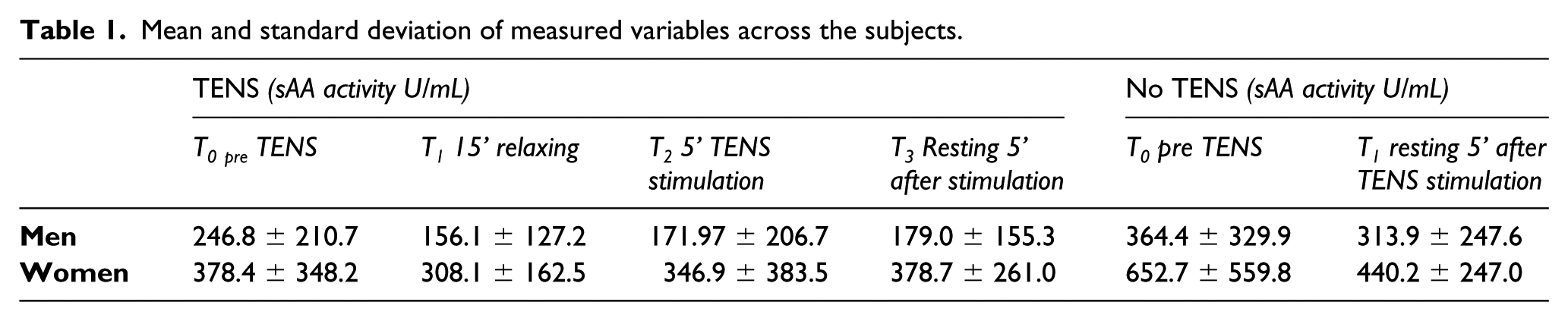
Patients in the control group were not submitted to TENS and the protocol was as follows: T0 = First saliva sample taken T1 = Second saliva sample taken after 20 min of autonomous relaxation
Saliva was collected using a sterilized glass tube manufactured by Salimetrics (SalivaBio Oral Swab (SOS) Method), following the manufacturer’s instructions. Immediately afterwards, within 2 h of collection, all samples were defrosted at −20°C. During the analysis phase, samples were defrosted and the spectrophotometric analysis was carried out following the manufacturer’s instructions supplied with the kit (Salimetrics 1-1902 – Alpha Amylase Salivary Immunoassay Kit – http://www.salimetrics.com/assay-kit/Alpha-Amylase-Salivary-Kinetic-Reaction-Kit).
Statistical analysis
Statistical analysis was performed using the R program v 3.0.3 library ‘stats’. Shapiro-Wilk test revealed a normal distribution of data and parametric statistical analysis was assessed. Analysis of variance (ANOVA) and t-test were used for differential statistics. A generalized linear model was tested assumed sAA activity as dependent variable and gender, treatments (TENS or no TENS), and time of treatment as independent variables (Table 1).
Results
Overall statistics showed that sAA activity was statistically different between gender P = 0.03. TENS and/or relaxing does not affect the sAA activity. However, sAA activity tends to be lower after TENS stimulation, as represented in Figure 1. Interestingly sAA activity remains consistently higher in women compared to men for all experimentation settings.

Comparison of sAA activity in the test and control groups.
Discussion
Data obtained from this pilot study should be interpreted carefully. First of all, the results seem to indicate that women have a lower ability to relax both in spontaneous conditions and with TENS stimulation, compared to men, in both the test and control groups (Figure 1). Many authors in the recent literature suggest that the levels of sAA are perhaps related to hormone levels. Experiments on both men and women under stress have shown that women have a greater amount of sAA.21,22 Moreover, some authors focused on the locus coeruleus-noradrenergic neuromodulation system, 23 one of the main centers regulating arousal, vigilance, alertness,24–26 pain, and sensory afferents, and is probably involved in a variety of disorders. Opioids reduce the activity of the locus coeruleus.27,28 Low-frequency TENS causes the generation of endorphins from ventral–lateral PAG and the activation of the opioid descending path to RVM. In addition, PAG fibers meet the pericoeruleus region in which opioids are generated on the dendritic system that comes from locus coeruleus neurons. 29 For this reason, perhaps low-frequency TENS causes a lowering of the locus coeruleus frequency tone firing thus reducing simultaneously the state of arousal, perception of pain, and state of relaxation. In particular, TENS stimulation increases relaxation, especially in men (Table 1). The stress influence on sAA has already been reported by several authors. 30 This result may prove that TENS could reduce stress, as other scientific studies have already reported. However, it is important to notice that this study has some limitations: the small size of the sample analyzed and the lack of randomization of the two groups. In future, it would be highly desirable to carry out a larger number of analyses recruiting a larger sample size, possibly more homogeneous; and also analyzing the salivary cortisol levels strictly correlated with the sAA levels.
By doing this, authors will have more solid and accurate evidence for scientific literature. Finally, the results of this pilot study seem to indicate that analysis of sAA, through a non-invasive saliva sample, could be an efficient aid to understand the functions of the autonomic nervous system. In future, additional studies will be needed with further analysis and additional combinations of stress-markers, to gain a complete and full-scale understanding of the mechanisms that control all our bodily functions.
Footnotes
Declaration of conflicting interests
The author(s) declared no potential conflicts of interest with respect to the research, authorship, and/or publication of this article.
Funding
This research received no specific grant from any funding agency in the public, commercial or not-for-profit sectors.