
Abstract
Background:
Cancer is one of the major heterogeneous disease with high morbidity and mortality with poor prognosis. Elevated levels of reactive oxygen species (ROS), alteration in redox balance, and deregulated redox signaling are common hallmarks of cancer progression and resistance to treatment. Mitochondria contribute mainly in the generation of ROS during oxidative phosphorylation. Elevated levels of ROS have been detected in cancers cells due to high metabolic activity, cellular signaling, peroxisomal activity, mitochondrial dysfunction, activation of oncogene, and increased enzymatic activity of oxidases, cyclooxygenases, lipoxygenases, and thymidine phosphorylases. Cells maintain intracellular homeostasis by developing an immense antioxidant system including catalase, superoxide dismutase, and glutathione peroxidase. Besides these enzymes exist an important antioxidant glutathione and transcription factor Nrf2 which contribute in balancing oxidative stress. Reactive oxygen species–mediated signaling pathways activate pro-oncogenic signaling which eases in cancer progression, angiogenesis, and survival. Concomitantly, to maintain ROS homeostasis and evade cancer cell death, an increased level of antioxidant capacity is associated with cancer cells.
Conclusions:
This review focuses the role of ROS in cancer survival pathways and importance of targeting the ROS signal involved in cancer development, which is a new strategy in cancer treatment.
Keywords
Background
Cancer is one of the major heterogeneous diseases with high morbidity and mortality. Despite extensive research and considerable efforts for developing targeted therapies, it is still an alarming condition with a poor prognosis and high mortality. Numerous studies have provided evidence that change in redox balance and deregulation of redox signaling are common hallmarks of cancer progression and resistance to treatment. It is well known that cancer cells show persistently high levels of reactive oxygen species (ROS) due to oncogenic transformation including alteration in genetic, metabolic, and tumor microenvironment.
1
Recent studies have demonstrated that cancer cells are highly adapted to elevated levels of ROS by activating antioxidant pathways. Thus, targeting the ROS signaling pathways and redox mechanisms involved in cancer development are new potential strategies to prevent cancer. Reactive oxygen species are constantly generated during the metabolic process, and their nature of existence in the form of free radicals, ions, and molecules with a single unpaired electron confers them high reactivity.
2
Broadly, ROS are grouped as oxygen-free radicals which include hydroxyl radical (·OH), superoxide (O2·−), organic radicals (R·), alkoxyl radicals (RO·), nitric oxide (NO·), peroxyl radicals (ROO·), disulfides (RSSR), thiyl peroxyl radicals (RSOO·), sulfonyl radicals (ROS·), and thiyl radicals (RS·). Other forms of ROS include singlet oxygen
List of free radicals and their biological damage.

Cellular Generation of ROS
During an aerobic cellular metabolism, ROS are constantly generated from oxygen. Mitochondria contribute the maximum in the generation of ROS as it consumes approximately 80% of molecular oxygen during oxidative phosphorylation as shown in Figure 1. It is well known that the electron transport chain (ETC) encompasses 5 complexes, namely, complexes I-IV and adenosine triphosphate (ATP) synthase on the inner membrane of mitochondria. During the process of cellular respiration, electrons are transported through a series of mitochondrial complexes to the terminal electron acceptor, molecular oxygen (O2). In the process of cellular metabolism, the electrons released from the ETC react with O2 to produce superoxide

Mitochondrial generation of ROS.
Another important signaling associated with ROS production is the membrane-bound enzyme NADPH oxidases (NOXs). However, the NOX-mediated release of ROS, by the mechanism of the oxidative burst, is commonly associated with the immune response and mediated by cells of immune system, such as macrophages and neutrophils, and by inflammatory reactions. The NOX-mediated mechanism involves various stages such as activation of NOX genes and transmembrane proteins for the transport of electrons across biological membranes where there is a reduction of molecular oxygen into superoxide by NOX as a part of redox signaling. 14
Oxidative Stress–Induced ROS Generation
Free radicals’ contribution is multifaceted in carcinogenesis and the malignant progression of tumor cells, which may be considered as a unique characteristic of cancer. In general, low concentration of ROS acts as the mitogens and promotes cell proliferation and survival, whereas intermediate concentration leads to a transient or permanent cell cycle arrest and induces cell differentiation. At high concentration, ROS can produce oxidative damage, especially in the DNA, causing mutations which eventually lead to cancer. 15 Oxidative damage caused by ROS also contributes to alteration of proteins; one such example is the mechanism of redox signaling involving H2O2-mediated oxidation of cysteine residues within proteins. Cysteine residues exist as a thiolate anion (Cys-S−) at physiological pH and are more susceptible to oxidation compared with the protonated cysteine thiol (Cys-SH). During redox signaling, H2O2 oxidizes the thiolate anion to sulfenic form (Cys-SOH) causing allosteric changes within the protein that alter its function. The sulfenic form can be reduced to thiolate anions by the antioxidant enzymes such as disulfide reductases, thioredoxin (Trx), and glutaredoxin (Grx) to recover its original state and functioning of the proteins. Hence, the oxidation of cysteine residues within proteins serves as a reversible signal transduction mechanism when occurring at low nanomolar concentration of a range of H2O2. At higher levels of peroxide, the oxidized thiolate anions form sulfinic (SO2H) or sulfonic (SO3H) species. Unlike sulfenic modifications, sulfinic and sulfonic forms are irreversible which results in permanent oxidative damage of the protein. This redox signaling is balanced by peroxiredoxins and glutathione peroxidases. 16
Reactive oxygen species can induce oxidative stress followed by altering cell membrane lipid bilayer by the process of lipid peroxidation of polyunsaturated fatty acids. This causes the generation of lipoperoxyl radical (LOO·), which, in turn, reacts with a lipid to yield a lipid-based radical and a lipid hydroperoxide (LOOH). These LOOHs are unstable, and they generate new peroxyl and alkoxy radicals and decompose to secondary products. Free radicals produced during lipid peroxidation have very minute and local effects because of their short life. However, the breakdown products of lipid peroxides such as aldehydes, malondialdehyde, hexanal, 4-hydroxynonenal (HNE), or acrolein serves as “oxidative stress second messengers” due to their prolonged half-life and their ability to diffuse from their site of generation. Among the products of lipid peroxidation, HNE is chemically reactive because of its highly electrophilic nature and it easily reacts with glutathione, proteins, and DNA which leads to the covalent modifications on macromolecules. 17
ROS in Genomic Instability and Cancer Development
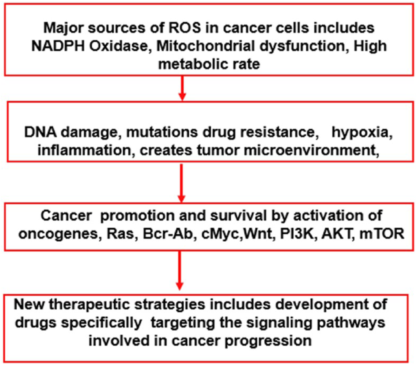
Cancer cells show a persistent metabolic oxidative stress compared with normal cells, which is mainly due to inherent mitochondrial dysfunction and NOX activation. As a part of metabolic reactions, high levels of ROS are generated and unregulated levels can lead to oxidative damage such as DNA mutation–causing cancer initiation and progression. These oxidative damages comprise a mixture of DNA lesions including base damage, DNA single-strand breaks, and DNA double-strand breaks, rearrangement of DNA sequence, base modification, DNA miscoding lesions, gene amplification, and the activation of oncogenes. 4
Elevated levels of ROS during cancer transformation are mainly due to high metabolic rate in mitochondrial, endoplasmic reticulum, and cell membranes. These changes are marked by respiratory dysfunction, low coupling efficiency of the mitochondrial ETC, raising electron leakage and increased ROS generation. 5 Cancer cells maintain their high energy levels through a high rate of glycolysis followed by lactic acid fermentation even in the presence of abundant oxygen; this is called aerobic glycolysis, also termed the Warburg effect, followed by oxidation in mitochondria. This metabolic switch is essential for the cancer cells to adapt to hypoxic conditions with less mitochondrial defect and ROS production. 18 In responsive to mitochondrial ROS (mROS) in cancer cells, hypoxia-inducible factors (HIFs) are activated to cancer cells to adapt to their diminished oxygen microenvironment which is essential for cell survival, growth, and proliferation. 19
Reactive oxygen species play a vital role at every stage of cancer development, including initiation, promotion, and progression. Increase in intracellular ROS levels may result in the activation of oncogenes and oncogenic signals including constitutively active mutant Ras, Bcr-Abl, and c-Myc which are involved in cell proliferation and inactivation of tumor suppressor genes, angiogenesis, and mitochondrial dysfunction. High metabolism in cancer induces Wnt signal, specifically Wnt/β-catenin pathway where c-Myc is regulated by Wnt/β-catenin and consequently, it can attribute greater metastatic potential. 20
ROS as a Signaling Molecule in Cancer Survival
H2O2 reversibly oxidizes cysteine thiol groups of phosphatases such as phosphatase and tensin homolog (PTEN), protein-tyrosine phosphatase 1B (PTP1B), and protein phosphatase 2 (PP2) which cause loss of their activity and promote the activation of the PI3K/Akt/mTOR survival pathway. Moreover, H2O2-mediated oxidation of prolyl hydroxylase domain protein 2 (PHD2) causes the stabilization of HIF1 during hypoxia, which is important for cancer metastasis. 21 The possible mechanism involved in promoting targeted protein oxidation by H2O2 may involve the ability of ROS-scavenging enzymes such as glutathione peroxidase to measure and transduce the H2O2 signal which is called as redox-relay mechanism. Another mechanism proposed is called as floodgate model in which oxidation causes inactivation of the ROS-scavenging enzymes by hyperoxidation or phosphorylation causing localized increases in H2O2 leading to protein oxidation. 22
Studies have demonstrated that H2O2 can promote the activation of Ras and growth factor signaling which in turn activates PI3K/Akt/mTOR, MAPK/ERK and inactivates PTEN signaling cascades. Recently, it has been demonstrated that breast cancer–associated mitochondrial DNA haplogroup promotes neoplastic growth via ROS-mediated AKT activation. 23 Oncogenic mutations in Ras can lead to increased ROS production through NOX isoform (NOX4) which enhances cell proliferation. In a recent study, it was demonstrated that Kras-derived mROS-activated nuclear factor κ-light-chain-enhancer of activated B cells (NF-κB) through polycystin-1(PKD1) to upregulate epidermal growth factor receptor (EGFR) pro-proliferative signaling. Moreover, ROS promote angiogenesis and metastasis by stabilizing HIF and activating 5′-adenosine monophosphate–activated protein kinase (AMPK) and one-carbon metabolism pathways to enhance NADPH production and maintain redox balance. 24 Hypoxia-induced mROS stabilize the oxygen-sensitive HIF-α subunit by dimerization with HIF-β and induce the expression of pro-angiogenic genes. Under normoxic conditions, with sufficient oxygen, HIF-α is degraded following PHD2-mediated hydroxylation and subsequent recognition by the E3 ubiquitin ligase von Hippel-Lindau protein. To raise the antioxidant capacity and prevent ROS-mediated tumor cell death, AMPK is activated which promotes NADPH production and prevent anabolic processes that require NADPH consumption. In addition, in hypoxia, HIF-dependent upregulation of the one-carbon metabolism enzyme serine hydroxymethyltransferase (SHMT2) promotes mitochondrial serine catabolism and NADPH production. 25
It is a well-known fact that metastasis requires endothelial mesenchymal transition, loss of cell-cell adhesion, dissociation of cancer cell from the primary site, and damage of basement membrane. Moreover, ROS have been shown to regulate numerous signaling pathways (eg, the MAPK and PI3K/Akt pathways) and transcriptional activities (eg, HIF and Snail) to enhance cancer cell migration and invasion. Furthermore, ROS-dependent oxidation of v-Src causes enhancement in the invasion potential and anchorage of Src-transformed cells. Reactive oxygen species has been reported to confer anoikis resistance to cancer cells through the oxidation and activation of Src, leading to constitutive, ligand-independent EGFR activation and prosurvival signaling. Elevated ROS levels resulting from mutations in mitochondrial DNA, which impair the complex I activity, have also been shown to promote the metastasis 26 (Figure 2).

Role of ROS in signal transduction.
Cancer Progression and Effect of ROS in Metabolic Pathways
Recent evidence suggested that alteration and deregulation of redox signaling are prominent hallmarks of cancer and can be strongly compromised in malignancy and drug resistance. The cancer cells exhibit persistently high levels of ROS as a consequence of genetic, metabolic, and microenvironment-associated instability. This high level of ROS is compensated by increased antioxidant ability by the cancer cells. Although it is contradictory, this pro-oxidant shift enhances tumor growth and activates an inflammatory response, stabilizing the HIF-14 and eventually reprogramming the metabolism.27–29 Due to the persistent high ROS microenvironment, cancer cells adapt to an efficient mechanism of ROS detoxification by showing high dependency on antioxidant system for their survival. Thus, different strategies have to be built up to disrupt the functional cross talk or elevating the burden of oxidative stress in the presence of selective metabolic inhibitors which might induce lethality to cancer cells. 6 Evidence suggest that cancer progression involves numerous alterations in specific metabolic pathways involved in synthesis of proteins, lipids, and nucleotides. Besides this, there is an increase in the generation of NADPH and GSH, an antioxidant, and redox cofactors such as NADH and FADH. There is a reciprocal cross talk between metabolism and redox balance of cancer cells, with a particular emphasis on the role of glycolysis, glutaminolysis, fatty acid oxidation, one-carbon metabolism, and the pentose phosphate pathway. 30
Glycolysis is an essential pathway through which glucose is transformed to pyruvate with the generation ATP and NADH. Otto Warburg in 1924 reported that cancer cells extensively use glycolytic pathway regardless the presence of sufficient partial pressure of oxygen; this phenomenon is known as the Warburg effect. 31 Various studies have reported that oncogenic activations and loss of tumor suppressor genes cause a proglycolytic shift which benefits the cancer cells to sustain growth and proliferation by providing macromolecules and reducing equivalents, mainly pentose phosphate pathway–derived NADPH or glutaminolysis-derived GSH which are important to overcome oxidative stress in cancer cells. Recent studies showed that cancer cells in glucose deprivation increase glucose metabolism to restrict the burden of ROS and prevent cell death. It is also noticed that inhibition of lactate dehydrogenase also impaired the cancer cell progression by decreasing the intracellular ATP levels and inducing oxidative stress. However, inhibition of glycolysis has proven to represent a successful strategy in selectively increasing cytotoxicity in pancreatic and breast cancer cells but not in normal cells. 32
Fatty acid oxidation occurs in the mitochondria with the generation of NADH, FADH2, and acetyl-CoA to support biosynthetic pathways and produce ATP. However, in cancer cells, a consistent fraction of the acetyl-CoA enters into the tricarboxylic acid cycle and generates citrate, which is therefore exported into the cytosol and enters into metabolic reactions catalyzed by the malic enzyme and the isocitrate dehydrogenase, which eventually produces NADPH. Cancer cells use these reducing equivalents as a preventive measure against cell death under loss of matrix adhesion and metabolic stress conditions. Etomoxir is a drug found to impair NADPH production and promote oxidative stress–induced cell death in human glioblastoma cells associated with profound ATP depletion and to strengthen the proapoptotic effect of cytotoxic agents in human leukemia cells. 33
Pentose phosphate pathway is a major catabolic pathway of glucose through which cancer cells produce large amounts of ribose 5-phosphate, a precursor of nucleotide synthesis and NADPH, and promote both ROS generation (NOXs) and ROS detoxification (by replenishing the reduced GSH and TRX pools). Activation of the PPP represents a key hallmark of many cancers where this metabolic pathway is found at the crossroad between glycolytic activity, unrestricted proliferation, and scavenging of excessive ROS. 34
Immune Evasion of ROS Through Antioxidant Defense in Cancer
Antioxidants are the first line of defense against free radicals and other oxidants by either neutralizing or halting the formation of free radicals. There are set of enzymes which are responsible for transforming free radicals into stable and less damaging molecules which includes catalase (CAT), superoxide dismutase (SOD), and glutathione peroxidase (GPx), etc. 35 Some of them are mentioned in the following sections.
Glutathione
Glutathione has an indispensable role in maintaining intracellular redox homeostasis usually during hypoxia and high production of ROS and NO·. Glutathione exists in reduced (GSH) and oxidized (glutathione disulfide, GSSG) states. In its reduced state, it sequestrates ROS, which is transformed and recycled by the action of the glutathione reductase enzyme (GRd). The electron source used by this enzyme is NADPH, which is mainly derived from the pentose phosphate pathway. GSH is also an essential cofactor for the enzyme GSH peroxidase, which is involved in detoxification of peroxides, including the H2O2 generated in cell membranes that react with GSH (Figure 3A). Peroxides have a dual role in carcinogenesis; about 90% of total glutathione exists in reduced form GSH and less than 10% in disulfide form GSSG; change in the ratio indicates oxidative stress. 7

Immune evasion of ROS through antioxidant defense in cancer survival. (A) H2O2 generated in the matrix can oxidize biomolecules including proteins, lipid membrane, and DNA-generating alkoxy radical by Fenton reaction. H2O2 generated can be converted to HO2 by catalase, glutathione peroxidase. Glutathione exists in reduced form as GSH in the enzyme glutathione peroxidase and gets oxidized (GSSG) in the process of reduction of H2O2. Oxidized glutathione (GSSG) is reduced by glutathione reductase, which obtains its equivalents NADPH from HMP shunt. (B) Nrf2-ARE pathway activation takes place when cell is subjected to oxidative stress. In the cytoplasm, Nrf2 is constitutively bound to Keap1 form in the form of dimer—Nrf2-Keap1. During oxidative stress, Nrf2 is released from Keap1, hence allowing the transcriptional factor Nrf2 to translocate to the nucleus. Nrf2 in MAF family proteins binds with ARE-regulated genes. This activates antioxidant enzymes, pro-inflammatory response, and cell survival. ARE indicates antioxidant responsive element; HMP, hexose monophosphate; ROS, reactive oxygen species.
Nuclear factor erythroid 2–related factor 2
Nuclear factor erythroid 2–related factor 2 (Nrf2) is the basic region leucine-zipper transcription factor and one of the most important master regulator of antioxidant pathways. In normal conditions, Nrf2 is bound to the endogenous inhibitor Kelch-like ECH-associated protein 1 (Keap1). Keap1 is a cytosolic protein that inhibits Nrf2 signaling by promoting Nrf2 degradation through proteasomal pathway. When ROS react with redox reactive cysteines in Keap1, Nrf2 is released from Keap1, hence allowing the transcriptional factor Nrf2 to translocate to the nucleus. In the nucleus, Nrf2 dimerizes with basic leucine-zipper partners (bZip) such as small Maf-family proteins and binds to antioxidant responsive element (ARE), which is located in the promoter of the phase II and antioxidative genes. It is a regulatory enhancer region within gene promoters. c-Jun is then supposed to act mainly as transcriptional activator, whereas the small Mafs as well as c-Myc inactivate gene transcription after Nrf2 binding. 8 Nrf2-ARE binding regulates the expression genes involved in the cellular antioxidant and anti-inflammatory defense such as phase 2 detoxification enzymes including glutathione, superoxide dismutase, glutamate-6-phosphate dehydrogenase, heat shock proteins and ferritin, and pro- and anti-inflammatory enzymes such as cyclooxygenase-2 (COX-2), inducible nitric oxide synthase (iNOS), and heme oxygenase 1; it also regulates mitochondrial biogenesis. 36 Some studies have proven that activation of Nrf2 may lead to the inhibition of pro-inflammatory responses of Cox-2 and iNOS expression. The defense against the stress may be probably due to the concentration of glutathione content and Nrf2 which further provides cytoprotective effects against Fas-mediated apoptotic pathways. 37 Nrf2 is inhibited by modifications of cysteine residues of Keap1 that apparently alter the interaction of Keap1 with Nrf2 and lead to its relocation to the cytoplasm where it is subsequently degraded by the ubiquitin proteasome. 38 Therefore, Keap1 and Nrf2 act as a cellular sensor for damage caused by free oxygen radicals by the constant shuttling of Keap1 between the nucleus and the cytoplasm under normal conditions. 39 Karyopherin-6 (KPNA6) is a protein which facilitates nuclear import and attenuates Nrf2 signaling, clearance of Nrf2 protein from the nucleus, and restoration of the Nrf2 protein to basal levels. These findings suggest that KPNA6-mediated Keap1 nuclear import plays an essential role in modulating the Nrf2-dependent antioxidant response and maintaining cellular redox homeostasis. 40 Nrf2 can be activated by cigarette smoke, infection, oxidative stress, or inflammation. Impairment of Nrf2/ARE pathway leads to oxidative stress, inflammation, and mitochondrial dysfunction. 41 Nrf2 is also considered as tumor suppressor because of its cytoprotective functions against oxidative stress. However, hyperactivation of the Nrf2 pathway creates an environment that favors the survival of normal as well as malignant cells, protecting them against oxidative stress, chemotherapeutic agents, and radiotherapy imparting it an oncogenic property, and Nrf2 can be a powerful putative therapeutic target in cancer treatment (Figure 3B).42–45 Carbonyl reductase 1 is another important enzyme that regulates the expression of Nrf2 during oxidative stress and helps to detoxify ROS. 46
It has been demonstrated that ROS are likely to participate as immunosuppressive agents in cancer microenvironment and facilitate tumor invasion, metastasis, and resistance. Studies have demonstrated that ROS play a crucial role in inhibitory activities of tumor-induced immunosuppressive cells. Therefore, ROS are not only mediators of oxidative stress but also players of immune regulation in cancer development. The ROS-mediated signaling can be regulated by antioxidant defense.47–49
ROS Regulate Metastasis via Matrix Metalloproteinases
Matrix metalloproteinases (MMPs) play a pivotal role in the processes of cancer invasion and metastasis. Metastasis is a cascade process, including cell invasion, degradation of basement membranes, and stromal extracellular matrix, eventually causing invasion and metastasis. The MMPs are a family of related enzymes that degrade extracellular matrix, which are considered to be the important factors in facilitating tumor invasion. It has been reported that increased expression of MMPs is predictive of tumor aggressiveness, metastasis, and poor patient survival. Recently, MMPs have been considered to be an important factor in triggering epithelial-mesenchymal transition (EMT). Expression of MMP-2, 3, 9, and 28 in EMT by the loss of intact E-cadherin increased motility and invasiveness, downregulation of epithelial markers, and upregulation of mesenchymal markers. Studies demonstrated that there is an involvement of Rac signaling for cytoskeletal rearrangement and in mediating integrin signaling. Several reports have demonstrated that ROS can be generated by integrin-Rac pathway, resulting in tumor cell migration and invasion. MMP-3–induced EMT appears to be mediated via induction of ROS and increased expression of the Rac1b. The ROS-quenching agent N-acetyl cysteine (NAC) effectively inhibited the MMP-3–induced EMT. These results clearly suggest that the treatment with MMP-3 stimulates the expression of Rac1b, which increases intracellular ROS, leading to the induction of EMT, suggesting that MMP-3 inhibitor or the inhibitors of ROS could be useful in the reversal of EMT or the killing of EMT-type cells or cancer stem cells leading to decreased tumor aggressiveness (Figure 4).50–56
Overview of ROS in cancer progression and specific target in cancer therapy. ROS indicate reactive oxygen species.
Conclusions
Several substantial research studies have shown that altered cell metabolism has a crucial role in the development of different types of cancer. The exact nature of the impact of oxidative stress on cancer initiation, progression, and/or response to therapy requires further investigation. Reactive oxygen species exhibit a double-edged sword, both sides of which have been exploited for potential therapeutic benefits. There is still considerable controversy as to whether ROS modulation by either antioxidant supplementation or inhibition is clinically beneficial or detrimental for cancer treatment. Antioxidants provide crucial survival and proliferation signals to cancer cells; cancer cells depend on an increased antioxidant capacity to counteract elevated ROS levels; and antioxidant inhibitors represent a promising therapeutic strategy in anticancer.
Footnotes
Acknowledgements
The authors would like to thank DST-FIST and GITAM University for providing lab facilities.
Funding:
The author(s) disclosed receipt of the following financial support for the research, authorship, and/or publication of this article: The review was supported by UGC, New Delhi, for Post-doctoral for woman (No. F.15-1/2013-14/PDFWM-2013-14-GE-AND-12376 (SA-II).
Declaration of conflicting interests:
The author(s) declared no potential conflicts of interest with respect to the research, authorship, and/or publication of this article.
Author Contributions
SK conceived of the presented idea. AB, SNG and MG developed the idea. RRM has verified and all authors have verified the final manuscript.