
Abstract
Hepatocellular carcinoma (HCC) is one of the common cancers with a high incidence and mortality. The human replication factor C (RFC) family contains 5 subunits that play an important role in DNA replication and DNA damage repair. RFCs are abnormally expressed in a variety of cancers; some of them are differentially expressed in HCC tissues and related to tumor growth. However, the expression, prognostic value, and effect targets of the whole RFC family in HCC are still unclear. To address these issues, we performed a multidimensional analysis of RFCs in HCC patients by Oncomine, UALCAN, GEPIA, Human protein atlas, Kaplan-Meier plotter, cBioPortal, GeneMANIA, String, and LinkedOmics. mRNA expression of RFCs was significantly increased in HCC tissues. There was a significant correlation between the expression of RFC2/3/4/5 and tumor stage of HCC patients. Besides, high mRNA expression of RFC2/4 was associated with worse overall survival (OS). Moreover, genetic alterations of RFCs were associated with worse OS in HCC patients. We found that genes co-expressed with RFC2/4 were mainly involved in biological processes, such as chromosome segregation, mitotic cell cycle phase transition, and telomere organization and they activated the cell cycle and spliceosome pathways. The gene set is mainly enriched in cancer-related kinases AURKA, ATR, CDK1, PLK1, and CHEK1. E2F family members were the key transcription factors for RFCs. Our results suggest that differentially expressed RFC2 and RFC4 are potential prognostic biomarkers in HCC and may act on E2F transcription factors and some kinase targets to dysregulate the cell cycle pathway. These efforts may provide new research directions for prognostic biomarkers and therapeutic targets in HCC.
Introduction
Hepatocellular carcinoma (HCC) is the most common pathological type of primary liver cancer and ranks as the sixth most common malignancy and the fourth leading cause of cancer-related death worldwide. 1 Generally, HCC patients have no typical clinical symptoms in the early stage and have been diagnosed in the middle and late stage when the symptoms are obvious. 2 Despite advances in detection methods and various targeted drugs for the diagnosis and treatment of HCC, the 5-year survival rate remains less than 5%.3,4 Therefore, it is of great clinical importance to explore diagnostic and prognostic biomarkers with higher sensitivity and specificity and new therapeutic targets.
The human replication factor C (RFC) family is a complex containing 1 larger subunit (RFC1/140 kDa) and 4 small subunits (RFC2/40 kDa, RFC3/38 kDa, RFC4/37 kDa, and RFC5/36 kDa), and they are highly conserved throughout evolution.5,6 RFCs act as clamp loaders, which has a crucial role in loading proliferating cell nuclear antigen (PCNA) onto primed DNA to elongate the DNA chain. In addition, RFCs can bind to cell cycle checkpoint proteins to initiate signal transduction downstream of DNA damage checkpoints, thus participating in mismatch repair and excision repair of damaged DNA.7,8 The expression of RFCs is upregulated in various cancers, including breast cancer, nasopharyngeal carcinoma, colorectal cancer, gastric cancer, prostate cancer, lung cancer, and hepatocellular carcinoma.9-15
Previous studies have found abnormal expression and functions of some members of the RFC family in HCC. Yao et al 9 found that the expression level of RFC3 was significantly upregulated in HCC tissues and cells and, when the HCC cells with RFC3 gene were knocked down, both cell viability and proliferation were effectively inhibited. Downregulation of RFC3 expression led to the arrest of HCC cell cycle in the S phase. Arai et al 15 also found that HCC cell proliferation decreased and that levels of apoptosis and sensitizing of chemotherapeutic drugs increased when the expression of RFC4 was downregulated in HCC cells. However, the differences in expression levels of mRNA and proteins, genetic alterations, biological functions, prognostic significance, and therapeutic targets of RFCs in HCC have not yet been completely identified. Therefore, we conducted a comprehensive and integrated bioinformatics analysis to explore the expression, prognostic values, and therapeutic targets of RFCs in HCC.
Materials and Methods
Oncomine analysis
Oncomine database (https://www.oncomine.org/) is a cancer microarray database and a platform for integrating data to mine cancer gene information. To date, the database has collected 715 gene expression data sets and 86 733 samples of cancer and normal tissues. 16 It was used to compare the transcription levels of RFCs between HCC and normal liver tissues. Student’s t test was used to analyze the difference in the expressions of RFCs in HCC. The Cut-off of P value and fold change were listed as follows: P value: .05, fold change: 1.5, gene rank: 10%, data type: mRNA.
UALCAN analysis
UALCAN (https://ualcan.path.uab.edu/index.html) is a simple, fast, and effective website tool for TCGA data mining analysis, which can directly query the expression of multiple genes in the tumor and the relationship with the prognosis. 17 In this study, UALCAN was used for analyzing the transcriptional expressions of RFCs in HCC and normal liver tissues. Difference of expression was compared by students’ t test and the cutoff of P value was .01.
Gene expression profiling interactive analysis (GEPIA) database
GEPIA (http://gepia.cancer-pku.cn/index.html) is an interactive analysis website developed by Peking University, containing RNA sequencing expression data of 9736 tumors and 8587 normal samples from The Cancer Genome Atlas (TCGA; https://tcga-data.nci.nih.gov/tcga/) and the Genotype-Tissue Expression project (GTEx; https://www.gtexportal.org/home/index.html). It provides customizable functions such as tumor/normal differential expression analysis, profiling according to cancer types or pathological stages. 18 In this study, the “Multiple Gene Comparison” module was used for the multi-gene comparison analysis of RFCs. In addition, the “Single Gene Analysis” module of GEPIA was used for exploring the relationship between RFCs expression and pathological stage. Student’s t test was used to generate P value for differential expression or pathological stage analysis. The cutoff of P value was .05.
The human protein atlas database
The human protein atlas (https://www.proteinatlas.org/) contains information about protein expression in human tissues that has been found, which can be used for screening and verifying immunohistochemical results, as well as the expression location of various proteins. 19 In this study, immunohistochemical images were used for directly comparing the protein expression of different RFCs in HCC and normal liver tissues.
Kaplan-Meier plotter analysis
The Kaplan-Meier plotter (https://www.kmplot.com/) is an online database of the relationship between mRNA expression level and clinical outcomes in cancer patients. 20 In this study, HCC patients were divided into the high-expression and the low-expression group according to the autoselect best cutoff, the overall survival (OS), and relapse-free survival (RFS) of HCC patients were analyzed by Kaplan-Meier survival plots, risk ratio (HR), 95% confidence intervals (CI), and log-rank P values. Only the probe sets with best JetSet scores for RFCs were selected to produce Kaplan-Meier plots. A statically significant difference was considered when P value <.05.
cBioPortal database
cBioPortal (https://www.cbioportal.org/) is an open-access, user-friendly, and interactive website resource for exploring and visualizing multidimensional cancer genomics dataset. 21 In this study, we analyzed the genomic profiles of RFCs in 373 HCC samples from the TCGA database, which contained mutations, putative copy-number alterations from GISTIC and mRNA expression z-score (RNA-seq V2 RSEM). The z-score threshold was ±1.8. The correlation between genetic alterations of RFCs and overall survival (OS) and disease-free survival (DFS) of HCC patients was analyzed by Kaplan-Meier plot, and the significance of survival difference was confirmed by log-rank test. The cutoff of P value was .05.
GeneMANIA analysis
GeneMANIA (http://www.genemania.org/) is an online analysis tool that searches for related expressed genes and constructs gene interaction networks through a large number of correlation data including protein and genetic interactions, pathways, co-expression, co-localization, and protein domain similarity. 22 We used it to construct RFCs gene co-expression and pathway networks and to predict their potential function.
String database
String (https://string-db.org/) is a database of known and predicted protein-protein interactions, and covers 24584628 proteins from 5090 organisms. 23 We used String to construct the protein-protein interaction network of RFCs and explore the interaction among the RFCs.
LinkedOmics analysis
The LinkedOmics (http://www.linkedomics.org/login.php/) is an online analysis platform that provides multi-omics data of 32 TCGA cancers types for multi-dimensional analysis. 24 In the study, using the “LinkedFinder” module in linkedOmics, we identified co-expressed genes associated with RFC2/4 in hepatocellular carcinoma, analyzed them using Pearson’s correlation coefficient, and the results were shown by volcano and heat maps. And utilizing the “LinkedCompare” module in linkedOmics, we identified overlapped genes associated with RFC2/4 in hepatocellular carcinoma, and the results were shown by Venn plot. In addition, we used the “LinkedInterpreter” module to conduct biological analysis of RFC2/4 and related overlapped genes by Gene Set Enrichment Analysis (GSEA), mainly including enrichment analysis of biological process, Kyoto Encyclopedia of Genes and Genomes (KEGG) pathways, kinase-target, miRNA-target, and transcription factor-target. Weighted set coverage was employed to reduce redundancy in the enrichment results. The rank criterion was a false discovery rate (FDR) <0.05, the minimum number of genes was 3, and 500 simulations were performed.
Result
Transcription levels RFCs in HCC patients
The Oncomine database was used to analyze and compare RFCs mRNA transcription levels in more than 20 cancer tissues and normal tissues (Figure 1). In Roessler Liver 2 dataset, RFC1/2/3/4/5 mRNA over-expression were found in HCC tissues compared with normal liver tissues with fold change of 2.021 (P = 6.58E−61), 1.645 (P = 8.11E−44), 2.629 (P = 6.6E−62), 4.600 (P = 1.15E−88), and 1.787 (P = 1.77E−35), respectively 25 (Table 1). The result from Roessler Liver dataset showed that RFC1/3/4/5 mRNA expression were 1.832-fold (P = 1.15E−05), 1.963-fold (P = 9.47E−09), 3.315-fold (P = 4.58E−08), and 1.806-fold (P = 1.08E−05) increase in HCC tissues, respectively 25 (Table 1). And Chen et al 26 observed 1.820-fold (P = 8.93E−20) increase in RFC1 mRNA expression and 2.031-fold (P = 5.28E−19) increase in RFC4 mRNA expression (Table 1). However, Wurmbach et al 27 found that only RFC1 mRNA expression was 5.178-fold (P = 8.71E−06) increased in HCC tissues compared with normal liver tissues (Table 1). Next, the expression of RFCs family members in HCC was assessed on the UALCAN website with RNA sequencing data based TCGA database. As was shown in Figure 2A to E, the mRNA expression of RFC1 (P = 1.62E−12), RFC2 (P < 1E−12), RFC3 (P = 1.62E−12), RFC4 (P = 1.62E−12), and RFC5 (P < 1E−12) significantly elevated in HCC tissues compared with normal liver tissues. We also compared the relative expression levels of RFCs in HCC tissues, and found that among all RFCs, the expression of RFC2 was the highest, and the expression of RFC3 was the lowest (Figure 2F).

mRNA expression of RFCs in different types of cancers (Oncomine). Color is determined by the highest gene rank percentile gene based on log fold change; red represents upregulation and blue represents downregulation. The values in each square represent the number of databases that meet our screening criteria.
The significant difference of RFCs expressions in transcription level between HCC and normal liver tissues (Oncomine).
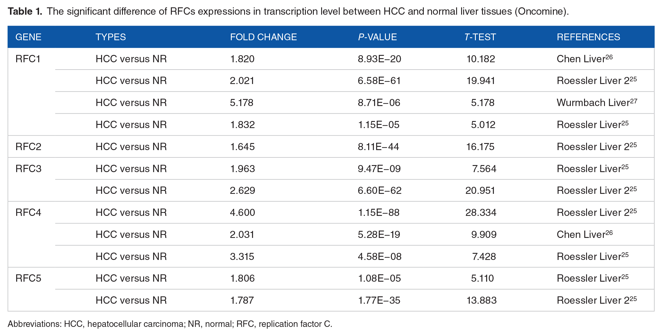
Abbreviations: HCC, hepatocellular carcinoma; NR, normal; RFC, replication factor C.
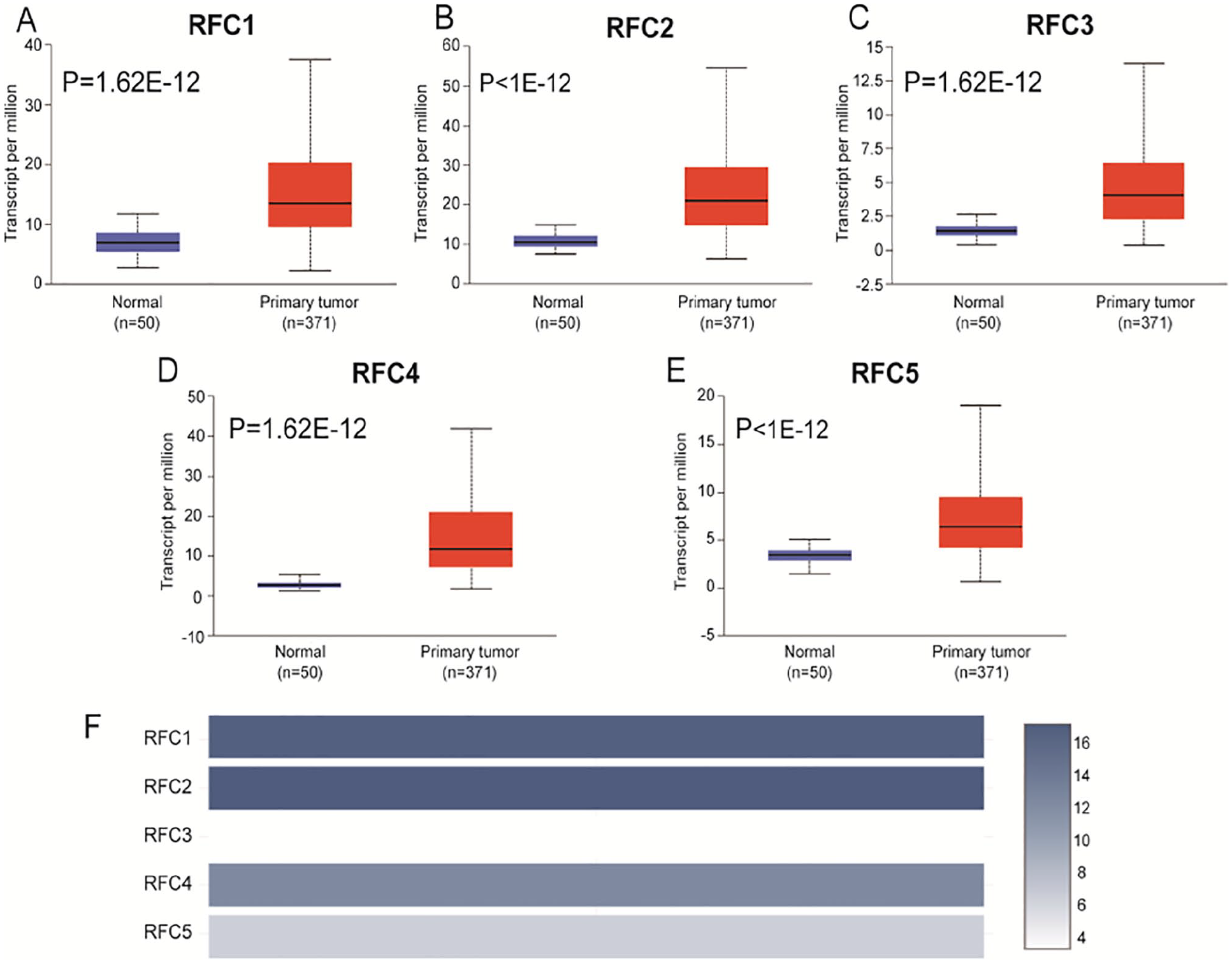
mRNA expression of RFCs in HCC tissues and normal liver tissues (UALCAN and GEPIA): (A-E) the mRNA expressions of RFCs were found to be over-expressed in HCC tissues compared to normal tissues; data were presented as the mean ± standard error; transcript per million was used to measure the expression and (F) the relative level of RFCs in HCC.
Relationship between mRNA expression of RFCs and clinicopathological characteristics of HCC patients
To evaluate the predictive value of differentially expressed RFCs in the progression of HCC patients, we analyzed the relationship between mRNA expression of RFCs and clinicopathological characteristics of HCC patients by GEPIA database. The results show that the expression levels of RFC2 (P = .000384), RFC3 (P = .0206), RFC4 (P = .000102), and RFC5 (P = .000557) were significantly correlated with tumor stage, and there was no significant statistical difference between RFC1 and tumor stage (P = .5) (Figure 3). As the tumor progressed, the expression levels of RFC2, RFC3, RFC4, and RFC5 increased in HCC patients. The result suggests that RFCs may play a predictive role in the tumorigenesis and progression of HCC.

Correlation between mRNA expression of RFCs and tumor stages of HCC patients (GEPIA): (A) RFC1, (B) RFC2, (C) RFC3, (D) RFC4, and (E) RFC5. The white dots, the black bars, the black lines, and the width of the blue shapes represent the median, the 95% confidence intervals, the interquartile range, and the density of distribution, respectively. Pr(>F) < .05 was considered statistically significant.
After analyzing the mRNA expression of RFCs in HCC, we also explore the protein expression of RFCs in HCC tissues by the human protein atlas. The result was showed in Figure 4, RFCs proteins were not detected in the normal liver tissues, and high and medium protein expressions of RFC2 and RFC4 were observed in the HCC tissues, respectively (Figure 4B and D). In addition, there was low protein expression of RFC1/3 in HCC tissues (Figure 4A and C), and protein expression of RFC5 was not detected in HCC tissues (Figure 4E).

Representative immunohistochemical image for protein expressions analysis of RFCs in HCC tissues and normal liver tissues (human protein atlas). Proteins expression levels of (A) RFC1, (B) RFC2, (C) RFC3, (D) RFC4, and (E) RFC5 in HCC and normal liver tissues. Microscopic magnification of all samples in 200 μm. Different antibody types and staining intensities are showed in the images.
The prognostic value of mRNA expression of RFCs in liver cancer patients
We further explored the critical efficiency of RFCs in prognostic value, and the Kaplan-Meier Plotter database was used to evaluate the relationship between expression levels of RFCs and the survival time in liver cancer patients. Relapse-free survival curves are shown in Figure 5, the high mRNA expression of RFC2 (P = .0068), RFC4 (P = .0029), and RFC5 (P = .00096) were significantly associated with shorter relapse-free survival (RFS) (Figure 5B, D, and E), and there was no significant correlation between the expression level of RFC1 (P = .37) and RFC3 (P = .17) and RFS (Figure 5A and C). We also evaluate the value of differently mRNA expression of RFCs in the overall survival (OS) of liver cancer patients and found that high mRNA expression of RFC2 (P = .0086) and RFC4 (P = .001) was significantly associated with shorter overall survival (Figure 6B and D); mRNA expression of RFC1 (P = .099), RFC3 (P = .2), and RFC5 (P = .12) showed no significant correlation with overall survival (Figure 6A, C, and E). These results indicated that mRNA expression of RFC2 and RFC4 were significantly associated with the prognosis of liver cancer.
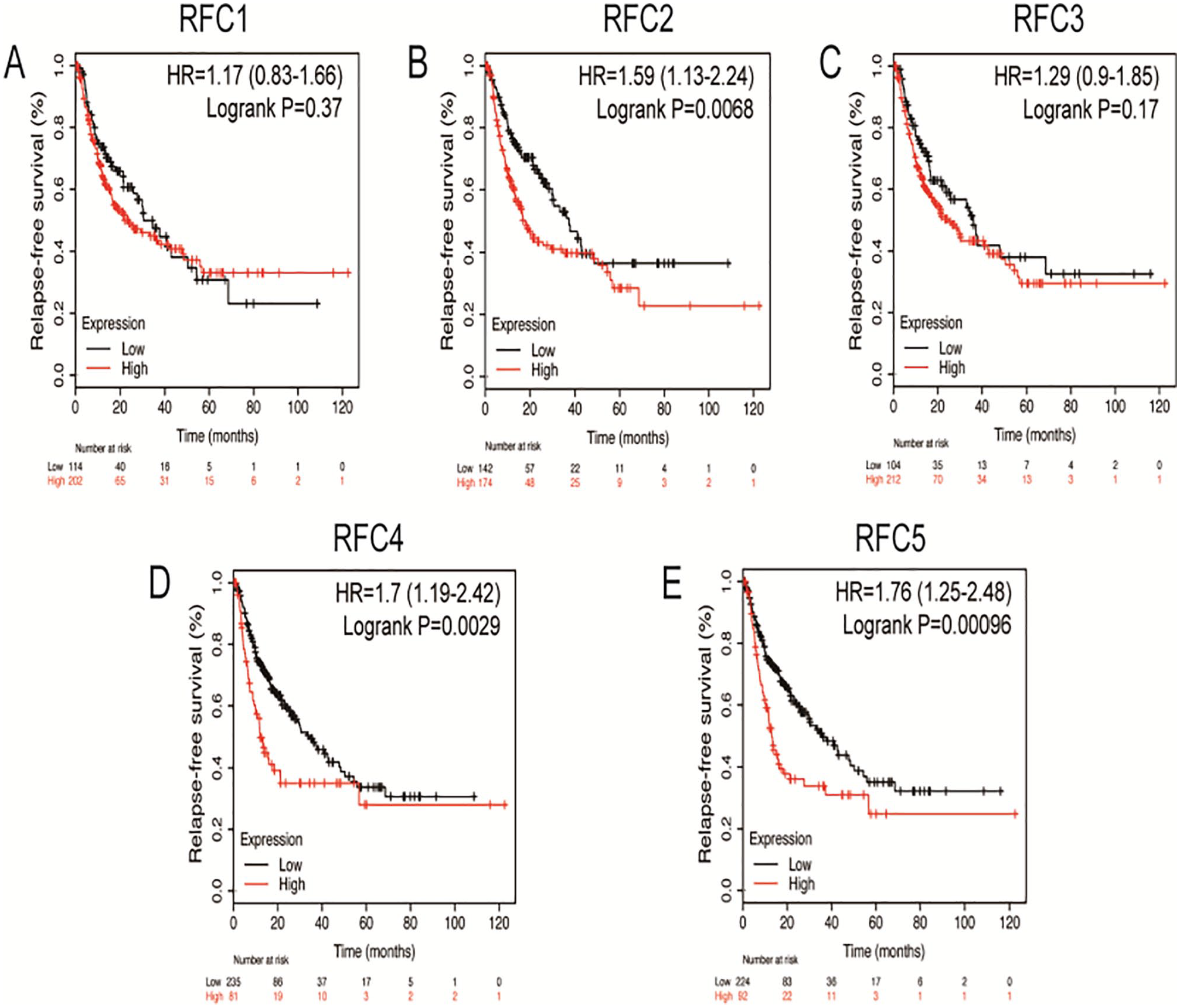
Relationship between RFCs expression and RFS in liver cancer patients (Kaplan-Meier Plotter). The RFS curves of (A) RFC1, (B) RFC2, (C) RFC3, (D) RFC4, and (E) RFC5 in liver cancer patients (n = 316). Data were presented as the HR with a 95% confidence interval. P < .05 was considered statistically significant.

Relationship between RFCs expression and OS in liver cancer patients (Kaplan-Meier Plotter). The OS curves of (A) RFC1, (B) RFC2, (C) RFC3, (D) RFC4, and (E) RFC5 in liver cancer patients (n = 364). Data were presented as the hazard ratio with a 95% confidence interval. P < .05 was considered statistically significant.
Genetic alteration, prognostic value, co-expression network, and interaction analyses of RFCs in HCC patients
Next, we analyzed genetic alteration of RFCs, and their associations with overall survival (OS) and disease-free survival (DFS) in HCC patients. We could found high alteration rate of RFCs in HCC patients, as was shown in Figure 7A and B, RFC1, RFC2, RFC3, RFC4 and RFC5 were altered in 15%, 11%, 6%, 12%, and 8% of the queried HCC samples, respectively. Frequency of enhanced mRNA expression was the highest alteration in these samples. In addition, we could observe that the genetically altered group was significantly associated with shorter OS (P = .0160) (Figure 7C) and were not significantly associated with DFS (P = .0721) from results of the Kaplan-Meier plot and log-rank test (Figure 7D). Therefore, the genetic alteration of RFCs may be the prognosis mark of HCC patients. We also calculated the correlation of RFCs mRNA expression with each other by cBioPortal tool in HCC patients, and Pearson’s correlation test was used. The results showed that RFC2 was correlated with RFC4 and RFC5, RFC3 was correlated with RFC4 and RFC5, and the highest correlation was observed between RFC4 and RFC5 (Figure 7E).
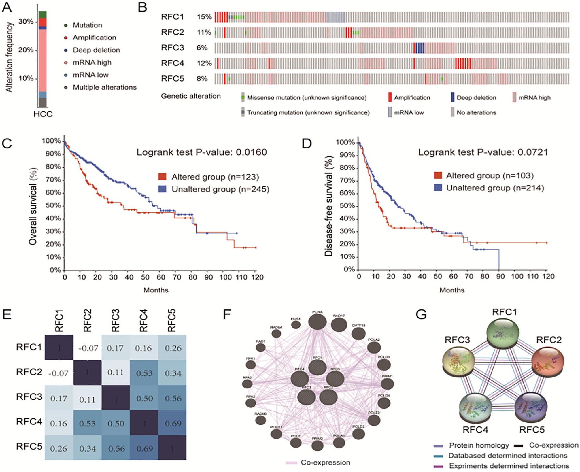
Genetic alteration, prognostic value, and interaction networks analyses of RFCs in HCC patients (cBioPortal, GeneMINIA, and String): (A) summary of frequencies of RFCs in HCC, (B) genetic alteration of each RFCs in HCC patients, (C, D) the relationship between genetic alteration and OS and DFS, (E) heat map of Genetic correlations of RFCs in HCC by Pearson’s correlation coefficients, and (F, G) co-expression network and protein-protein interaction network of RFCs.
Moreover, we constructed the gene-gene interactive network to explore the related co-expression genes and their involved functions (Figure 7F). These results revealed that RFCs were co-expressed interactively with PCNA, RAD17, CHTF18, POLA2, POLD3, PRIM1, POLD4, POLE2, POLD2, POLA1, PRIM2, POLE, POLD1, RAD9B, RPA3, RPA2, RPA1, RAD1, RAD9A, and HUS (Supplemental Table S1). And the functions of RFCs were primarily related to telomere maintenance, DNA replication, mitotic recombination, nucleotide-excision repair, and DNA gap filling. We also conducted a protein-protein interaction (PPI) network of RFCs with String to explore the potential interaction among them. Several nodes of 5 and edges of 10 were obtained in the PPI network (Figure 7G). The functions of RFCs were associated with mismatch repair, DNA replication, and nucleotide excision repair.
Biological process and KEGG pathway analysis of co-expression gene correlated with RFC2/4 in HCC patients
The results of previous analyses showed that the expression levels of RFC2 and RFC4 were strongly correlated with the prognosis of HCC patients, so we conducted further analyses for these 2 genes. First, we used the LinkedOmics to analyze the mRNA sequencing data from 371 HCC patients in the TCGA, to explore co-expression genes of RFC2/4. As was shown in Figure 8A, the expression of 3110 genes (red dots) was significantly positive correlated with RFC2, while 2702 genes (green dots) were significantly negative correlated with RFC2 (Pearson’s correlation coefficient >0.2, FDR <0.01). And Figure 8B and C show the top 50 positively and negatively correlated genes. Similarly, Figure 8D showed 4033 genes (red dots) of positive correlations and 2189 genes (green dots) negative correlations with RFC4 (Pearson’s correlation coefficient>0.2, FDR<0.01), and the top 50 positively and negatively correlated genes were showed in Figure 8E and F, respectively. As was shown in Figure 8G and H, 4476 overlapped positive genes were correlations with RFC2 and RFC4, while 2972 overlapped genes were negative correlations with RFC2 and RFC4 (FDR <0.05). Next, we performed biological process and KEGG pathway analysis of these genes correlated with RFC2/4 by GSEA. The result showed that the overlapped genes participate primarily in chromosome segregation, mitotic cell cycle phase transition, telomere organization, and chromatin assembly or disassembly (Figure 9A). KEGG pathway analysis showed that these overlapped genes activate these pathways of cell cycle and spliceosome, and inhibit PPAR signaling pathway and peroxisome (Figure 9B and C).

Genes differentially expressed in correlation with RFC2/4 in HCC (LinkedOmics): (A) correlations between RFC2 and differentially expression of genes in HCC by Pearson test, (B, C) heat maps of 50 genes positively and negatively correlated with RFC2 in HCC, (D) correlations between RFC4 and differentially expression of genes in HCC by Pearson test, (E, F) heat maps of top 50 genes positively and negatively correlated with RFC4 in HCC, and (G, H) Veen plots of overlapped positively and negatively genes associated with RFC2/4 in HCC.

Significantly enriched BP and KEGG pathway of RFC2/4 in HCC (LinkedOmics): (A, B) the significantly enriched biological processes and KEGG pathway of RFC2/4 in HCC; dark blue and orange indicate FDR ⩽ 0.05, light blue and orange indicate FDR > 0.05, (C) KEGG pathway annotations of the cell cycle pathway; red marked nodes are associated with the Leading Edge Gene.
Kinase targets, transcription factor targets, and miRNA targets of RFC2/4 in HCC
We also analyzed the kinase, transcription factor, and miRNA target of overlapped co-expression gene set to explore the targets of RFC2/4 in HCC by GSEA. As was shown in Table 2, the top 5 most significant kinase targets of overlapped correlated gene set of RFC2/4 related mainly to the aurora kinase A (AURKA), ATR serine/threonine kinase (ATR), cyclin-dependent kinase 1 (CDK1), polo-like kinase 1 (PLK1), and checkpoint kinase 1 (CHEK1). We found that transcriptional levels of these kinase genes were significantly elevated in HCC tissues. And also found that expression levels of these kinase genes were significantly associated with the OS in HCC patients (Figure 10).
The kinase target, transcription factor target, and miRNA target of RFC2/4 in HCC (LinkedOmics).
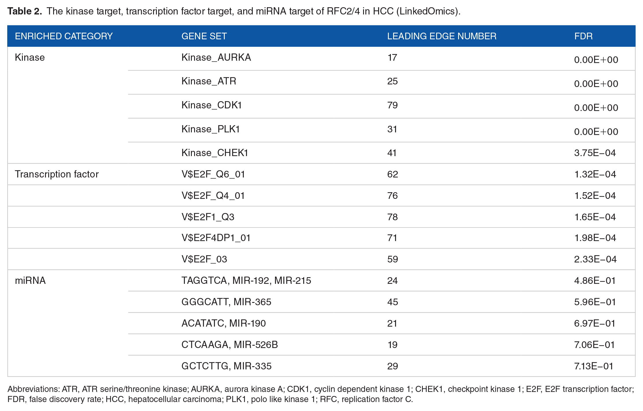
Abbreviations: ATR, ATR serine/threonine kinase; AURKA, aurora kinase A; CDK1, cyclin dependent kinase 1; CHEK1, checkpoint kinase 1; E2F, E2F transcription factor; FDR, false discovery rate; HCC, hepatocellular carcinoma; PLK1, polo like kinase 1; RFC, replication factor C.

Expression and survival analysis of the related targets of RFC2/4 (UALCAN and Kaplan-Meier plotter): expression levels of (A) AURKA, (B) ATR, (C) CDK1, (D) PLK1, (E) CHEK1, and (F) E2F1 in HCC tissues and normal liver tissues. Correlation analysis between overall survival and mRNA expression levels of (G) AURKA, (H) ATR, (I) CDK1, (J) PLK1, (K) CHEK1, and (L) E2F1 in HCC patients. Data were presented as the mean ± standard error and the HR with a 95% confidence interval. Transcript pre million was used to measure the expression. P < .05 was considered statistically significant.
The enrichment of transcription factors for overlapped co-expression gene was mainly the E2F transcription factor family, including E2F_Q6_01, E2F_Q4_01, E2F1_Q3, E2F4DP1_01, and E2F_03 (Table 2). Previous studies have confirmed that E2F driven transcription was associated with the development and progression of HCC. 28 We also analyzed the expression of E2F1 in HCC and its effect on prognosis and found that E2F1 was significantly highly expressed in HCC and was associated with poor prognosis (Figure 10). No significantly associated miRNA target was enriched for overlapped co-expression genes of RFC2/4 (Table 2).
Discussion
RFC is a structure-specific DNA-binding protein that acts as a primer recognition factor for DNA polymerase and plays an important role in DNA replication and repair and regulation of cell cycle checkpoints.29,30 There is no doubt that DNA replication is a key process in the infinite proliferation of tumor cells. 31 In previous studies, RFCs abnormal expression has been reported in some cancers,9-15 and some RFCs have been identified as activators of tumorigenesis and prognosis in several cancers.9,14,32 However, the prognostic value and biological function of RFCs in HCC have yet to be comprehensively explored.
We first analyzed the mRNA expression of RFCs and their correlation with the clinicopathological characteristics in the HCC. We found that all RFCs were significantly highly expressed in HCC tissues compared with normal liver tissues. We also found that protein expression of RFC2 and RFC4 was increased in HCC tissues. Moreover, the expression level of RFC2, RFC3, RFC4, and RFC5 was significantly correlated with the tumor stage. HCC patients with high expression of RFC2 and RFC4 were significantly associated to worse overall survival. The results suggest that the RFC2 and RFC4 may be potential diagnostic and prognostic markers. Several studies have found that RFC3 and RFC4 are highly expressed in HCC cells and promote cell proliferation and growth.9,15 However, we found that the expression of RFC3 was not significantly related to the prognosis of HCC patients.
Next, to understand the role of RFCs gene alterations in HCC progression, we also explored molecular characteristics of RFCs and their prognostic values in HCC patients. There were frequent genetic alterations of RFCs in HCC, and elevated mRNA expression was the most common alteration. In addition, genetic alteration was significantly associated with worse overall survival in HCC patients. Research has shown that the accumulation of genetic alterations is thought to drive the progression, invasion, and metastasis of tumors. 33
In our study, we found that RFC2 and RFC4 can be considered as potential prognostic markers, so we further explored the biological processes and pathways in which RFC2 and RFC4 were involved. Our results suggest that the functional network of RFC2 and RFC4 participated mainly in pathways of cell cycle and spliceosome. The cell cycle is controlled by the signaling pathway comprising cyclins, cyclin-dependent kinases, cyclins kinase inhibitors, and the related regulators.34,35 Disruption of cell cycle pathways may lead to cell cycle arrest and is associated with the prognosis of human cancers. 36
We also sought to characterize the kinase targets, transcription factor targets, and miRNA targets of the RFC2 and RFC4 in HCC. They mainly enriched to the cancer-related kinase AURKA, ATR, CDK1, PLK1, and CHEK1. These kinases regulate the cell cycle and genomic stability.37-41 Overexpression of AURKA has been found to enhance tumor proliferation and promotes cancer metastasis and cancer stem cells in HCC. 42 A causal relationship between CDK1/PLK1 and HCC has been established, and inhibitors of CDK1 and PLK1 are effective in inhibiting HCC growth and are being tested in clinical trials.43-45 Our results also found that transcriptional levels of these kinase genes were significantly elevated in HCC tissues, and significantly associated with the prognosis of HCC patients. In addition, we found that the E2F family members are key transcription factors for RFCs. Among them, E2F1 is one of the key links in the cell cycle regulation network, and the abnormal expression of E2F1 is involved in the occurrence and development of HCC, and upregulation of E2F1 expression was found to be associated with poor prognosis in HCC patients.46,47 Our results are consistent with this view. No relevant miRNA targets were enriched for RFCs, we suspect that RFCs may be involved in the mRNA spliceosome pathway, and keeping away from miRNA.
There are some limitations to our study. First, although high expressions of RFC2 and RFC4 were significantly related to worse prognosis in HCC patients, all the data analysis in our study was based on the online databases; more independent HCC patients are needed to confirm our results. Second, we found that genetic alterations of RFCs were associated with mRNA upregulation, amplification, and deletion. These alterations were significantly associated with worse OS in HCC patients, but the potential molecular mechanism remains undefined and requires further exploration.
In conclusion, our study showed multidimensional evidence of the importance of RFCs and prognostic value of RFC2/4 in HCC. In addition, the results of our RFC2/4 targets analysis indicated that they may act on E2F transcription factors and cell cycle-associated kinases, which dysregulate cell cycle pathways in HCC. These efforts may provide new research directions to identify prognostic biomarkers and therapeutic targets for HCC.
Supplemental Material
sj-pdf-1-evb-10.1177_1176934321994109 – Supplemental material for Exploration of Prognostic Biomarkers among Replication Factor C Family in the Hepatocellular Carcinoma
Supplemental material, sj-pdf-1-evb-10.1177_1176934321994109 for Exploration of Prognostic Biomarkers among Replication Factor C Family in the Hepatocellular Carcinoma by Jianxiong Deng, Fangyan Zhong, Weiguo Gu and Feng Qiu in Evolutionary Bioinformatics
Footnotes
Funding:
The author(s) received no financial support for the research, authorship, and/or publication of this article.
Declaration of Conflicting Interests:
The author(s) declared no potential conflicts of interest with respect to the research, authorship, and/or publication of this article.
Author Contributions
FQ, FZ, and JD designed and performed the study; FZ and JD acquired, analyzed, and interpreted the data; JD drafted the manuscript; FZ, XD, WG, FQ, and JD reviewed the manuscript for the important intellectual content. All authors read and approved the final version of the manuscript.
Availability of Data and Materials
All data generated or analyzed during this study are included in this article.
Supplemental Material
Supplemental material for this article is available online.
References
Supplementary Material
Please find the following supplemental material available below.
For Open Access articles published under a Creative Commons License, all supplemental material carries the same license as the article it is associated with.
For non-Open Access articles published, all supplemental material carries a non-exclusive license, and permission requests for re-use of supplemental material or any part of supplemental material shall be sent directly to the copyright owner as specified in the copyright notice associated with the article.