
Abstract
Objectives
The aim of the study was to compare CT with video-otoscopy for detecting external ear canal masses in cats and to describe CT features of feline external ear disease.
Methods
This retrospective study evaluated the external ear canals of cats that underwent both CT and video-otoscopy. Two blinded observers reviewed the CT scans and their findings were compared with video-otoscopy results evaluated by one blinded observer.
Results
Of the 32 examined ears, 13 had an external ear canal mass detected by both CT and video-otoscopy. No false positives were identified. All 19 ears without masses were correctly identified as true negatives, resulting in perfect agreement for mass detection (kappa = 1.000). On histopathology, 9/13 masses were diagnosed as polyps, 1/13 masses was diagnosed as carcinoma, 2/13 did not undergo histopathology and 1/13 masses seen on video-otoscopy was diagnosed as otitis externa with granulomatous tissue formation. Of the 16 cats examined, 12 had a mass in the external ear canal diagnosed by video-otoscopy: six domestic shorthairs, five Maine Coons and one Oriental Shorthair cat. Of these cats, 11 had a unilateral mass and one had a bilateral mass. There was very low agreement between CT and video-otoscopy for ear canal wall enhancement and erythema (kappa = 0.033), as well as for ear canal thickening and stenosis (weighted kappa = 0.056). There was no significant difference between chronic and non-chronic clinical signs in terms of the presence of ear canal wall mineralisation (P = 0.223).
Conclusions and relevance
There is high agreement between video-otoscopy and CT in detecting external ear canal masses in cats. However, there is very low agreement between CT and video-otoscopy for ear canal wall enhancement, erythema, thickening and stenosis. The presence of external ear canal mineralisation did not correlate with the chronicity of clinical signs.
Introduction
Feline otitis externa is a common dermatological disease in small animal practice, defined as inflammation of the external ear canal outside the tympanic membrane. 1 The external auditory meatus has vertical and horizontal parts, ending at the tympanic membrane. 2 CT findings of otitis externa are defined as a narrowing of the external ear canal, soft tissue material within the lumen and mineralisation of the external ear canal. 3
The prevalence of otitis externa in cats has been reported to be in the range of 2–10%, which is significantly less than 10–20% in dogs.4,5 It can present in acute or chronic forms, caused by various primary and secondary factors. 6 This multifactorial aetiology makes it a complex disorder, requiring precise identification of the cause for successful management.6,7 According to Trower et al, 8 during otoscopic examination, only two-thirds of cases had a visible tympanic membrane, making differentiation between otitis media and externa challenging without additional signs such as sympathetic or facial nerve dysfunction. 9
The most common mass in the feline external ear canal is a non-neoplastic, polypoid growth called a feline nasopharyngeal polyp (FNP), originating from the tympanic membrane, auditory tube or nasopharyngeal mucosa. The aetiology of FNPs is unknown but is considered either congenital or inflammatory, often linked to ongoing infections. Although aural polyps usually affect young cats, they can be found in cats of all ages.10–12 CT findings of nasopharyngeal polyps include homogeneous, ill-defined, hypo- to iso-attenuating masses on pre-contrast images and well-defined, homogeneous stalk-like structures with strong peripheral rim enhancement on post-contrast images. 13
Malignant neoplasms of the ear canal are rare in cats, typically found in the external ear canal, with squamous cell carcinoma and apocrine/sebaceous carcinomas being the most common. 14 Radiological findings of feline ear neoplasia are not well described. Both aural polyps and neoplasms are often associated with secondary infections and have non-specific clinical presentations.10,14
In recent years, video-otoscopy has revolutionised the practice of veterinary otology. It has become widely used, as its advantages over handheld otoscopes are unquestionable. It provides better visualisation of the ear structures owing to superior magnification and lighting, complete visualisation of the ear canal and tympanic membrane, and digital documentation. 15
CT and video-otoscopy are commonly used techniques to assess the external ear in cats; however, there is no information on the agreement between the two. In addition, literature on the CT features of external ear disease in cats is limited.
The aim of this study was to compare the results of CT and video-otoscopy for the presence and severity of external ear disease in cats. The hypotheses were as follows: high agreement between video-otoscopy and CT in detecting external ear canal masses and otitis externa; high agreement between CT ear canal wall enhancement and erythema on video-otoscopy; poor agreement between CT external ear canal thickening and ear canal stenosis on video-otoscopy; and ear canal wall mineralisation would be more prevalent in cats with chronic otitis externa.
Materials and methods
The clinical programme was searched for cats that were presented to the veterinary dermatology and surgical departments at the University of Ghent in Belgium between 2012 and 2023 with clinical signs related to ear diseases (eg, otitis externa/media/interna, head shaking, vestibular syndrome, pruritis, ear pain, ear discharge, and so on), if they underwent both video-otoscopy and CT of the head within a 3-week period. Data, such as signalment and clinical presentation (age, breed, sex, clinical signs), were recorded.
CT techniques and image evaluation
All patients had CT helical acquisitions of the head from the nasal planum to at least the first cervical vertebrae under general anaesthesia in sternal recumbency using a 320-slice single-source CT scanner (Aquilion ONE Vision; Canon Medical Systems). Contrast was injected intravenously via the cephalic vein at a dose of 700 mg/kg of non-ionic iodinated contrast medium (Omnipaque 350; Iohexol) with a power injector (OptiVantage, LF). No standardised CT protocol was used as a result of the retrospective nature of the study (slice thickness was in the range of 0.5–1.25 mm). The images were viewed using a soft tissue and bone algorithm on a computer workstation (Apple Mac; Apple) with DICOM viewer software (Osirix v.13.0.1). Multiplanar reconstruction (MPR) was used as needed.
A total of 16 cats, presented to the small animal department and that had undergone video-otoscopy examination and head CT, were included in the study. Two radiologists, a second-year diagnostic imaging resident and European Congress of Veterinary Diagnostic Imaging diplomate, evaluated the CT studies retrospectively, blinded to the results. The results were compared and the final diagnosis was obtained.
Images were viewed at both the bone and soft tissue windows.
The evaluated structures on CT were as follows:
External ear canal wall mineralisation (present, absent) on standardised bone window (WL: 300, WW: 1500), transverse view (Figure 1)
Horizontal ear canal wall thickness (mild, moderate, severe) on standardised soft tissue window (WL: 40, WW: 350), transverse view. Subjective assessment of the external ear canal wall thickness on pre-contrast images (Figure 2)
External ear canal wall contrast enhancement, standardised soft tissue window (WL: 40, WW: 350), transverse view. Attenuation values were measured in the subjectively most enhanced part of the horizontal external ear canal on pre- and post-contrast images
External ear canal mass (present/absent) on standardised soft tissue window (WL: 40, WW: 350), transverse view
If the external ear canal mass was present, pattern of contrast enhancement of the mass was assessed (peripheral, diffuse, absent, mild, moderate, severe) on standardised soft tissue window (WL: 40, WW: 350), transverse view. Attenuation values were calculated in the centre and in the outer margins (rim enhancement) of the mass and the numbers were compared (Figure 3)
Fluid/mucous in the external ear canal (present, absent) on standardised soft tissue window (WL: 40, WW: 350), transverse view
Mass mineralisation (present, absent) on standardised bone window (WL: 300 WW: 1500), transverse view
Tympanic bullae (thickening, thinning) on standardised bone window (WL: 300, WW: 1500), transverse view
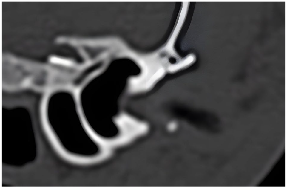
Transverse CT reconstruction images of ear canal wall mineralisation, displayed in a bone window

Pre-contrast transverse CT reconstruction images showing (a) mild, (b) moderate and (c) severe wall thickening of the horizontal ear canal, displayed in a soft tissue window

Post-contrast transverse CT reconstruction images of the mass with (a) peripheral and (b) diffuse contrast enhancement, displayed in a soft tissue window
Video-otoscopy studies were conducted by a dermatology resident, with data collected retrospectively by a board-certified dermatologist. Evaluated structures included:
Fluid/mucous/dry exudate (present/absent)
Erythema (present/absent)
Ear canal stenosis (absent/mild/moderate/severe)
Ear canal ulceration (present/absent)
External ear canal mass (present, absent)
CT findings and results were compared with video-otoscopy results. CT wall thickening was correlated with wall canal stenosis on video-otoscopy. CT wall contrast enhancement was correlated with erythema/ulceration on video-otoscopy, external ear canal mass presence on CT with presence of the external ear canal mass on video-otoscopy. The accuracy and reliability of CT and video-otoscopy in detecting ear disease were compared. All calculations were performed using a video-otoscopy as gold standard.
Statistical analysis
To quantify the agreement between CT mass and video-otoscopy mass, and between CT ear canal wall enhancement and erythema on video-otoscopy, we used Cohen’s kappa. We used the weighted kappa to assess the agreement between CT external ear canal thickening and ear canal stenosis on video-otoscopy. Finally, chronic vs non-chronic cats were compared for the absence/presence of ear canal wall mineralisation using the Wilcoxon rank-sum test.
Results
A total of 16 cats (32 ears) were included, comprising eight domestic shorthairs, six Main Coons, one British Shorthair and one Oriental Shorthair. The population included 11 male castrated cats, one female entire cat and four female spayed cats, aged 7 months to 18 years (median 6, mean 7).
Of the cats, 12 had external ear canal masses, with 11 being unilateral. Radiologists identified 13 ear canal masses: 7/16 external ear canal masses in domestic shorthairs; 5/12 in Maine Coons; 1/2 in Oriental Shorthairs; and 0/2 in British Shorthairs. All external ear canal masses identified on CT were also diagnosed on video-otoscopy reaching very high agreement (kappa = 1.000). On CT, 7/13 external ear canal masses presented moderate peripheral contrast enhancement, 3/13 strong peripheral contrast enhancement and 3/13 strong diffuse contrast enhancement. Out of 13 external ear canal masses, 11 had histopathology examinations. Nine masses were diagnosed as inflammatory polyps and one as carcinoma. Two masses were presumed benign based on characteristics observed on CT and video-otoscopy. One ear was diagnosed as an inflammatory polyp on video-otoscopy, but histopathology showed otitis externa with proliferative granulation tissue.
All (13/13) ear canal masses were associated with abnormal tympanic bullae on CT. None of the external ear canal masses showed evidence of mineralisation on the CT.
Of the 32 ears, three (9%) were diagnosed with ear canal stenosis upon examination via video-otoscopy, while 25 (78%) presented with ear canal wall thickening on CT. Two ears corresponded to chronic presentation and moderate ear canal thickening on CT and one to acute presentation and severe ear canal wall thickening. Agreement was very low for CT external ear canal thickening and ear canal stenosis on video-otoscopy (weighted kappa = 0.056).
Ear canal enhancement on CT was seen in 13/19 (68%) chronic cases, 6/6 (100%) acute cases and 2/7 (29%) asymptomatic cases. The presence of erythema on video-otoscopy was seen only in one ear and this corresponded to chronic presentation and increased contrast uptake on the CT. Agreement was very low for CT ear canal wall enhancement and erythema on video-otoscopy (kappa = 0.033).
On CT, 21/32 (66%) ear canals had evidence of fluid/mucous/exudate; however, on video-otoscopy, fluid/mucous/exudate were noted in 20/32 (63%) ear canals.
CT ear canal wall mineralisation was detected in 3/6 (50%) acute cases, 6/19 (31%) chronic cases and 3/7 (42%) asymptomatic cases. This did not achieve statistical significance. No difference between chronic and non-chronic cats for absence/presence of ear canal wall mineralisation was calculated (P = 0.223).
Otitis externa was diagnosed on CT as well as video-otoscopy in 21/32 (66%) cases. Diagnoses regarding otitis externa on CT vs video endoscopy were conflicting in 11/32 (34%) cases. In total, 10 ears were diagnosed as otitis externa on CT with no evidence on the video-otoscopy. One ear was diagnosed as normal on CT, while video-otoscopy showed signs of otitis externa. All ears with the presence of external ear canal mass had evidence of otitis externa on CT; however, two masses seen on video-otoscopy were not associated with otitis externa.
On video-otoscopy, no external ear canal showed evidence of ulceration.
Discussion
The most commonly presented breeds for CT and video-otoscopy evaluation of external ear disease were domestic shorthair and Maine Coon. Otitis externa was diagnosed in all cats with a mass in the external ear canal on CT and in 85% of cases on video-otoscopy.
We observed a lower prevalence of external ear canal abnormalities on video-otoscopy compared with CT in symptomatic cats, possibly due to prior antimicrobial therapy. The excessive use of antimicrobials can mask underlying conditions and obscure the detection of otitis externa. 16 In cats, recurrent ear infections are frequently linked to underlying pathologies, with inflammatory polyps being a common cause. 16
The aetiology of feline inflammatory polyps remains debated, with hypotheses including chronic viral infection, congenital origin or prolonged inflammation in the middle ear or upper respiratory tract.17–22
In our study, male cats were more frequently affected, and the most common pathology was external ear canal mass, with a prevalence of 75%. The median age of cats diagnosed with external ear canal polyps was 6–7 years, which significantly exceeds the age reported in the existing literature, which was typically around 1.5 years.10,17–19,22–24 A polypoid mass in old cats raises the suspicion of a tumour; therefore, histopathology is recommended. 16 Although no breed or sex predisposition has been reported for aural polyps, it has been noticed by many authors that polyps are more commonly seen in certain breeds. 16 According to Stanton et al, 25 feline inflammatory polyps might be hereditary, congenital or associated with a genetic predisposition to the disease. The authors identified cases of feline inflammatory polyps in siblings of Maine Coons from the same bloodline but raised in different environments. The diagnosis of feline inflammatory polyps in siblings raises the possibility of a congenital origin for the disease. 25 In our study, Maine Coons and domestic shorthairs were overrepresented, comprising 38% and 50% of the population, respectively.
Our study found a weak correlation between external ear canal contrast enhancement on CT and evidence of erythema on video-otoscopy, suggesting these modalities may not always provide consistent results when assessing external ear canal inflammation. Cats with acute presentations were more likely to show evidence of external ear canal contrast enhancement on CT compared with those with chronic presentations or asymptomatic cases. This implies that the severity or duration of signs may influence the imaging findings. The study suggests that CT may be more sensitive in detecting inflammation of the external ear canal than video-otoscopy. This is because CT can visualise deeper structures and may be better at detecting subtle changes indicative of inflammation. In contrast, video-otoscopy primarily assesses the superficial aspect of the external ear canal, which may limit its ability to detect deeper inflammation. We speculate that the use of anti-inflammatory medications before examination may have masked inflammation on video-otoscopy, leading to a discrepancy between the findings of CT and video-otoscopy. In addition, pigmentation of the external ear canal could hinder the ability of the operator to visualise inflammation on video-otoscopy, further highlighting its limitations. While both CT and video-otoscopy are valuable tools for evaluating external ear canal inflammation in cats, CT may offer greater sensitivity in detecting inflammation, especially in cases where video-otoscopy findings are inconclusive or influenced by factors such as prior medication use or pigmentation.
Poor agreement was found between CT ear canal wall thickening and stenosis on video-otoscopy, likely due to the small size of the feline ear canal, making otoscopic examination challenging. 16 Anatomical variations in the ear canal may also contribute to differing interpretations of wall thickening or stenosis. 26
The association between external ear canal mineralisation on CT and chronic otitis externa in cats has been documented previously. 27 According to Foster et al, 26 it is possible that external ear canal mineralisation is a normal variation. In our study, we established no correlation between chronicity of the clinical signs and ear canal wall mineralisation. This suggests that mineralisation in the ear canal can occur in cases that are either acute or acute-on-chronic. We imply that the presence of mineralisation may not necessarily correlate with chronic conditions but also with acute exacerbations or instances where there is a sudden change in the condition of the ear canal. Our other hypothesis is that mineralisation develops without producing overt clinical signs, so even though mineralisation might be occurring, it may not manifest noticeable signs that would indicate its presence. This could potentially complicate diagnosis and treatment since the clinician may not be aware of the extent of mineralisation without further diagnostic procedures. Furthermore, anatomical variations might also contribute to differences in how mineralisation manifests clinically or how it responds to treatment. Overall, these points highlight the complexity of diagnosing and managing ear canal mineralisation, particularly in relation to the chronicity of clinical signs. The lack of agreement between the two suggests that mineralisation may not always correlate with the duration or severity of clinical signs, and other factors, such as acute exacerbations and anatomical variations, may play significant roles.
In our study, CT and video-otoscopy were equally effective in identifying external ear canal masses, with complete agreement between both techniques. This agreement could streamline the diagnostic process, as clinicians can choose the imaging modality based on factors such as availability, cost, patient comfort and the specific clinical scenario without sacrificing diagnostic accuracy. It also underscores the importance of using multiple diagnostic tools in conjunction to ensure thorough evaluation and accurate diagnosis in cases of external ear masses.
CT is particularly useful for detecting masses or abnormalities within deeper structures of the ear, such as the inner/middle ear, surrounding soft tissue structures and tympanic bullae.28,29 It offers high-resolution images that can reveal the size, location and characteristics of any masses present. Detailed cross-sectional views allow for the comprehensive evaluation of the internal ear anatomy and pathology.28,29 Therefore, CT imaging surpasses video-otoscopy, facilitating more precise prognosis and treatment planning for cats suspected of having tympanic or nasopharyngeal masses. It helps in surgical planning for complex ear conditions requiring precise localisation of lesions or abnormalities.
The advantages of video-otoscopy are real-time visualisation of the inside of the external ear canal and the eardrum, with detailed images due to magnification and illumination of the ear canal and eardrum, as well as immediate feedback during examination allowing for prompt assessment and patient education. Furthermore, video-otoscopy may be used in conscious patients. The other advantage of video-otoscopy is the possibility of medical intervention, such as ear flushing, retrieving foreign objects, biopsies or mass removals, at the same time as diagnosis. 30
On CT scans, a feline inflammatory polyp is often characterised by the presence of a dense, oval-shaped structure with uniform density, clearly delineated borders and peripheral enhancement.13,28,31–34 In contrast, aggressive neoplastic conditions and infections may show both tympanic bullae bone proliferation and tympanic bulla lysis with marked heterogeneous contrast enhancement of the mass.3,13,28,32 It has been reported that it is challenging to differentiate inflammatory polyps from otitis media or early onset of neoplasia.3,18,24,28,34–36
In the current study, all external ear canal masses but one were benign, and their characteristics were in agreement with the literature (Figure 4). Feline inflammatory polyps had moderate to strong peripheral contrast enhancement and only about 20% of feline inflammatory polyps presented a diffuse contrast-enhancement pattern. One mass diagnosed on histopathology as severe otitis externa with granulation tissue formation had strong and diffuse contrast enhancement.

Inflammatory polyp shown on (a) post-contrast transverse CT reconstruction images, displayed in a soft tissue window, and (b) video-otoscopy
Most inflammatory polyps are reported to occur unilaterally. 16 In our study, all cats presented with a unilateral mass apart from one, which was diagnosed with a bilateral mass.
The limitations of the present study include its retrospective nature, lack of standardised protocols, absence of histopathology in two cases and small sample size, potentially limiting generalisability and introducing bias.
Conclusions
We observed high agreement between video-otoscopy and CT in detecting external ear canal masses, but very low agreement between CT ear canal wall enhancement and erythema on video-otoscopy as well as between CT external ear canal thickening and ear canal stenosis on video-otoscopy. These findings highlight the complementary nature of these diagnostic modalities and underline the importance of utilising both techniques for a comprehensive assessment of ear diseases in feline patients. Moreover, the presence of external ear canal mineralisation did not correlate with the chronicity of clinical signs, highlighting the complexity of diagnosing and managing this pathology in feline patients.
Footnotes
Author note
This paper was presented in part at the 2023 ECVDI conference.
Conflict of interest
The authors declared no potential conflicts of interest with respect to the research, authorship, and/or publication of this article.
Funding
The authors received no financial support for the research, authorship, and/or publication of this article.
Ethical approval
The work described in this manuscript involved the use of non-experimental (owned or unowned) animals. Established internationally recognised high standards (‘best practice’) of veterinary clinical care for the individual patient were always followed and/or this work involved the use of cadavers. Ethical approval from a committee was therefore not specifically required for publication in JFMS. Although not required, where ethical approval was still obtained, it is stated in the manuscript.
Informed consent
Informed consent (verbal or written) was obtained from the owner or legal custodian of all animal(s) described in this work (experimental or non-experimental animals, including cadavers, tissues and samples) for all procedure(s) undertaken (prospective or retrospective studies). No animals or people are identifiable within this publication, and therefore additional informed consent for publication was not required.