
Abstract
Objectives
The aim of this study was to verify whether a single oral dose of gabapentin (100 mg) or melatonin (3 mg) given 60 mins before a cardiac evaluation would reduce anxiety without interfering with heart rate (HR), systemic blood pressure (SBP), electrocardiogram (ECG) and echocardiographic indexes.
Methods
Seventy-five client-owned healthy cats underwent two sets of cardiac assessments 60 mins apart, randomly divided into gabapentin, melatonin and placebo groups. The interval between treatment and the second ECG and SBP measurement was 60 mins, and 70 mins for echocardiography. A compliance score (CS) classified the behavior, focusing on the ease of handling.
Results
Most variables did not change between the examinations. The placebo group showed more significant changes (SBP, tricuspid annular plane systolic excursion, HR during echocardiography, aortic flow velocity, S′ wave from lateral mitral annulus), but they were not considered to be hemodynamically relevant. Gabapentin and melatonin significantly increased the cats’ compliance without interfering with cardiac assessment. Eight cats presented with mild sedation, seven after gabapentin and one after melatonin. No major side effects were observed.
Conclusions and relevance
Gabapentin tranquilized the cats when it was given 60 mins prior to ECG and SBP measurement, and 70 mins prior to echocardiography, without interfering with systolic echocardiographic indexes. Melatonin also decreased the CS, but without sedation in most cases. The waiting period may have relaxed the cats in the placebo group, resulting in lower SBP measurements. However, this tranquility did not last as some echocardiographic changes signaled a sympathetic predominance.
Introduction
Cardiomyopathies are frequently diagnosed in cats, although overt clinical signs may be absent until they are at an advanced stage.1,2 Thus, preventive cardiac evaluation is often indicated, especially in older animals or in those of breeds at higher risk. 1 Because the examinations require restraint, a prior tranquilizer may be recommended to guarantee animal welfare and facilitate the performance of the procedure. Domestic cats frequently react aggressively or fearfully in veterinary settings, 3 inhibiting echocardiography or systolic blood pressure (SBP) measurement and handling. The stress generated by physical restraint often leads veterinarians to be suspicious of SBP measurements, speculating whether an elevated SBP value is due to a sympathetic response, the white-coat effect or is evidence of an actual disease. 4 In this context, previous use of a substance to reduce anxiety without compromising cardiac assessment is convenient because it does not confound the interpretation of the examination, increases the cat’s welfare and provides better veterinary care.
Gabapentin is a drug known to facilitate both the transport and examination of cats.5,6 Recently, its oral administration 2 h before echocardiography proved to be effective in tranquilization, although it decreased some systolic echocardiographic parameters. 7 In feline medicine, melatonin is best known for acting in the reproductive field, 8 effectively and reversibly inhibiting endogenous ovarian activity, 9 and is often recommended to suppress estrus in cats, administered orally or as an implant.10,11 Based on a recent search on two medical database platforms (PubMed and ProQuest), unlike gabapentin, melatonin has not been indicated as a preappointment substance for veterinary use. However, a meta-analysis reported that melatonin has an anxiolytic effect in humans, with high-grade evidence, reducing preoperative anxiety. 12 In veterinary medicine, melatonin provides a beneficial calming effect for anxious dogs 90 mins after its administration and reduces the dose of propofol required for anesthesia induction in ‘trustful’ dogs. 13
Both gabapentin and melatonin take around 1 h to achieve maximum plasma concentration (Tmax),9,14 which permitted studying them in the same trial. Based on this, the objective of this investigation was to ascertain whether a single dose of gabapentin or melatonin would reduce anxiety without interfering with heart rate (HR), SBP, electrocardiography (ECG) parameters and echocardiographic indices. Also, despite the maximum effect of gabapentin being reached approximately 2 h after oral administration, 15 we sought to investigate if a shorter interval would be sufficient to tranquilize the cats for cardiac examination.
Materials and methods
This study was designed as a prospective, randomized, double-blind, placebo-controlled investigation and was carried out between July and December 2020 at a veterinary teaching facility. All procedures were previously approved by the Institutional Animal Care and Use Committee (protocol 014/2019) and complied with the National Institutes of Health Guide for the Care and Use of Laboratory Animals.
Animals
Client-owned cats were recruited for the study after a complete physical examination was performed. Inclusion criteria comprised a young or mature adult cat (1–10 years old), 16 normal auscultation and an ECG trace without arrhythmias. Cardiac diseases were also ruled out by standard transthoracic echocardiography, which required a normal myocardial thickness during diastole, 17 and normal-sized cardiac chambers, systolic indices within reference intervals and normal diastolic function parameters. 1
Exclusion criteria were established to select only cats that appeared healthy after checking their medical history and performance of a physical examination. Animals previously diagnosed with a disease or receiving any medication were not admitted.
Procedures
The algorithm in Figure 1 describes the procedures initiated with the clinical history and physical examination, followed by baseline SBP, ECG, echocardiography and behavioral evaluation by compliance score (CS). Animals that did not meet the inclusion criteria were excluded at each stage. The following actions were randomization, treatment and a waiting interval of 60 mins preceding the second series of the same protocol. The schedule was rigorous in terms of exactitude, allowing a maximum of 5 mins of delays. Most of the examinations were performed during the mornings.
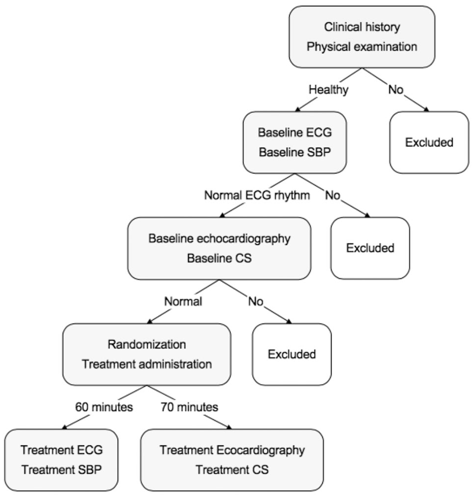
Algorithm describing the sequence of procedures, exclusion method and interval between examinations.
We used the Doppler technique to indirectly measure the SBP; 18 it was repeated five times and averaged to give the baseline SBP. Cats with a high baseline SBP remained in the experiment whenever it was considered an influence of sympathetic tone due to stress, as both clinical history and echocardiographic measurements revealed no evidence of chronic systemic hypertension.
Subsequently, a 2 min recording of a computer-based ECG was performed (baseline ECG). The only exclusion criterion based on the ECG was the presence of arrhythmias, with both sinus rhythm and sinus tachycardia considered normal (Figure 1). The ECG variables were HR, mean cardiac axis, QT interval and T wave polarity. On the sequence, the first echocardiography was performed (baseline echocardiography). Both ECG recording and SBP measurements were conducted in a single room, whereas echocardiography was executed in another, which required transporting the cats between them.
The cats were randomly assigned to receive a manipulated capsule of the same shape and size, packaged in numbered bottles (1, 2 and 3), containing 100 mg of gabapentin (median dose = 20 mg/kg), 3 mg of melatonin (median dose = 0.65 mg/kg) or a placebo (lactose powder). Locked randomization was used to ensure a balance between the groups according to the predetermined 1:1:1 ratio, producing groups of the same size. 19 To avoid biases about the timing of capsule administration (early morning or around midday) or the order of examination (first or last examined cat), six pieces of paper with all possible sequences from 1 to 3 were placed in an envelope. One of the authors (MFS) selected the sequence, unaware of the capsule’s contents, randomly varying the order of treatments each examination day and not repeating the sequence until all the papers were removed from the envelope. The oral capsule was administered immediately after the baseline echocardiography, flushing with 2 ml of water from a syringe to help with swallowing. The cats were kept in a silent, dark room between the two sets of procedures.
Sixty minutes later, another SBP measurement (treatment SBP) and ECG recording (treatment ECG) were performed. The second echocardiography was performed precisely 70 mins after capsule administration. A further author (MJGRP) assessed the behavior during echocardiography.
Echocardiography
Both echocardiographs were performed by the same operator (GLRT), with the cat positioned following the recommendations of the Echocardiography Committee of the Specialty of Cardiology of the American College of Veterinary Internal Medicine. 20
The echocardiographic evaluation included the variables from the right transversal parasternal view: the M-Mode measurements of the left ventricle, the left atrium (LA) and aorta ratio (LA:Ao) measured at the maximum LA diameter, 21 and the fractional shortening of the left atrium using anatomic M-mode. 22 The maximum diameter of the LA was measured right after the T wave using right parasternal long-axis four-chamber images. 23
The mitral annular plane systolic excursion from the free wall and interventricular septum (IVS), respectively, the tricuspid annular plane systolic excursion (TAPSE) and the mitral annular velocities were obtained as described elsewhere.24,25
Two speckle-tracking echocardiography (STE) variables were applied to assess the left ventricle’s systolic function: longitudinal strain (LSt) and tissue motion annular displacement (TMAD). Each parameter was measured at AP2 and AP4 images, using the aortic valve closure time calculated from the beginning of the QRS complex to the end of the aortic valve spectra. 26
The offline calculation of LSt required selecting three regions of interest (ROIs) by the operator: the septal and lateral mitral valve annulus and the epicardial region of the LV apex. 27 Myocardial tracking was automatically performed by the equipment software (QLAB Software; Auto Cardiac Motion Quantification). Manual corrections were made whenever the automatic tracking was obviously incorrect.
TMAD measurement required the selection of the same three ROIs already described for LSt. 28 The software automatically tracked the displacement of the two points at the mitral ring towards the apex (mm). Also, a virtual midpoint (MP) between the two annular ROIs was automatically created, and its displacement towards the left ventricular apex was tracked (TMAD MP). Lastly, the proportional displacement of this midpoint to the total length of the LV was calculated (TMAD MP%).
Compliance score and sedation score
For behavioral assessment, we followed the previously described CS (Table 1), 5 focusing on reducing signs of stress and aggression and increasing adherence in cats during the echocardiographic examination. Although the terms ‘tranquilizer’ and ‘sedative’ are often used interchangeably, the present study adopted a differentiation between them. Tranquilization was defined as a reduction in anxiety, while sedation had more potent central nervous system (CNS) depression, compromising performance and environment perception. 30 When observed, sedation was classified according to a score developed for cats, 29 described as sedation score (SS) (Table 1).
Compliance score and sedation score

Statistical analysis
A priori power analysis was conducted to determine the minimum sample size needed to ensure adequate power to detect a clinically significant effect. The sample size calculation was executed in GPower 3.1, 31 using a continuous outcome with 32% of expected effect for the F test, 80% of sample power, 5% error, two measurements (baseline and treated) and three groups (placebo, melatonin and gabapentin) – all estimated parameters – and resulted in a sample size of 75 animals, 25 in each group (gabapentin, melatonin and placebo).
Analysis was conducted in R 4.0.5 (R Core Team 2021). 32 Statistical analysis was conducted to test the null hypothesis that there would be no difference between therapies with gabapentin, melatonin and placebo concerning the assessment of CS, SS, SBP, ECG, HR and echocardiographic indices.
First, a descriptive analysis of the data was carried out with an estimate of simple and relative frequency and a 95% confidence interval (CI) for all qualitative variables. The mean, median, SD, interquartile range and 95% CI were calculated for all quantitative variables. The Shapiro–Wilk test of normality examined whether quantitative variables followed a normal distribution.
The differences between groups and moments were evaluated with different methods for parametric and non-parametric approaches. For parametric variables (normality test P >0.05), A two-way ANOVA was performed to verify differences between groups (ANOVA), moments (paired t-test) and interactions (F-test) was performed. For non-parametric variables, a non-parametric approach combining Wilcoxon signed ranks test (between moments) and Kruskall–Wallis (between groups) with the package ‘nparLD’ was carried out. 33 This approach considers corrections for type I errors and also interactions. Corrected P values were calculated within each group (baseline and treatment) and between groups, comparing them at baseline and after treatment. A P value of <0.05 was considered to indicate a statistically significant difference.
The analysis of the baseline and treatment CS was performed with Fisher’s exact test. Sedation was evaluated by classifying the animals as ‘sedated’ or ‘non-sedated’, using a corrected χ2 test.
Results
A total of 75 cats were enrolled in the study. Of these, 42 (56%) were female and 33 (44%) were male. The majority were mixed breed (96%), and there were two Persians (2.7%) and one Bengal (1.3%). The mean ± SD age was 5.1 ± 2.5 years (range 1–10). The heaviest cat (11 kg) was considered an outlier, but it did not substantially affect the average body weight (BW), which was 4.76 ± 1.3 kg (range 2.5–11) with this outlier included and 4.68 ± 1.1 kg (range 2.5–7.4) with it excluded. The same occurred with body surface area, which was 0.281 ± 0.051 m2 (range 0.184–0.495) when all the cats were included and 0.278 ± 0.045 m2 (range 184–0.380) without the outlier.
Demographic data are summarized in Table 2. Without adding the heaviest cat, the mean BW of the gabapentin group fell from 5.1 ± 0.97 to 4.8 kg, increasing the mean dose from 19.7 to 20.7 mg/kg.
Mean and SDs and ranges of age, body weight, body surface area and drug doses in gabapentin, melatonin and placebo groups

The distribution of sex between groups was similar: the gabapentin group had 14 females (56%), the melatonin group 13 (52%) and the placebo group 15 (60%). The three purebred cats were randomly assigned to receive gabapentin.
The continuous variables from the two sets of examinations in each group are provided in Table 3. Most did not present a normal distribution (23/42). Figure 2 displays the graphic distribution of the variables that were significantly altered after treatment: five from the placebo group (SBP, TAPSE, HR during echocardiography, aortic flow velocity, S′ wave from lateral mitral annulus), three from the gabapentin treatment group (E-wave velocity, septal A’ wave and the IVS thickness during diastole [IVSd]) and two from the melatonin group (HR during ECG and LA:Ao).
Mean and SDs of systemic blood pressure measurements and variables from electrocardiography and echocardiography, before (baseline) and after treatment, in gabapentin, melatonin and placebo groups

P <0.05 (statistically significant)
Kruskal–Wallis test comparing between groups and interactions; absence of a symbol means Wilcoxon signed ranks test comparing baseline and treated corrected for interactions
Paired t-test with Bonferroni correction
ANOVA comparing between groups and interactions
SBP = systolic blood pressure; ECG = electrocardiography; HR = heart rate; bpm = beats/min; QT = time from the beginning of the QRS complex to the end of the T wave; ECHO = echocardiography; LA = left atrium; Ao = aorta; FS = fractional shortening; LAD = LA diameter; IVSd = interventricular septum thickness at end-diastole; LVIDd = left ventricular internal dimension at end-diastole; LVPWd = left ventricular posterior wall thickness at end-diastole; IVSs = interventricular septum thickness at end-systole; LVIDs = left ventricular internal dimension at end-systole; LVPWs = left ventricular posterior wall thickness at end-systole; EF = ejection fraction; mitral E wave = peak velocity of early diastolic transmitral flow; mitral A wave = peak velocity of late transmitral flow; E:A ratio = ratio of mitral E wave to mitral A wave; IVRT = isovolumetric relaxation time; MAPSE = mitral annular plane systolic excursion; FW = free wall; IVS = interventricular septum; TAPSE = tricuspid annular plane systolic excursion; STE = speckle tracking echocardiography; AP2 = apical two-chamber image; LSt = longitudinal strain; TMAD MP = tissue motion annular displacement of a virtual midpoint between the interventricular and free wall mitral annulus towards the left ventricle apex; TMAD MP% = the proportion of midpoint displacement toward the left ventricular apex to the total length of the left ventricle; AP4 = apical four-chamber image

Box plots representing cardiovascular variables with a significant difference between baseline and treatment with placebo, oral gabapentin (14.3–24.9 mg/kg) or melatonin (0.5–0.9 mg/kg). Aortic flow = peak velocity of aortic flow; Echo HR = heart rate during echocardiography; Lat S’ = peak velocity of systolic mitral annular motion as determined by pulsed wave Doppler, measured at the lateral annulus; SBP = systemic blood pressure; TAPSE = tricuspid annular plane systolic excursion; E-wave = peak velocity of early diastolic transmitral flow; IVSd = interventricular septum thickness at end-diastole; Lat A’ = peak velocity of diastolic mitral cm/s annular motion as determined by pulsed-wave Doppler, measured at the lateral annulus; ECG HR = heart rate during electrocardiography; LA/Ao = ratio of the left atrial dimension to the aortic annulus dimension
The behaviour assessment using CS before (baseline) and after treatment is provided in Table 4. No cat was categorized as a score of 3 (ie, extreme struggling with or without urination or defecation). According to the CS results, gabapentin was the only substance that managed to drop the score from 2 to 0, in 4/25 cats (16%), bypassing the intermediate score between them. Only eight cats were considered sedated (ie, they produced sufficient CNS depression to cause muscle relaxation and an apparent unawareness of the environment) 30 – seven were in the gabapentin group (28%), including the Bengal and the outlier cat weighing 11 kg, and one was from the melatonin group (4%). Therefore, gabapentin statistically significantly provoked more sedation than the other treatments (P = 0.004). Finally, the sedation in all patients was considered to be mild (SS score 1, Table 1).
Behavioral assessment using the compliance score in gabapentin, melatonin and placebo groups before (baseline) and after the treatment

Data are presented as n (%)
P value <0.05 is statistically significant
Side effects were only documented in cats given gabapentin. Mydriasis was observed in one cat (4%), whereas nine cats (36%) presented drowsiness at home. Of note was a single cat treated with gabapentin that changed the polarity of the T wave on the ECG trace (negative to positive), whereas most maintained the baseline polarity.
Discussion
Restraint in a veterinary environment generates stress and fear, activating the sympatho-adrenal and the hypothalamic–pituitary–adrenal pathways, 34 which are both affected by gabapentin and melatonin by distinct mechanisms.35–37
Minimal cardiovascular effects were observed after the administration of gabapentin, melatonin or placebo to cats in a hospital environment. Interestingly, there were more changes in the second set of examinations in the placebo group than in those receiving an active substance (Table 3, Figure 2). Nonetheless, these differences had no hemodynamic implications that could impair the characterization of cardiac function.
Although changes in the placebo group were deemed irrelevant to cardiovascular function, it is worth speculating why they occurred. They must be a consequence of the procedures performed, including the interval between the two sets of examinations. Interestingly, SBP significantly decreased on the second measurement, suggesting that waiting in a dark, silent room can relax cats and decrease sympathetic tone. Additionally, a dark-stimulated melatonin release could have contributed to this finding. In rats, long-term exposure to continuous darkness for 10 days led to an increase in serum melatonin, 38 but little is known about the consequence of being in a dark room for only 1 h during daytime.
On the contrary, most of the changes on the second echocardiography in the placebo group showed a predominance of sympathetic tone (Figure 2). Transportation of the animals from one room to another may have triggered a stress response, increasing HR during echocardiography, aortic flow velocity and the systolic annular velocity (S′ wave). As these cats were not given a tranquilizer, this group might have better illustrated the nuisance of being carried, taken in and out of the cat carrier, and being handled and restrained repetitively. Fewer variables altered significantly after the administration of gabapentin, and none was directly related to systolic function. Gabapentin caused an increase in the IVSd and mitral E′ wave velocity, and a decrease in the mitral annular A′ wave velocity (Figure 2). As already mentioned, these insubstantial changes had no implications on the overall cardiac assessment. In contrast, a previous study with a much longer interval between drug administration and subsequent echocardiography (120 mins) observed a modest reduction in systolic function in healthy cats, highlighted by an increase in the left ventricle diameter during systole and a decrease in two-dimensional FS. 7 In this investigation, the contraction force was preserved, emphasized by STE, with the LSt and TMAD retaining their values after gabapentin was administered. It is possible that the smaller interval adopted, almost half of the previous one, contributed to the preservation of systolic function. As reported previously, gabapentin maintained all parameters within the reference intervals for healthy cats. 7
Melatonin interfered less with cardiovascular physiology (Table 3). An acute soporific effect of melatonin given in the daytime was observed in humans, independently of the time of application. 39 Owing to the darkness, endogenous melatonin may also have been secreted. The summation of endogenous and exogenous melatonin induced these cats into a deeper drowsy state. The sudden withdrawal from the dark environment may have activated sympathetic tone, 40 increasing the ECG-derived HR. Subsequently, the HR dropped to its normal range in a hospital environment, despite being higher than observed at home. 41 The other significant difference observed was a minor decrease in the LA:Aorta ratio (from 1.3 to 1.2) on the second echocardiography, which was also observed in dogs with mitral myxomatous degeneration after 4 weeks of melatonin supplementation (2 mg/kg). 42
We chose to perform the behavioral assessment during the echocardiography because this examination requires more prolonged restraint than SBP measurement or ECG recording. The search to reduce anxiety aims to allow better echocardiographic images, enabling diagnosis. The CS was chosen for the evaluation because the goal is to have greater compliance. Another alternative to classifying behaviour would be the seven-level cat stress score (CSS), 43 based on the cat’s assessment score, 44 and designed to interpret the cat’s behavior without manipulation. Despite being widely used, there is no evidence that the CSS is more effective in stress analysis, as there is no correlation between its scores and the urinary cortisol:creatinine ratio. 45 Furthermore, the CS classification was a straightforward way to assess the cats as it only has four levels (Table 1).
Interestingly, all cats tended to be more relaxed during the second set of examinations, even when treated with a placebo. As already speculated, dark-induced melatonin secretion may have helped tranquilize the cats in the placebo group. However, only cats in the gabapentin and melatonin groups experienced a significantly reduced CS (Table 4). Unfortunately, most cats were already easy to handle at baseline (score 0). A sample with more animals classified as having a higher CS might have helped to differentiate the effects of the two substances. However, gabapentin as a preappointment medication has already been amply proven. Regarding melatonin, its use in dogs before anesthesia reduces the induction dose of propofol. 13 Additionally, melatonin’s action may be more potent with a longer interval, a higher dose, 46 combined with gabapentin 47 or with a preceding administration.
The proportion of sedated cats after gabapentin administration was lower than in previous investigations (n = 7/25 [28%]). A 2021 study demonstrated sedation in 5/10 (50%) cats 60 mins after the oral administration of a higher dose of gabapentin (27.9 ± 2.6 mg/kg). 7 Another investigation mentioned sedation as an at-home side effect observed by the owners in 12/20 (60%) cats. 5 In contrast, an experiment comparing gabapentin (50 mg and 100 mg) and placebo in community cats kept in cages without handling found no difference in sedation scores between treatments, even though there were variable signs of relaxation. 15 It is worth mentioning that many papers use the terms sedation and tranquilization synonymously, which might have influenced the conclusions mentioned above. Additionally, there remains no consensus on whether gabapentin really causes sedation: some cats became ataxic and slower but not overtly sedated as they would with a combination of acepromazine and butorphanol, or acepromazine, butorphanol and ketamine. 48
Melatonin achieved mild sedation in only one cat. As mentioned previously, it was not expected that gabapentin or melatonin would cause proper sedation in animals when administered alone. However, melatonin-induced sleep provided an alternative to conventional sedation in human pediatric patients (aged <4 years) submitted to MRI, causing 65% of them to sleep when it was given 30 mins before the procedure. 49 Another investigation described melatonin-induced sleep as an excellent alternative to sedation, especially in children younger than 3 years. 50 Therefore, it is more likely that the cat in the present study was not under actual sedation but was almost asleep.
The interval between treatment administration and the CS classification in this study was based on Tmax. In rats, gabapentin readily crosses the blood–brain barrier (BBB) and concentrates in brain tissue via an active transport process, achieving maximum brain interstitial fluid concentration at approximately 1 h. 51 It accumulates intracellularly in brain tissue and has a low degree of binding to plasma proteins (3%), resulting in similar drug concentrations in cerebrospinal fluid and blood plasma. 52 Therefore, this drug’s pharmacokinetic/pharmacodynamic relationships might explain why the sedation was achieved coincidently with its Tmax. However, the Tmax of oral gabapentin in cats varies across publications, ranging from 63 mins 14 to 100 mins 53 after an oral dose of 10 mg/kg. The variation can be explained by higher bioavailability or differences in sampling sites and times. 53 Coincidentally, a previous study with gabapentin proved that 60 mins was enough to reach maximum sedation, while the highest stress reduction was only achieved after 2 h. 15 In our experiment, the opposite occurred, with most cats being tranquilized 70 mins after gabapentin and only a few exhibiting sedation. Of note, excessive handling and transport from the ECG room to the echocardiography room might have interfered with the degree of CNS depression.
The amphiphilic nature of melatonin allows it to easily cross cellular and morphophysiological barriers, including the BBB, 54 which would justify consideration of the maximum tranquilizer effect coinciding with its Tmax. However, little is known about melatonin’s pharmacokinetics in cats, and the investigation available focuses on its reproductive action. 9 For this reason, it is impossible to specify the precise moment tranquilization will take place. In a study in dogs, oral melatonin administration required 90 mins to calm participants in preoperative circumstances. 13
The cats in this study tolerated the tested substances well, and no notable side effects were documented. Drowsiness was the only alteration reported by owners during the day of gabapentin treatment, as seen in people 55 and cats. 6 Also, mydriasis was noted in one cat during the second echocardiography. Pupillary diameter was unchanged in dogs given gabapentin orally for 3 days, 56 and mydriasis is recognized as a rare side effect in humans and is considered a psychophysical indicator of CNS depression. 57
An interesting finding was the change in T wave polarity between the ECG tracings of a cat receiving gabapentin. A previous investigation with dexmedetomidine – another sedative – also showed inversion on T wave polarity in 2/11 (18%) cats. 58 Drug-induced T wave inversion is uncommon, and its explanation is unclear. 59 Although this modification is not alarming in this species as positive, biphasic or negative T waves are considered normal, 60 its clinical importance warrants further understanding.
The limitations of this study need to be acknowledged. First, assuming that high baseline SBP measurements were caused by stress alone may have included chronically hypertensive cats, despite their clinical history and normal echocardiographic indexes. Regarding compliance, the adopted CS classification was not widely validated in behavioral assessment. Besides, it is a categorical variable, most likely to be biased and might require larger samples. Most of the included cats accepted handling during the first echocardiography, and none was classified as having the maximum CS. If the CS level had been higher, the effects of the treatments might have been even more evident, or one substance might have overlapped the other. Consequently, the experiment could not clarify whether the tested substances would allow for the management of cats that are extremely struggling. Finally, melatonin is not subject to the same standardization required for approved drugs, which may interfere with future results.
Conclusions
Oral gabapentin or melatonin given to cats 70 mins before echocardiography effectively increased compliance without causing substantial changes in HR, SBP, ECG and most echocardiographic variables, including surrogates for systolic function. The shorter interval adopted for gabapentin is a considerable advantage, as most studies with this drug advise waiting 120 mins to achieve the maximum effect. Our findings facilitate the practice of feline cardiology, allowing the administration of gabapentin in the hospital once the waiting interval is shorter than previously recommended.
Although melatonin improved compliance with fewer changes in echocardiographic variables than other treatments, this was the first study of its use as a preappointment medication. According to evidence-based medicine, further investigations should be conducted to confirm the present findings. If confirmed, melatonin has the benefit of being an over-the-counter nutraceutical in most countries, making it easier to obtain.
Footnotes
Conflict of interest
The authors declared no potential conflicts of interest with respect to the research, authorship, and/or publication of this article.
Funding
The authors received no financial support for the research, authorship, and/or publication of this article.
Ethical approval
This work involved the use of non-experimental animals (owned or unowned) and procedures that differed from established internationally recognised high standards (‘best practice’) of veterinary clinical care for the individual patient. The study therefore had ethical approval from an established committee as stated in the manuscript.
Informed consent
Informed consent (verbal or written) was obtained from the owner or legal custodian of all animal(s) described in this work (experimental or non-experimental animals, including cadavers) for all procedure(s) undertaken (prospective or retrospective studies). No animals or people are identifiable within this publication, and therefore additional informed consent for publication was not required.