
Abstract
Objectives
The main objective of this study was to utilise a large database from a UK-based, commercial veterinary diagnostic laboratory to ascertain the prevalence of different forms of nasal disease within the feline population. Further objectives included using this database to detect any breed, sex or age predilections, or associations between the degree of brachycephalism, and the different conditions diagnosed.
Methods
Records from the laboratory were searched for feline submissions received between 31 May 2006 and 31 October 2013. For all samples taken from the nasal cavity, the diagnosis was recorded together with the breed, age, sex and neuter status of the cat, whether the clinical presentation was uni- or bilateral and whether a nasal discharge was present. Pedigree breeds were further subclassified according to skull conformation into brachycephalic, mesocephalic and dolichocephalic. Logistic regression models were constructed to assess the adjusted magnitude of association of significant risk factors with each disease, and each disease was also used as a potential independent risk factor for each other disease.
Results
The most prevalent nasal disease was rhinitis, followed by neoplasia and polyps. The most commonly diagnosed neoplasm was lymphoma, followed by adenocarcinoma and undifferentiated carcinoma, with benign tumours being very uncommon. No significant association was found between skull conformation and nasal diseases. The only statistically significant association was polyps being more likely to arise in younger male cats, with a mesocephalic skull conformation and no nasal discharge.
Conclusions and relevance
No significant association was found between skull conformation and nasal diseases, contrary to what might be expected. The only significant association found between any of the potential risk factors and various forms of nasal disease was polyps being more likely to arise in younger cats; other identified associations are only likely to be weak.
Introduction
Feline nasal disease can be frustrating both for owners and for clinicians, and diagnosis can be difficult as many of the most common conditions present with similar clinical signs.1,2 Common clinical signs include nasal or ocular discharge, sneezing, upper respiratory tract noise and dyspnoea.1–3 The most common causes of feline nasal disease include various viral and/or bacterial infections often leading to chronic rhinitis, as well as trauma/post-traumatic changes, anatomical issues such as stenosis, foreign bodies and nasal or nasopharyngeal polyps, together with various forms of neoplasia, of which lymphoma is the most commonly diagnosed.1,4,5
There is growing concern surrounding the breeding of brachycephalic (BC) pets within the veterinary community with regard to their welfare, and the degree of brachycephaly in popular breeds such as the Persian cat has become accentuated over time. 6 A study in dogs suggested the clinical signs for BC obstructive airway syndrome (BOAS) and rhinitis overlap, 7 but no such comparison has been made in cats, despite BOAS being documented in some cat breeds such as the Persian and Exotic. 8 The more extreme the skull conformation, the narrower the nasal passages and nasal cavity become, 9 allowing for potential disruption of airflow, which in dogs results in increased inspiratory effort, leading to oedema and inflammation. 10 It has also been shown that severe brachycephaly correlates with decreased drainage of the nasolacrimal ducts in cats, causing delayed fluid drainage in the nasal cavity. 9 The published literature on brachycephalism in cats is less extensive than in dogs; however, it has been shown that BC cats are more likely to have upper respiratory tract issues similar to those in BC dogs, which includes anatomical changes to the nasal cavity and turbinates, and upper airway swelling. 8 As yet, it is not known whether skull conformation is a factor in the development of chronic nasal conditions such as rhinitis or polyps in cats.
The diagnosis of nasal disease relies on various tests, including cytology and histopathology performed on nasal biopsies, microbial culture, rhinoscopy, radiography and advanced imaging techniques such as CT. The aims of the present study were to ascertain the prevalence of the most commonly diagnosed nasal conditions, based on cytology or histopathology, in a predominantly first-opinion, UK-based cat population, and to detect any breed, sex or age predilections, or associations with the degree of brachycephalism, and the different conditions diagnosed.
Materials and methods
Records from a large, UK-based commercial diagnostic laboratory (Finn Pathologists, Diss, UK) were searched for feline submissions received between 31 May 2006 and 31 October 2013, including samples submitted for various blood tests, cytology and histopathology. Samples taken from the nasal cavity, including nasal biopsy, nasal flush and cytology, were then searched for according to the diagnosis made by the pathologist originally reporting the case. Cases submitted from cats based outside of the UK were excluded. For all cases included in this study, and where the data were available from the original submission form, the breed, age, sex and neuter status of the cat were recorded, as well as whether the clinical presentation was uni- or bilateral, whether a nasal discharge was present, and the diagnosis/diagnoses given by the original reporting pathologist.
A total of 14 different feline breeds were recorded. Domestic shorthair (DSH), domestic longhair (DLH) and unspecified breed (‘not stated’) were amalgamated under the term ‘non-pedigree’; all others were considered pedigree. Pedigree breeds were then further subclassified according to skull conformation as ‘brachycephalic’ (BC; Persian and Persian crossbreeds, Burmese, Birman and Birman crossbreeds, British Shorthair, British Blue), ‘mesocephalic’ (MC; DSH, DLH, not stated, Maine Coon, Ragdoll, Bengal, Tonkinese) and ‘dolichocephalic’ (DC; Oriental, Siamese). Sex was recorded as one of the following: male, female or unknown.
The breed of cats in the study population was compared with the breed prevalence of a sample from the background population (n = 3771). This sample from the background population was based on the stated breed on submission forms accompanying fixed tissue samples received by the laboratory throughout the study period and with any diagnosis. 11
Diagnoses were recorded as either one or multiple of: (1) polyp (including nasal, nasopharyngeal or oropharyngeal); (2) neoplasia (including lymphoma, adenocarcinoma, undifferentiated carcinoma or adenoma); or (3) rhinitis (including ‘infectious’, such as bacterial or fungal; ‘suspected infectious’, including suppurative; ‘allergic’, ie, eosinophilic rhinitis; ‘idiopathic’, including ‘chronic-active rhinitis’, ‘ulcerative rhinitis’, ‘lymphoplasamcytic rhinitis’ or ‘rhinitis of unknown origin’).
Statistical analysis
To determine whether the breed distribution of the nasal samples differed from that of the underlying population, proportions were compared using the χ2 test. This test was also used to determine whether a significant association occurred between each disease (rhinitis, neoplasia or polyps) and each risk factor (age, sex, skull conformation, breed, purebred status), unless any cell contained ⩽5 cases, in which case Fisher’s exact tests were used. For these analyses, age was dichotomised by splitting at the median. Logistic regression models were then constructed to assess the adjusted magnitude of association of significant risk factors with each disease. Each disease was also used as a potential independent risk factor for each other disease. A manual step-wise forward model building process was used. A P value <0.05 was considered to be significant and two-tailed tests were used.
Results
The total number of feline submissions to the laboratory from 31 May 2006 to 31 October 2013 was 219,083, including blood samples, cytology and histopathology submissions. 12 Of these, samples arising from the nasal cavity comprised 0.18% (n = 405).
Of the 405 submitted samples, 133 cases of neoplasia, 215 of rhinitis and 81 cases of nasal polyps were identified. Six samples were positive for both rhinitis and neoplasia, 17 for both rhinitis and polyps and two samples were positive for both polyps and neoplasia. One sample was positive for all three diagnoses. The breed distribution in the nasal samples was significantly different from the distribution in the underlying population (P <0.001), with a greater proportion of purebred cats in the current sample.
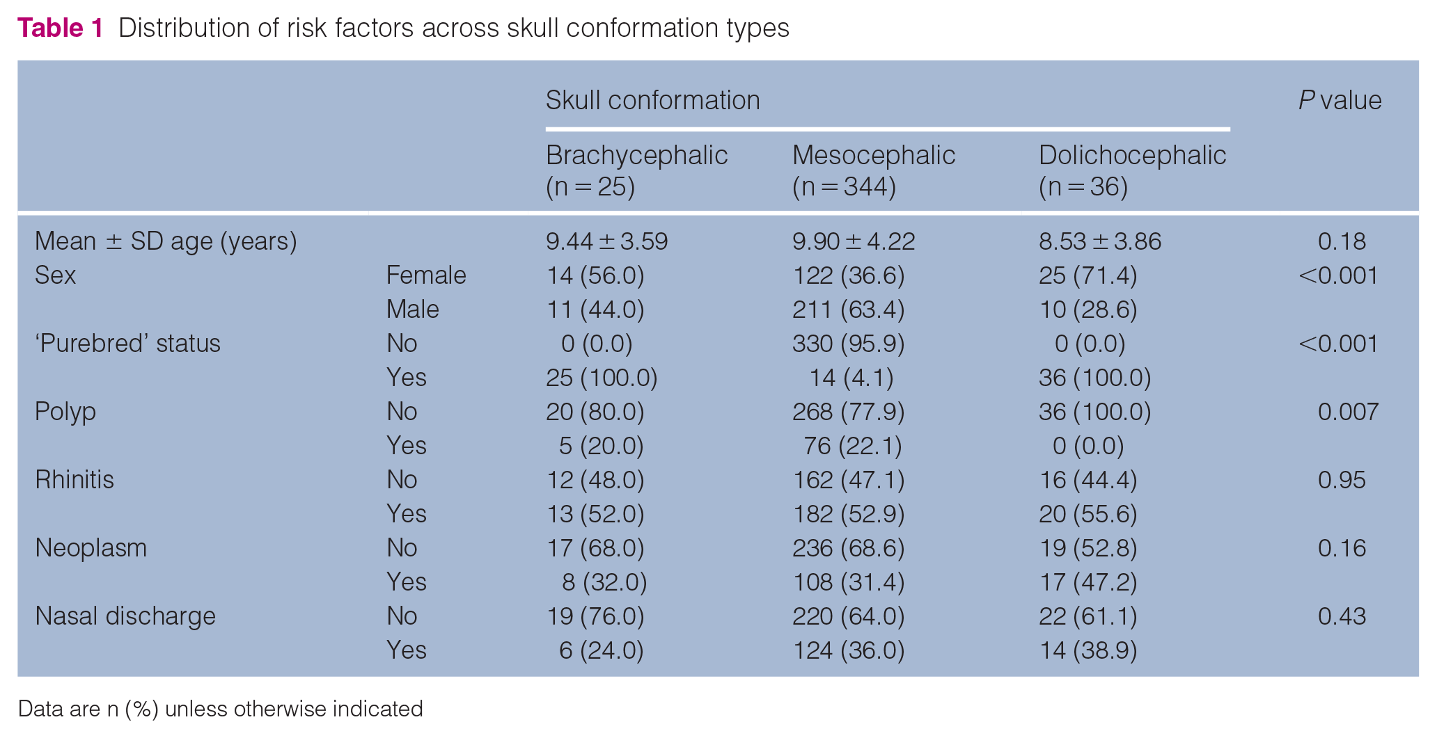
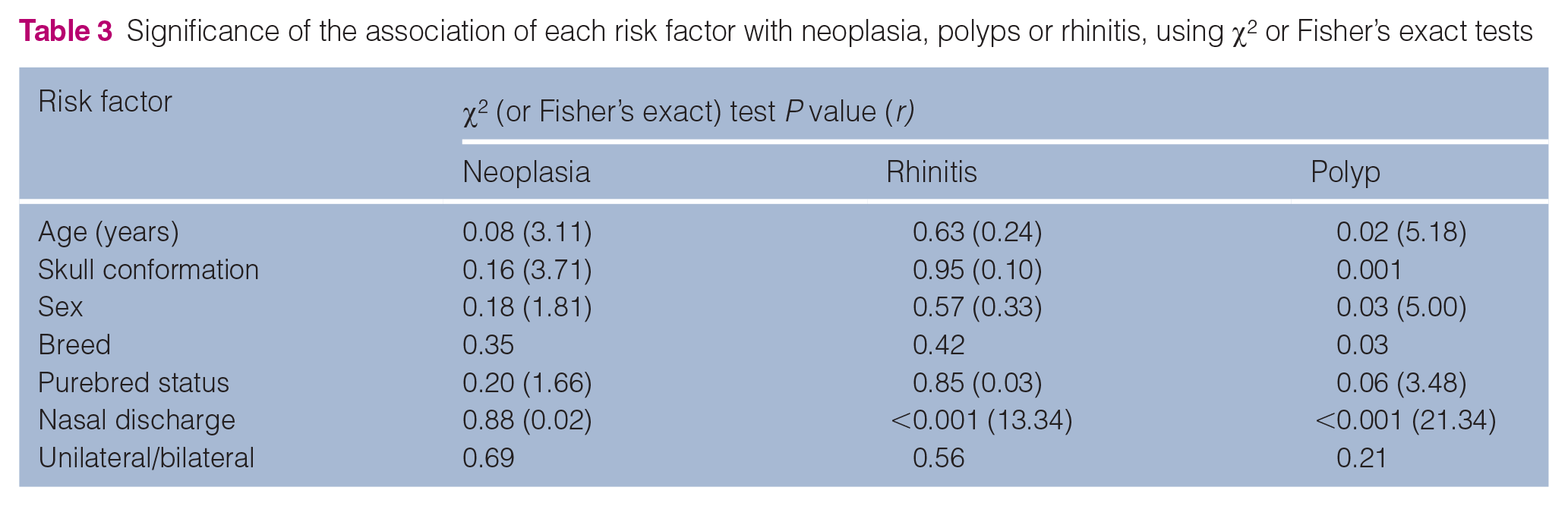
χ2 tests for the significance of association between neoplasia, polyps or rhinitis and each potential risk factor (age, breed, purebred status, sex, skull conformation, uni-/bilateral presenting signs, history of nasal discharge) revealed no significant associations between any risk factor and neoplasia or rhinitis (Tables 1–3). However, the presence of polyps was more likely in younger, male cats with MC skull conformation but without nasal discharge.
Distribution of risk factors across skull conformation types
Distribution of risk factors across samples with detected neoplasia, rhinitis or polyps

Data are n (%) unless otherwise indicated
BC = brachycephalic; MC = mesocephalic; DC = dolichocephalic
Significance of the association of each risk factor with neoplasia, polyps or rhinitis, using χ2 or Fisher’s exact tests
Multivariable logistic regression modelling
After including other significant variables in a logistic regression model describing the presence/absence of polyps, skull conformation, sex and nasal discharge were no longer statistically significant. Polyps were more likely to be diagnosed in nasal samples from younger cats, and in those where there was no concurrent diagnosis of neoplasia or rhinitis (Table 4).
Logistic regression model of polyps (n = 81) among 405 feline nasal samples received by a commercial veterinary diagnostic laboratory between 31 May 2006 and 31 October 2013

CI = confidence interval
All nasal disease
Of the 405 cats included in the study, 133 had neoplastic disease either in isolation or in combination with another pathological process. Neoplastic diseases diagnosed included lymphoma, adenocarcinoma, undifferentiated carcinoma and adenoma. In total, 126 cats had neoplasia as a single diagnosis, five had neoplasia with concurrent rhinitis and one had neoplasia (adenoma) in combination with a polyp. In total, 215 cats had a diagnosis of rhinitis, 193 as a single diagnosis and 16 cases had a concurrent polyp. Eighty-one cats had a diagnosis of polyp; in 63 cats this was the only diagnosis. One cat had concurrent neoplasia (lymphoma), rhinitis and a polyp (Figure 1).
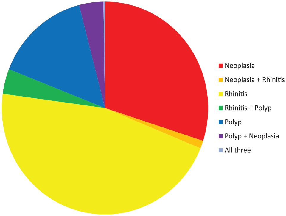
Proportion of cats with nasal disease, with the various diagnoses either alone or in combination
For 371 of these 405 cats, the age was stated on the submission form, giving a median age of 10 years (range 0.05–20 years; Figure 2). The sex was specified for 394 cats, with 161 females (40.9%), of which 122 were neutered, and 233 males (59.1%), of which 185 were neutered. Of the 405 cats, 18.5% were pedigree (n = 75) vs 11.4% of the background population. Of these, there were 35 Siamese (8.6%), 15 Persian and Persian crossbreeds (3.7%), six Maine Coons (1.5%), four Ragdolls (1.0%), two each of British Shorthair, Birman/Birman crossbreed, Bengal and Tonkinese (0.5%), and one each of the British Blue and Oriental breeds (0.2%). When subclassified according to skull conformation, 25 of these cats were considered BC (6.4%), 36 cats were classified as DC (8.9%) and the remainder as MC (n = 344 [84.9%]; Table 5).

Age of the cats presenting with various diagnoses
Breed and skull conformation in the background and study populations

Breed and skull conformation of the background (control) and study populations, with the study population shown also according to diagnosis
Non-pedigree includes domestic shorthair (DSH), domestic longhair (DLH) and not stated
Includes crossbreeds
BSH = British Shorthair; BC = brachycephalic (includes Persian cats and crossbreeds, Burmese, Birman and crossbreeds, BSH and British Blue); MC = mesocephalic (MC includes DSH, DLH, not stated, Maine Coon, Ragdoll, Bengal and Tonkinese); DC = dolichocephalic (includes Oriental and Siamese)
Neoplastic disease
Of the 133 cats diagnosed with neoplasia, age was stated on the submission form for 123 individuals, with a median age of 11 years (range 2–20 years; Figure 2). Sex was specified in 129 cats, with 59 females (45.7%) and 70 males (54.3%). Twenty-nine were pedigree (21.8%), of which 16 were Siamese (12.0%). When subclassified according to skull conformation, eight cats were considered BC (6.0%), 17 were classified as DC (12.8%) and the remainder as MC (n = 108 [81.2%]; Table 5). A nasal discharge was described in the clinical history provided for 48/133 cats with neoplastic disease (36.1%); of these, 15 were described as having epistaxis (11.3%). Clinical signs were described as bilateral in 13 cases (9.8%), and another 54 cats were described as having unilateral presenting signs (40.6%; data not specified in the remaining cases).
Neoplastic disease: lymphoma
Lymphoma was diagnosed in 68 cats, with a median age of 9 years (range 2–16 years; Figure 2) and with a nasal discharge described in 27 (39.7%). Of these 68 cats, 20 were pedigree (29.4%), with 14 Siamese (20.6%). BC breeds accounted for three cases (4.4%), while 15 were classified as DC (22.1%) and the remaining 50 as MC (73.5%; Table 5). Sex was recorded for 65 cats: 31 females (47.7%) and 34 males (52.3%).
Neoplastic disease: malignant epithelial neoplasia
Adenocarcinoma was the diagnosis given in 51 cases, with undifferentiated carcinoma diagnosed in a further 12 cats. Of all cats diagnosed with a malignant epithelial neoplasm (adenocarcinoma or undifferentiated carcinoma), a nasal discharge was recorded in 21 cases (33.3%). The median age for cats diagnosed with malignant epithelial neoplasia was 12.5 years (range 5–20 years; Figure 2). Of these 63 cats, nine were pedigree (14.3%). BC breeds accounted for five cases (7.9%), while two were classified as DC (3.2%) and the remaining 56 as MC (88.9%; Table 5). Sex was recorded for 62 cats, with 27 females (43.5%) and 35 males (56.5%).
A diagnosis of adenoma was seen in just two cases, one in a 12-year-old neutered male DSH cat with a concurrent polyp, and the second in a neutered female DSH cat, aged 18 years.
Rhinitis
Of the 215 cats diagnosed with various forms of rhinitis, either alone or concurrent with other nasal disease, the age was stated on the submission form for 197 cats, with a median age of 6 years (range 0.1–18 years; Figure 2). Sex was specified in 207 cats, with 82 females (39.6%) and 125 males (60.4%). Of these 215 cats, 41 were pedigree (19.1%). When subclassified according to skull conformation, 13 cats were considered BC (6.0%). Twenty cats were classified as DC (9.3%) and the remainder as MC (n = 182 [84.7%]; Table 5).
A nasal discharge was described in the clinical history provided for 94/215 cats with a diagnosis of rhinitis (43.7%); of these, 12 were described as having epistaxis. Clinical signs were described as bilateral in 20 cases, and another 82 cats were described as having unilateral presenting signs (data not specified in the remaining cases). Five cats had rhinitis concurrent with neoplasia; of these five cases, four had a diagnosis of lymphoma and one of adenocarcinoma.
Polyps
Eighty-one cats were diagnosed with a polyp originating from the nasal, nasopharyngeal or oropharangeal cavities, either alone (n = 63) or concurrent with other nasal disease. Age was stated on the submission form for all except eight of these cats, with a median age of 8 years (range 0.05–16 years; Figure 2). Sex was recorded for 80 of these cats, with 24 females (30.0%) and 56 males (70.0%). Of these cases, nine were pedigree (11.1%), and when subclassified according to skull conformation, five cats were BC (6.2%). No cats were classified as DC, with the remainder considered MC (n = 76 [93.8%]; Table 5). A nasal discharge was described in the clinical history provided on the submission form for 11 of these cats with a diagnosis of polyp; of these seven had concurrent rhinitis. Clinical signs were described as bilateral in three cases, and another 29 cats were described as having unilateral presenting signs (not specified in the remaining cases).
Discussion
This study examined the prevalence of different diseases arising in the feline nasal cavity, as based on cytological and histopathological diagnoses, from a large cohort of UK-based cats with samples predominantly submitted from first-opinion practices.
As such, one limitation of the current study is that this necessarily excludes those nasal diseases where the diagnosis is made via other modalities, such as imaging, and including stenosis and other anatomical defects, foreign bodies and trauma/post-traumatic injuries. However, some of the most common diseases are definitively diagnosed via cytology and histopathology, namely the various forms of rhinitis, tumours and polyps.
In this study, the most common category of disease diagnosed was rhinitis, either in combination with other pathology, or as a sole diagnosis. This was followed by neoplasia and then polyps. Concurrent diseases were often recognised on histopathology, with one patient diagnosed with lymphoma, rhinitis and a polyp. This order of prevalence is different to that previously reported by Henderson et al, 1 who reported that neoplasia was more commonly diagnosed than rhinitis. This may reflect a difference between the two study populations; the population in the Henderson et al study was referral practice based, as opposed to primarily first-opinion practice based in the current study. This difference in study populations most likely also explains the variation in the numbers of polyps diagnosed between the two groups.
A greater proportion of pedigree cats were represented in the present study population compared with the background population, but this may simply reflect the greater likelihood of owners of pedigree cats pursuing investigation and diagnosis rather than a predisposition of pedigree cats to nasal disease per se. Potentially contrary to expectations, in the present study no significant association between skull conformation and the various forms of nasal pathology was detected. The only significant associations found between the various forms of nasal disease and the risk factors assessed was of polyps arising in younger, male, MC cats without nasal discharge. However, after the inclusion of other significant variables to a logistic regression model describing the presence/absence of polyps, skull conformation, sex and nasal discharge were no longer statistically significant. Polyps were more likely to be diagnosed in nasal samples from younger cats, and in those where there was no concurrent diagnosis of neoplasia or rhinitis. χ2 tests are a test of association, whereas logistic regression is a measure of association; therefore, any significant associations between polyps and being male, having a MC skull or nasal discharge are only likely to be weak.
Findings of significant associations between various factors, including age, sex and breed, differ between the various published studies,1,4,13,14 with some reporting increased risk for male, older and Siamese cats for various conditions, and others not, including the present study. It is difficult to compare these studies directly as they differ somewhat in terms of the study population and the diagnoses included.
The three categories of diagnoses, namely rhinitis, neoplasia and polyp, all have the potential to be diagnosed on the same sample – despite this, the logistic regression showed they were not likely to co-occur. This may simply be due to the nature of the biopsy samples obtained, that is, a biopsy sample is likely to be obtained from a polyp or distinct mass if visible within the nasal cavity, and not from the adjacent mucosa, and this targeting of samples may result in concurrent pathology not being represented in the biopsy material submitted for assessment. One limitation of the current study is the reliance on the biopsy samples to be fully representative of all the nasal pathology present in that individual.
For cats diagnosed with neoplasia of any histological type, the age at diagnosis in the current study ranged from 2 to 20 years, with a median of 11 years, similar to a previous study. 4 In the present study, from the ages of 2–4 years, lymphoma was the only histological type of neoplasm diagnosed, with the youngest cat diagnosed with carcinoma being 5 years old. This previous study 4 also reported an increased risk in males and in older cats, but these associations were not found in the current study, nor by Henderson et al. 1
In agreement with various previous studies, lymphoma was the most commonly diagnosed neoplasm in the nasal cavity of cats.1,4,5 The second most commonly diagnosed tumour was adenocarcinoma, followed by undifferentiated carcinoma. By comparison, benign neoplasms were infrequent, comprising only 1.5% of all tumours diagnosed. One previous study 4 reported a wider range of tumour types than the present study, despite a similarly sized population, and it may be that some of the less common nasal neoplasms (including various forms of sarcoma) were not detected in the database search as they may not have specified the site as nasal in either the diagnosis given or in the sample type submitted.
Other limitations of this study include its retrospective nature, and the absence of clinical outcome data or information regarding previous therapies. Furthermore, some breeds and some categories of skull conformation were small. Rhinitis is a non-specific diagnosis encompassing a wide range of processes, and in the present study, there was no differentiation between the various forms; for example, fungal, bacterial or allergic rhinitis.
Conclusions
In this large-scale study of nasal biopsy submissions from UK-based cats, no significant association was found between skull conformation and nasal diseases, contrary to what might be expected. The only significant association found, when multivariable logistic regression modelling was used, between any of the potential risk factors and various forms of nasal disease, was polyps being more likely to arise in younger cats. No other significant associations, including between breed, sex or age, and the different conditions, was found.
Footnotes
Acknowledgements
The authors would like to thank all of the staff at Finn Pathologists for their assistance with accessing the database. The authors would also like to thank Professor Danièlle Gunn-Moore, The University of Edinburgh, for her advice regarding the classification of feline breeds into BC, MC and DC categories. This research was performed as part of a final year research project (SF) supported by the Royal Veterinary College.
Conflict of interest
The authors declared no potential conflicts of interest with respect to the research, authorship, and/or publication of this article.
Funding
The authors received no financial support for the research, authorship, and/or publication of this article.
Ethical approval
This work involved the use of non-experimental animals only (owned or unowned), and followed established internationally recognised high standards (‘best practice’) of individual veterinary clinical patient care. Ethical approval from a committee was not necessarily required.
Informed consent
Informed consent (either verbal or written) was obtained from the owner or legal custodian of all animal(s) described in this work for the procedure(s) undertaken. No animals or humans are identifiable within this publication, and therefore additional informed consent for publication was not required.