
Abstract
Objectives
The aim of this study was to investigate the frequency of identification and to describe the CT features of the os penis in cats without genitourinary disorders.
Methods
CT studies from cats that underwent an abdominal or pelvic examination between October 2013 and May 2019 were reviewed retrospectively. Cats with no signs of urinary disease and with the external genitalia included in the scan were recruited. Length, width, height and pre- and post-contrast attenuation values of the os penis in soft tissue and bone algorithms were measured independently by two observers.
Results
Twenty-three cats met the inclusion criteria. A cylindrical bone-attenuating structure inside the glans penis compatible with the os penis was visible in 20/23 (87%) cats. Mean length, width and height values were 3.48 mm × 1.41 mm × 1.37 mm in the soft tissue algorithm, and 3.26 mm × 1.15 mm × 1.06 mm in bone algorithm. The size of the os penis was not significantly different in neutered vs intact cats, but it was significantly larger in soft tissue vs bone algorithm. Age and body weight did not influence os penis size. Pre-contrast mean ± SD attenuation was 216.7 ± 69.5 Hounsfield units (HU) for soft tissue and 320.1 ± 135.9 HU for bone algorithms. Post-contrast attenuation was 289.1 ± 68.8 HU for soft tissue and 383.4 ± 130.9 HU for bone algorithms. A significant correlation between the attenuation in bone algorithm with body weight was noted, where the os penis was less attenuating with increased body weight (pre-contrast: r = –0.479; P = 0.038).
Conclusions and relevance
The feline os penis is commonly seen on CT images, being more frequently detected than on radiographs. Its presence should not be mistaken for uroliths in the penile urethra.
Introduction
In the anatomical literature, the feline os penis has been reported as a mineralisation of the penile erectile tissue of the corpus cavernosum within the glans, and is approximately 5–8 mm in length, without a ventral groove.1–4 The os penis is considered a heterotopic bone that is derived from the connective tissue of the penis by condensation of mesenchymal cells in the glans that subsequently differentiate in chondrocytes and osteocytes. 5 The os penis in carnivores is hypothesised to aid intromission of the penis into the vagina, stimulation of the female and prevention of urethral collapse during mating. 6
Urethral obstruction is reported to occur more commonly in young male cats and urolithiasis has been reported to represent 12–23% of cases.7–10 Radiopaque uroliths in the urethra of the cat can be challenging to differentiate from the os penis. In a previous study, the os penis was identified in 19/50 cats with computed radiography. 11
Currently, CT is becoming more readily available and mineral attenuation structures can be easier to depict. Therefore, we hypothesised that the frequency of identification of the os penis would be higher with CT. To our knowledge, there is no available information on the ability to identify the feline os penis using CT. Because recognition of the os penis is important to avoid misdiagnosis of urolithiasis, the aims of this study were to investigate the frequency of identification and to describe the CT features of the os penis in cats without genitourinary disorders, and to relate the presence of the os penis with age, body weight and neuter status.
Materials and methods
Patient selection
This was a retrospective descriptive study. The electronic medical database of the Fundació Hospital Veterinary Clinic (Autonomous University of Barcelona) was reviewed to identify feline patients that underwent an abdominal or pelvic CT before and after intravenous (IV) contrast administration between October 2013 and May 2019. Males were included in the study if: (1) they were not affected by any urinary disease demonstrated by the absence of urinary clinical signs, absence of biochemical alteration that suggested urinary disease and normal urinalysis; and (2) if the external genitalia were completely included in the scan. Patient data, including age, neuter status, breed and weight, were recorded.
Imaging procedure
All cats underwent general anaesthesia. CT examinations were obtained in soft tissue and bone algorithms, before and after IV administration of iopromide (Ultravist 300 mg/ml; Bayer pharma) or iopamidol (Scanlux 300 mg/ml; Sanochemia Pharmazeutika) in the cephalic vein at a dose of 2 ml/kg. Scans were performed with a 16 slice helical CT scanner (General Electric Brivo CT 385) with a slice thickness of 0.625 mm or 1.25 mm, interval thickness of 0.625 mm, collimation pitch of 0.5625:1, 120 kV, 50–90 mA, field of view according to patient size, and a matrix of 512 × 512.
CT images were reviewed independently by two of the authors (MTR and RA) with an image archiving and communication system software (Centricity PACS-IW [GE Healthcare]; Horos DICOM viewer 2019 [The Horos Project]) using soft tissue (window width: 400 Hounsfield units [HU], window level: 40 HU) and bone (window width: 2500 HU, window level: 480 HU) algorithms. All measurements were performed in both algorithms. The length of the os penis was measured in sagittal images obtained with multiplanar reconstructions. It was defined as the largest dimension in the craniocaudal plane. Width and height were measured in transverse images. The width was measured at the thickest point in the laterolateral plane, and height was measured at the thickest point in the dorsoventral plane. Attenuation values were measured using a region of interest (ROI) that included as much as possible of the os penis but excluding surrounding tissue.
Statistical analysis
Statistical analysis was performed using SPSS version 21 (IBM). The mean values for size and attenuation of the os penis were calculated, averaging the result of the two observers to minimise error and improve accuracy.
Continous variables are presented as mean ± SD and categorical variables as n (%). The normality of the data was assessed with Shapiro–Wilk’s test. Interobserver repeatability was assessed by the intraclass correlation coefficient (ICC), which ranges from 0 (no agreement) to 1 (perfect agreement). An independent samples t-test was performed to identify relationships between neuter status and the size and attenuation values of the os penis. A dependent samples t-test was performed to evaluate differences between measurements in soft tissue vs bone algorithms. Pearson’s correlation was performed to assess relationships between dimensions and attenuation of the os penis and age and body weight.
Results
Initially, 24 CT scans of cats including the genitalia were found, but one presented chronic kidney disease and was excluded. Therefore, 23 domestic shorthair cats with CT scans including the genitalia and no signs of urogenital disorders were finally included. Reasons for CT examination were as follows: 13/23 cats were previously included in another unrelated prospective study; 12 7/23 cats had CT for cancer staging not related to the genitourinary system; and 3/23 cats had CT for orthopaedic reasons.
Both observers identified the os penis in 20/23 cats (87%), but it could not be identified in 3/23 cats (13%). For the identification of the os penis, the observers showed perfect agreement (ICC 1.0). Of the cats where an os penis was identified, 15/20 were neutered (75%) and 5/20 were intact (25%) males. Mean age was 5.2 ± 3.6 years and mean weight was 4.7 ± 1.2 kg. The three cats where an os penis could not be identified were neutered (aged 2.0, 2.8 and 13 years; weight 4.2, 4.0 and 3.0 kg, respectively).
All ossa penis were seen as cylindrical mineral-attenuating structures inside the glans penis (Figure 1). In the soft tissue algorithm, the os penis was easier to identify and its length (P = 0.000), width (P = 0.003) and height (P = 0.021) were significantly larger than in the bone algorithm (Table 1). There was no statistical difference for the visualisation of the os penis between intact (n = 5/5) vs neutered (n = 15/15) cats (P = 0.328). The mean length, width, height and attenuation values for both soft tissue and bone algorithms for the os penis are summarised in Table 1. No statistical difference or correlation with age, or intact/neutered status vs os penis size and attenuation values were found. The attenuation values obtained with the bone algorithm were significantly higher than those from the soft tissue algorithm in both pre-contrast (P = 0.002) and post-contrast (P = 0.009) series. A significant correlation was observed between the attenuation of the os penis measured in bone algorithm and the body weight, where the attenuation of the os penis gradually decreased as body weight increased (pre-contrast: r = –0.479; P = 0.038).
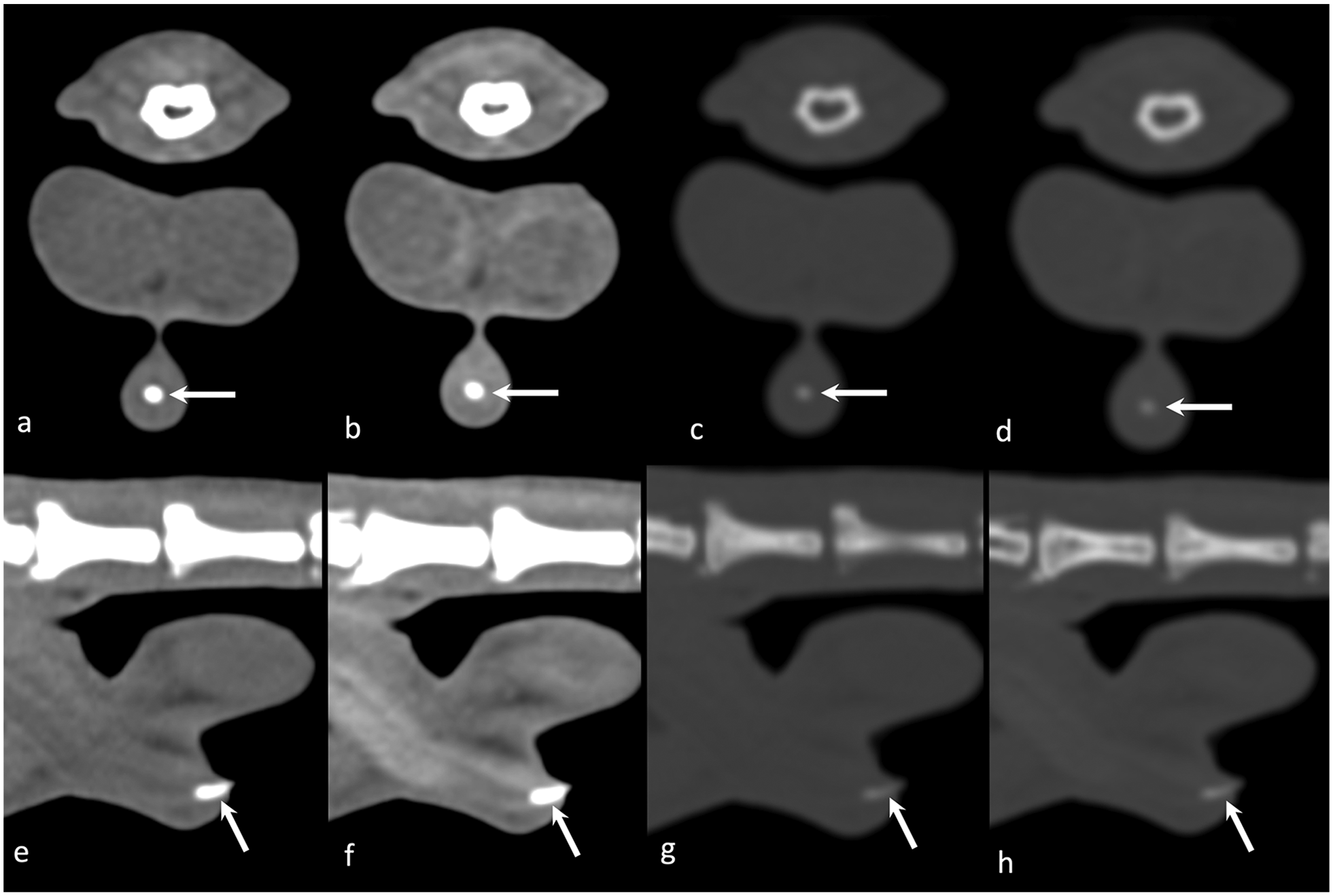
Feline os penis on CT images (arrows). (a–d) Pre- and post-contrast transverse images in (a,b) soft tissue and (c,d) bone algorithms. (e–h) Sagittal reconstruction in (e,f) soft tissue and (g,h) bone algorithms
Feline os penis size and attenuation values on CT images in soft tissue and bone algorithms

ICC = intraclass correlation coefficient; HU = Hounsfield units
According to the scale proposed by Bartko, 13 inter-observer agreement was poor for os penis width and height measured in soft tissue algorithm (ICC = –0.08 and 0.06, respectively). A good agreement was reached for the os penis length and pre- and post-contrast attenuation values measured with the soft tissue algorithm (ICC = 0.69, 0.76 and 0.62, respectively), and for os penis length, width and post-contrast attenuation measured with the bone algorithm (ICC = 0.79, 0.78 and 0.65, respectively). Very good agreement was reached for os penis height and the pre-contrast attenuation measured in bone algorithm (ICC = 0.88 and 0.81, respectively).
Discussion
The results of this study show that the os penis in cats was frequently identified on CT images, regardless of their neuter status, and this frequency is higher than in reports using radiography. In a previous study, the os penis was observed in 16% of analogue and 38% of computed radiographs. 11 The fact that the feline os penis is seen more frequently on CT images can be explained by the superior contrast resolution of CT when compared with radiography. 14
In the present study, cats showed, on average, a shorter cylindrical bone than in anatomical reports.2,4 The reason for this difference is unclear, but the lower sample size could have influenced this difference. However, no significant statistical differences were observed in the comparison between os penis size and neuter status.
In this study, the identification and assessment of the os penis was performed in soft tissue and bone algorithms. The characteristics of CT in the acquisition and reconstruction of images make dense structures, such as bones, appear thicker in soft tissue window owing to its low-frequency kernel and widest point spread function.15,16 These reconstruction characteristics, together with the slice selected and the placement of the electronic callipers by each observer for the measurements, could explain the poor interobserver agreement for os penis width and height in soft tissue algorithm.
The attenuation values of the os penis measured with the soft tissue algorithm were statistically significantly different from the values obtained in bone algorithm. These differences could be explained by the difficulty in determining the correct margins of the bone with the soft tissue algorithm, resulting in the inclusion of other penile tissues in the measurement. The fact that no statistical difference was found between age and neuter status with the attenuation values of the os penis may suggest that the mineralisation process continues even if the animals have been castrated. The reason for the significant negative correlation between the body weight and the HU values on bone algorithm is uncertain.
Slightly higher values in the attenuation of os penis in post-contrast images might be due to the presence of contrast medium in the vessels of the corpus cavernosum that contains the bone.2,17 Even though ROI measurements were placed within the os penis, avoiding small portions of the corpus cavernosum was challenging with both algorithms.
Urethral obstruction is reported to occur commonly in young male cats, and urolithiasis accounted for 12–23% of cases.7,9,10,18 Cats in this study had no signs or evidence of urinary tract disease. This is clinically relevant because the os penis might be misinterpreted as urethrolithiasis or dystrophic mineralisation of the urethral wall. 4
Owing to the retrospective nature of this study, limitations, including the small sample size and the lack of histopathological examination confirming the presence and/or dimensions of the reported os penis, are identified in this study.
Conclusions
The os penis was identified in 87% of male cats examined with CT, regardless of their neuter status, age or weight. This is a higher frequency when compared with previous radiography reports. Therefore, identification of the os penis should not be mistaken for urolithiasis in the penile urethra.
Footnotes
Acknowledgements
We are grateful to the staff of the Hospital Veterinary Clinic of the Autonomous University of Barcelona for their support.
Conflict of interest
The authors declared no potential conflicts of interest with respect to the research, authorship, and/or publication of this article.
Funding
The authors received no financial support for the research, authorship, and/or publication of this article.
Ethical approval
This work involved the use of non-experimental animals only (owned or unowned), and followed established internationally recognised high standards (‘best practice’) of individual veterinary clinical patient care. Ethical approval from a committee was not necessarily required.
Informed consent
Informed consent (either verbal or written) was obtained from the owner or legal custodian of all animal(s) described in this work for the procedure(s) undertaken. No animals or humans are identifiable within this publication, and therefore additional informed consent for publication was not required.