
Abstract
Objectives
The aims of the present study were to monitor, by radiographic examination, the skeletal development of the pelvis and the femorotibial joints of the domestic cat from the first week of life until the closing of the growth plates.
Methods
Radiographic examinations were collected from 15 domestic cats at weekly intervals during the first month and every 2 weeks from the second to the fourth month of age. After that, examinations were performed monthly until the age of 18 months.
Results
The ischiopubic growth plate closed at 2 months of age, followed by the fusion of the iliopubic, ilioischial, proximal femoral, greater trochanter and proximal fibular growth plates. The distal femur and proximal tibial growth plates were the last to close, with fusion occurring at 18 months. The mean time to closure of the iliopubic, ilioischial and distal femoral growth plates was shorter in females. The ossification centers first appeared, in ascending order, beginning with the lesser trochanter, followed by the greater trochanter, proximal fibular epiphysis, tibial tuberosity, patella, ischial tuberosity and lateral sesamoid of the popliteus muscle.
Conclusions and relevance
The complete closure of the growth plates of domestic cats occurs at approximately 18 months of age. Skeletal maturation at approximately 18 months of age is an important parameter to be considered in radiographic evaluation of certain skeletal changes, evolution of fractures and nutritional imbalance.
Introduction
Establishing the time of appearance of the ossification centers and the closure of the growth plates by radiographic examination is key to understanding skeletal evolution and allows the identification of abnormalities and estimation of patient age, which are all factors that may interfere with the course of clinical and surgical action and prognosis. Nevertheless, radiographic interpretation of the immature skeleton is challenging, 1 and the growth plates may be confused with fractures or pathological processes. Although a few references present images of the immature skeleton of domestic cats,2–4 and one shows schematic representations of animals <30 days old, these images are irregularly spaced through skeletal development, and information may be missing. 2
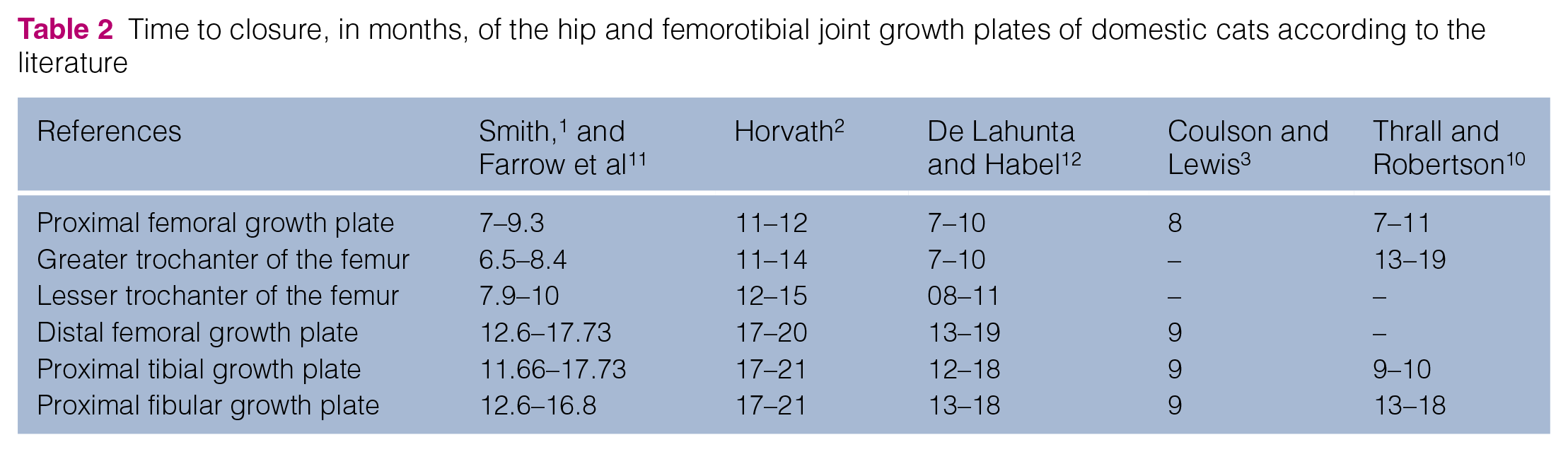
The factors that influence the closure of the growth plates are complex. In domestic cats, synchrony is observed between sexual maturity and the closure of the growth plates.5,6 The hormones estrogen and testosterone antagonize growth hormone and cease proliferation of the cartilaginous cells that make up the growth plate. 7 Female domestic cats reach sexual maturity at approximately 6–9 months of age, while males mature later, reaching sexual maturity between the age of 9 and 10 months. 8 The time to appearance of the ossification centers of a few bone structures and closure of the growth plates of domestic cats are presented in Tables 1 and 2, respectively.
Time to appearance, in days, of the ossification centers of hip and femorotibial joint structures in domestic cats according to the literature

Time to closure, in months, of the hip and femorotibial joint growth plates of domestic cats according to the literature
The goal of this study was to follow the development of the pelvis and the pelvic limbs of the domestic cat by radiographic evaluation from the first week of life until the closure of the growth plates.
Materials and methods
This study was approved by the Ethics Committee on Animals Use of the Federal University of Minas Gerais (UFMG; protocol number 82/2015). Fifteen unaltered domestic shorthair cats (nine males, six females) were kept at the Experimentation Center for Small Animals (CEPA) of the Veterinary School of UFMG for 2.5 years for radiographic examinations. The animals were born from 17 pregnant cats collected in the metropolitan area of Belo Horizonte; these cats were kept in CEPA to guarantee the radiographic examination of the offspring in the first week of life. After weaning, the adult cats and the offspring not used in this study were spayed or castrated and referred for adoption. To avoid genetic bias, each kitten was selected from a different litter.
The pregnant cats presented normal clinical status and were negative for feline immunodeficiency virus and feline leukemia virus. The animals were fed commercial cat food twice a day, and average daily intake in grams by the individual cats varied according to their age. The provided food had the recommended amounts of protein (30%), calcium (1–2%), phosphorous (0.85%), taurine (0.12%) and vitamin D3 (960 UI/kg). 13
The radiographic examinations were performed weekly during the first month of life, biweekly from the second to the fourth month of life, and monthly from the fifth month until the closure of the growth plates. We used the Viegas Moraes Industries (VMI), with a mean Kv of 50, mA of 100 and exposure time of 0.06 s. A small Styrofoam holder was used to keep the pelvis aligned. The cats were placed in dorsal decubitus with the pelvic limbs extended and rotated medially so that the patellae were superimposed to the sagittal plane of the femora. The pelvic limbs were positioned parallel to each other and to the spine. The obtained images were digitized in a Konica processor (Konica Regius, model 110) and analyzed using DICOM Image Viewer software.
For radiographic examinations during the first month of life, the kittens were removed from their litter, wrapped in the same cloth used in their housing and transferred to the radiography room. In addition to helping in the maintenance of body temperature, the cloths were used to minimize subsequent rejection by the mothers, as cats recognize each other through smell. Contact with other animals and people was avoided, and the radiographs were taken as quickly as possible. Before the kittens were returned to the litter, the cloth was brushed on the mother’s temples and then on the kitten to preserve the scent. The relationship between the kitten and the mother was observed for approximately 30 mins to ensure maternal acceptance.
After 4 months of age, the animals were tranquilized and anesthetized monthly for a ventrodorsal radiograph. They received 10 mg/kg ketamine (Cetamin; Syntec Vet) associated with 0.5 mg/kg midazolam (Midazolam; Eurofarma) and 0.3 mg/kg methadone (Mytedom, Cristalia), administered intramuscularly. After 15 mins, propofol (Propovet; Ouro Fino) was administered intravenously using a 22 G catheter fixed with tape.
In the radiographic evaluation, we recorded the time of appearance of the ossification centers of the ischial tuberosities, the proximal and distal femoral epiphyses, the greater and lesser trochanters, the tibial tuberosity and the proximal tibial epiphysis, the proximal fibular epiphysis, the patella, the lateral sesamoid of the gastrocnemius muscle and the sesamoid of the popliteus muscle.
Additionally, we recorded the closure of the ischiopubic, iliopubic, ilioischial, proximal and distal femoral, and proximal tibial and fibular growth plates. Growth plate closure was considered complete in the absence of a radiolucent line in the radiographic exams. 6 Femur length and pelvis width and height were measured in millimeters using DICOM Image Viewer software.
Femur length was measured by drawing a line from the proximal to distal end of the femur, through the center of the diaphysis. The femoral head and distal epiphysis were included in the femoral length, as they appeared.
To measure the width of the pelvis, we drew a line between the wings of the ilium (line A) and between the ischial tuberosities (line B). To determine the pelvic length, a line (line C) was drawn to measure the distance between the lines A and B (Figure 1).

Ventrodorsal radiograph of the pelvis of an 11-month-old domestic shorthair cat. The dotted lines show the width of the pelvis: (A) distance between the wings of the ilium and (B) distance between the ischial tuberosities. The solid line (C) shows the pelvic length
Descriptive analysis was performed for values of center (mean and median) and dispersion (SD and quartiles) of the mean time, in days, to the appearance of the ossification centers and closure of the growth plates. Next, a t-test was performed to infer the expected values of these variables for the studied population. The t-test was also used to determine differences between the sexes. 14
Results
Radiographic examination in the first month of life was challenging in some respects owing to the small size of the kittens and the possible rejection of the kittens by their mother. Four kittens were rejected, and re-introduction was performed gradually, by wiping a cloth on the mother’s temples and then on the kitten’s body. Nevertheless, three kittens were not accepted and thus required artificial feeding. Two of these were re-introduced to their litters the next day, and one of the kittens was permanently rejected and was introduced into another group. The temporary rejection rate was 13.3% and the permanent rejection rate was 6.7%.
The mean age of the domestic cats evaluated in the first week was 6 days (range 4–7 days), at which point the ilium, ischium, pubis, and femoral, tibial and fibular diaphyses were visualized. The youngest cat was 4 days old and already presented the abovementioned bones (Figure 2a). Determination of the exact time in which the ossification centers of these bones appeared was not possible.

Radiographs in ventrodorsal projection of the pelvis of domestic shorthair cats at (a) 4, (b) 13, (c) 21 and (d) 45 days of age. In (a) 1 = ilium; 2 = ischium; and 3 = femoral, 4 = tibial and 5 = fibular diaphyses. The ramus of the pubic bone (6) is visualized with difficulty. In (b) 6 = the ramus of the pubic bone; 2’ = ischial tuberosities; 3a = the femoral head; 3b = the distal femur epiphysis. In (c) 4a = proximal tibial epiphysis. In (d) 3c = greater trochanter; 3d = lesser trochanter. (e–g) Radiographs at 3, 12 and 18 months of age, respectively. In (e) 2b = ischial tuberosities; 5a = proximal fibular epiphysis; 7 = patella. In (f) 1a = absence of growth plates of the pelvis; 3a’ = proximal femur epiphysis; 3c’ = greater trochanter; 5a’ = proximal fibular epiphysis. In (g) 3b’ = distal femoral epiphysis; 4a’ = proximal tibial epiphysis
In the second week of the evaluation, the cats had a mean age of 12.2 days (range 9–14 days), and delimitation of the radiolucent spaces corresponding to the pelvic symphysis and the ischiopubic growth plate was identified, at which time the pubic ramus and the ischial tuberosities were present. The ossification center of the distal femoral epiphysis was predominantly observed at this stage, and the mean age of its onset was 13.6 days (Figure 2b).
In the third week of evaluation, the ossification centers of the proximal femoral and tibial epiphyses were radiographically visualized at a mean age of 16.07 and 18.27 days, respectively (range 11–24 days; Figure 2c). In two cats (13.33%), the proximal tibial epiphysis was observed in the second week of evaluation, at 11 and 13 days.
The mean age of appearance of the ossification centers varied between 38.07 and 190.27 days, depending on the bones (Figure 2d,e and Table 3). The lateral sesamoid of the gastrocnemius muscle was identified in all the cats, and the medial sesamoid was visualized in only one cat at 5.9 months of age.
Mean time, in days, for ossification center onset, 95% confidence interval (CI), SD, minimum value, maximum value, and 25%, 50% and 75% of the bones in 15 unaltered domestic shorthair cats (nine males and six females) from different litters

The ischiopubic growth plate was the one that fused earliest in cats, occurring at a mean age of 2.01 months. The growth plates of the greater trochanter and the proximal fibular epiphysis closed at 9.44 and 10.34 months, respectively (Figure 2f), and the fusion of the distal femoral and proximal tibial growth plates occurred at 13.56 and 18.02 months, respectively (Figure 2g and Table 4). The pelvic symphysis remained open in all cats until the end of the study. The tibial tuberosity was better visualized in the mediolateral projection (Figure 3).
Mean time, in days, for closure of the growth plates of the pelvis, femur, tibia and fibula, 95% confidence interval (CI), SD, minimum value, maximum value, 25%, 50% and 75% in 15 unaltered domestic shorthair cats (nine males and six females) from different litters


Radiograph in mediolateral projection of the pelvic limb of a domestic shorthair cat of 3 months of age; 4b shows tibial tuberosity
The mean time of appearance of 82% of the ossification centers of the proximal fibular epiphysis and the patella was significantly (P <0.05) shorter in the females compared with the males. The closure time of the iliopubic and ilioischial growth plates and of the distal femoral growth plate was also shorter in the females than in the males. The ischiopubic growth plate closed at the same age in both sexes. The proximal fibular growth plate was the only one that closed earlier in the males.
Regardless of sex, no significant difference was observed in the width of the pelvis measured by the distance between the wings of the ilium and between the ischiatic tuberosities. Pelvic length was 41.7% greater than its width at all times of radiographic evaluation, with a mean difference of 30 mm in the last examination, performed after the closure of the growth plates of the pelvis and of the hip and femorotibial joints (mean time 18.02 months).
The estimated daily growth rates of the femur length and pelvis width and length were obtained by the percentage variation of each measurement among radiographic evaluations, divided by the mean period among them. Figure 4 shows the growth rate by radiographic evaluation for the three associated and independent variables, respectively. The highest growth rate occurred between birth and the first 100 days of life. Then, the growth rate slowed until 300 days of age and remained relatively constant until 600 days of age, when it reached zero (Figure 4).

Estimated daily growth rate of femur length and pelvis width and length by radiographic evaluation in 15 domestic shorthair cats
Discussion
Understanding the skeletal evolution of the cat is essential for adequate clinical evaluation and diagnosis. Skeletal changes due to trauma or endocrine disorders may culminate in inadequate diagnosis and treatment of cats during the growth phase. Sometimes the non-visualization of the bones in radiographic examinations at early ages is misinterpreted as congenital defects or fractures. Emergence of the ossification centers and closure of the growth plates occur at different ages and vary from bone to bone. Evaluation from birth to adulthood is required to obtain these data. Nevertheless, evaluating kittens from birth demands an understanding of animal behavior. Removal of the kittens for examination, even for a short period of time, may result in rejection by the mother and interfere with the results.
Although the rate of permanent rejection of the kittens was small (6.7%), it highlights the need for caution when re-introducing kittens into the group following handling during the first month of life. Maintenance of the body temperature and of the characteristic smell is a challenge that can be controlled by keeping the kittens in the same cloth used in their housing. However, permanent rejection may still occur, as observed in one case in this study, and may require the introduction of the kitten into another litter.
The closure time of the ischiopubic growth plate of the cats in this study was similar to that reported in the literature, 3 while the iliopubic and ilioischial growth plates were closed at 168 days, a period intermediate to the 120 and 240–270 days previously reported.2,3 Therefore, different factors, such as climatic, hormonal and genetic conditions, 10 have been suggested to influence the speed of growth plate closure.
In general, the closure time of the proximal and distal femoral and the greater trochanter growth plates observed in this study is similar to that reported by some authors1,12 but differs from that reported by other authors. 2 The closure time of the proximal fibular growth plate in this study is within the range reported in the literature.1,2,12
According to the literature,1–3,11 variation occurs in the closing period of the aforementioned growth plates in domestic cats, and determining their time of fusion in the radiographic examination is challenging, as it requires the perfect alignment of the bones for appropriate evaluation of the absence of a radiolucent line for growth plate closure to be considered. In some animals, reminiscent cartilage centers may persist for years, even after histological growth is interrupted. 1
Thus, no standard for radiographic examination exists regarding the exact time of fusion of the growth plates. We also note the methodological variation among the mentioned studies, as well as environmental influences. The possibility of factors such as genetics, race, feeding and different litters influencing the evaluation of the closure time of the growth plates has already been described. 9
In general, the shorter time for development of the ossification centers and closure of the growth plates in females may be due to the later sexual maturity in males. 8 In the present study, the earlier growth plate closure (3 days) in males vs females, regarding the ossification of the greater and lesser trochanters, does not provide enough evidence to discard the hypothesis that these structures appear at the same time in both sexes.
The lack of differences between sexes regarding the closure of the ischiopubic growth plate may be real or, alternatively, a variation between the sexes could have been undetected, not because it is lacking, but because of overlapping structures hindering observation in the ventrodorsal projection.
Regarding the appearance of the proximal and distal femoral epiphyses, the greater trochanter of the femur, the proximal tibial and fibular epiphyses, the tibial tuberosity, the patella, the lateral sesamoid gastrocnemius muscle and the ischial tuberosity, the data from this study are similar to those reported in the literature.1,2 The appearance time of the ossification center is less variable when compared with the growth plate fusion time, 1 and this result was therefore expected. The observation of the medial sesamoid of the gastrocnemius muscle in only 1/15 cats in this study corroborates the literature reporting on its rarity.3,11
The factors that interfere in the closure of the growth plates and, consequently, in the growth rate, are not completely defined, but genetics and hormones are known to be important. In cats, the highest growth rate occurs until 100 days of age, which seems to be the natural growing pattern of the kitten. Next, a decline in velocity is observed in comparison with the first phase but is still more or less consistent until 250 days of age, the approximate time when females and males reach puberty. In this period, estrogen and testosterone antagonize the growth hormone, and proliferation ceases for the cartilaginous cells of the growth plate, 7 which leads to the gradual decline of growth until it ceases completely (Figure 4), as previously reported. 5
The determination of the exact moment at which cats reach puberty is not easy because of individual variation, which may explain the observation of earlier reproductive behavior in three females and three males in this study. This attainment of puberty may have contributed to the more pronounced decline in the growth rate at 7 months of age (Figure 4). The literature reports a period of sexual maturity in domestic cats between 6 and 9 months of age in females and 9 and 10 months in males. 8 The growth phase between birth and 100 days of age seems to be the critical moment in which environmental and nutritional factors must be considered and properly managed to avoid manifestation of nutritional and/or endocrinological diseases at a later phase.
The cats evaluated in this study were not neutered, which made it impossible to understand the effects of neutering on the closure of the studied growth plates. Likewise, the closing time of the tibial tuberosity growth plate was not quantified. Instead, it was only recorded as the moment when the tibial tuberosity ossification center appeared.
In general, neutering is known to delay the closure of growth plates in cats, but few data are available on its effect on each growth plate. The greater trochanter, femur, and distal and tibial tuberosity growth plates close later in neutered male cats than in non-neutered males, as reported in a retrospective study. 6 Although it does not provide information about the neutering age of the evaluated cats, the study provides rele-vant information because neutering, especially when performed early, results in a delay in the closure of growth plates, increasing the probability of fracture occurrence.
Conclusions
The closing time of the growth plates is shorter in females than in males. Skeletal maturation at approximately 18 months of age is an important parameter to be considered in radiographic evaluation of certain skeletal changes, evolution of fractures and nutritional imbalance. Although further studies with larger sample sizes are needed, the data presented herein contribute to a better understanding of the radiographic images of the skeleton of immature domestic cats.
Footnotes
Conflict of interest
The authors declared no potential conflicts of interest with respect to the research, authorship, and/or publication of this article.
Funding
The authors received no financial support for the research, authorship and/or publication of this article.
Ethical approval
This study involved the use of experimental or client-owned animal(s) outside of internationally recognized high standards (‘best practice’) of individual veterinary clinical patient care. The study therefore had ethical approval from an established committee as stated in the manuscript. The authors have stated in the ‘Materials and methods’ the nature of the institutional, national or international ethical review body used.
Informed consent
Informed consent (either verbal or written) was obtained from the owner or legal custodian of all animal(s) described in this study for the procedure(s) undertaken. No animals or humans are identifiable within this publication, and therefore additional informed consent for publication was not required.