
Abstract
Objectives
The objective of this study was to evaluate the techniques and short-term effects of cryopreservation of feline red blood cells (RBCs) in liquid nitrogen using glycerol or hydroxyethyl starch (HES) as a cryoprotectant.
Methods
Feline RBCs were manually mixed with either 20% glycerol or 12.5% HES and frozen for 24 h in liquid nitrogen. The samples were thawed and glycerolized samples were manually washed. Success of the freeze/thaw process was determined by recovery rate of RBCs and evaluation of morphological changes using scanning electron microscopy (SEM). A unit of canine packed RBCs was also subjected to the same methodology to evaluate the cryopreservation handling technique.
Results
Feline RBCs preserved with 20% glycerol had a high recovery rate (94.23 ± 1.25%) immediately after thawing. However, the majority of the cells were lost during the washing process, with a final packed cell volume of <1%. A recovery rate was unable to be assessed for samples preserved with HES owing to the high viscosity of the mixture. SEM revealed significant morphological changes after glycerol was added to the feline RBCs. Although these morphological changes were partially reversed after thawing, the majority of the RBCs were lost during the washing process. Minimal morphological changes were noted in the HES sample. Similar results were noted with the canine RBCs.
Conclusions and relevance
The described manual technique for cryopreservation using glycerol was not able to successfully preserve feline or canine RBCs. In the present study, it was difficult to make conclusions about the efficacy of HES. Further studies evaluating HES as a cryoprotectant are warranted.
Introduction
Administration of red blood cells (RBCs) is a crucial, life-saving therapy in both animals and humans. RBC products such as whole blood or packed RBCs (pRBCs) are commonly administered to treat blood loss, failure of erythropoiesis and hemolytic anemia.1,2 However, RBC transfusions are not without risk as administration of incompatible blood can result in life-threatening transfusion reactions.1–3
Cats pose a particular challenge as they possess naturally occurring alloantibodies against non-self blood types,1–3 making administration of compatible feline blood types imperative to minimize acute hemolytic reactions.1–3 However, access to donor blood of the less common blood types (B and AB) is limited, owing both to the scarcity of donors and limited availability of banked units. 3 As refrigerated RBC products are stored for a maximum of 30 days, 4 consistent stocking of less common blood types leads to waste of unused units. It also often takes time to acquire units of these rare blood types from blood banks. Xenotransfusion of cats with canine RBCs has been described in emergent situations, but canine RBCs are rapidly destroyed by feline antibodies within 4–7 days leading to a predictable hemolytic reaction of variable intensity. 5 Also, xenotransfusion is a one-time intervention because once a cat has developed anti-canine antibodies, it can never again receive canine blood. 5
To address similar issues with RBC availability and storage, cryopreservation has been used for human RBCs since the 1950s.6,7 Cryopreservation halts all biochemical processes and allows long-term storage of RBCs. Cryopreserved RBCs are used by the military and in other situations when the need for units of RBCs is unpredictable, in addition to preserving units of rare blood phenotypes.6–8 In order to minimize osmotic stress during the freezing and thawing processes, a cryoprotectant is added to RBCs prior to freezing.6–9 Currently, the US Food and Drug Administration has approved use of human RBCs suspended in 40% weight/volume (w/v) glycerol stored at −80°C or lower for up to 10 years.6–9 An alternative method employed in Europe uses 20% glycerol followed by rapid freezing in liquid nitrogen.6–9 Multiple in vivo studies in humans have established that administration of frozen RBCs is as safe and effective as transfusion of refrigerated RBCs.10–13 However, the processing time involved in freezing and thawing RBCs (in addition to the need to store the products properly) adds additional expense. The cost of cryopreserved RBCs using glycerol can be as much as 3–4 times that of a unit of refrigerated RBCs, 14 limiting the routine use of cryopreserved RBCs.
Glycerol penetrates the membranes of RBCs and prevents formation of ice crystals and subsequent membrane damage.6,7 Frozen RBCs must be de-glycerolized via multiple washing steps prior to transfusion to avoid post-transfusion osmotic hemolysis of the recipient’s RBCs and penetration of other biological cells upon transfusion.6–9 In humans, post-thaw cells must meet certain standards for transfusion such as minimal hemolysis with a recovery rate of >80% of RBCs and <1% hemolysis in vitro after removal of glycerol via washing.6–8 In addition, post-thaw RBCs must exhibit minimal changes in morphology and adenosine triphosphate (ATP) activity vs refrigerated cells, indicating preservation of RBC cellular functions.6–8,10–16
Hyperosmolar hydroxyethyl starch (HES; ie, ⩾11.5%) has been studied as an alternative to glycerol. 17 Hyper-osmolar HES dehydrates RBCs, reducing the incidence of intracellular ice formation. 9 Units of blood frozen with HES can be transfused directly to patients without the need for any post-thaw wash steps as the HES is not toxic to the recipient.8,13,18
Investigations into frozen canine RBCs have shown successful cryopreservation for up to 2 months using both glycerol and HES.18–22 Kim et al reported that 12.5% weight/weight (w/w) HES showed comparable cryoprotection of canine pRBCs to 20% glycerol based on hematologic and morphological parameters. 20 Cryopre-served canine pRBCs using 20% or 40% glycerol after thawing and washing, 19 and 11.5% (w/w) HES after thawing, 20 have been transfused into dogs with no transfusion reactions.19,20 At the time of the study design, no cryopreservation studies had been published using feline pRBCs.
The primary objective of this study was to evaluate and compare the in vitro effectiveness of glycerol and HES as cryoprotectants for feline pRBCs frozen for 24 h vs feline pRBCs refrigerated for 24 h. We hypothesized that glycerol and HES would both be acceptable cryopreservation solutions for feline blood; acceptable cryopreservation would result in an RBC recovery rate of >80%, <1% hemolysis in vitro and minimal changes in RBC morphology vs both pre-freezing samples and refrigerated samples.
Materials and methods
A total of six units (units #1–6) of feline type A pRBCs (20–30 ml/unit) were purchased from a commercial blood bank (Animal Blood Resources International). The units were shipped on the same day as they were collected, using overnight shipping; this made their age at arrival at our institution less than 24 h. All units were utilized within 48 h of receipt, making their ages between 24 and 72 h. Each unit was warmed to room temperature immediately before processing by immersion in a 37°C water bath for 10–20 mins to a surface temperature of 23°C. Temperatures were measured by a digital thermometer applied to the surface of the bag.
For pRBC units #1–3, each unit was divided into three aliquots of equal volume and only 20% glycerol was used as cryoprotectant as HES was not available at the beginning of the study (Figure 1). As units of pRBCs typically range from 20 to 30 ml in size from the blood bank, the aliquots ranged from 6 to 10 ml each. The first aliquot (aliquot 1) was used as a control for baseline analysis and was then stored in a refrigerator at 4°C for 24 h. The remaining two aliquots (aliquots 2 and 3) were used in the glycerol fast cooling technique (see below). For pRBC units #4–6, hypertonic HES became available, so each unit was divided into five aliquots (Figure 1). When the units were divided into five aliquots of identical volume, the aliquots ranged from 4 to 6 ml per aliquot depending on the size of the unit of pRBCs. The first aliquot was used as the control for baseline sample analysis and then was stored in a refrigerator at 4°C for 24 h. Aliquots 2 and 3 were used in the low glycerol fast cooling technique as before. Aliquots 4 and 5 were used in the HES cryopreservation technique (see below). The glycerol and HES aliquots were frozen for 24 h, thawed, and a washing step was then performed for glycerolized samples.

Experiment design. For units 1–3, each unit was divided into three equal aliquots. Aliquot 1 was used as the baseline control and was refrigerated for 24 h. Aliquots 2 and 3 were frozen with glycerol as the cryoprotectant. For units 4–6, each unit was divided into five equal aliquots. Aliquot 1 was used as the baseline control and was refrigerated for 24 h. Aliquots 2 and 3 were frozen with glycerol as a cryoprotectant. Aliquots 4 and 5 were frozen with hydroxyethyl starch (HES) as a cryoprotectant. The washing steps in the glycerolized samples included washing cells first using 3.2% NaCl followed by two washes with 0.9% NaCl + 0.2% dextrose. The cells were then re-suspended in isotonic 0.9% saline with 0.2% dextrose. For the glycerolized sample in unit 4 only, Anticoagulant Citrate Dextrose Solution, Solution A, USP (ACD-A) was added immediately after thawing to aliquot 2 and after the third wash in aliquot 3 using the ratio of 45:7 (in ml) of sample:ACD-A. The asterisk (*) indicates the time point where a sample was collected for packed cell volume and scanning electron microscope analysis
The described glycerol and HES techniques were modeled after those reported to be successful in cryopreservation of canine RBCs.17,19,20 To assess the impact of the handling technique on pRBC cryopreservation, a unit of canine pRBCs was purchased at the same time as feline unit 5 and assessed in parallel with feline unit 5 using the exact same techniques and materials.
Low glycerol-fast cooling technique9,10,19
A 40% glycerol solution was prepared by mixing 40 ml of 100% glycerol solution (Glycerol; Fisher Chemical) with 60 ml distilled water and autoclaving the resulting solution at a liquid setting. An equal volume of 40% glycerol was added to each aliquot of pRBCs in a stepwise fashion in a 50 ml conical tube. Mixing was performed gradually by adding one-quarter of the total volume of glycerol to the warmed pRBCs every 10 mins. After each addition, continuous manual mixing was performed either by using a 10 ml pipette (feline unit #1) or by gently rotating the tube up and down (feline units #2–6, canine unit). The final concentration of glycerol in each aliquot was 20% (w/v). Each glycerolized pRBC aliquot was then subdivided into two more aliquots (aliquots 2 and 3), transferred to long-term storage cryogenic tubes (Nalgene General Long-Term Storage Cryogenic Tube; Thermo Scientific) and submerged in liquid nitrogen for freezing (–130°C). All mixing was carried out under a sterile hood.
Frozen RBC units were thawed after 24 h to a surface temperature of 23ºC by manually agitating the cryogenic tube for 15–20 mins in a 42°C water bath. Glycerol was removed in a series of washing procedures using first 3.2% NaCl and then two washes with 0.9% NaCl + 0.2 % dextrose solutions.9,10,18 Each wash solution was added in an equal volume to that of the pRBC solution with gentle mixing and allowed a 5 min equilibration period prior to centrifugation at 1000 g for 5 mins. The supernatant fluid was discarded after each step and the RBCs were re-suspended in the next washing solution. After the last centrifugation step, each aliquot was re-suspended in an equal volume of 0.9% NaCl + 0.2% dextrose. For the glycerolized sample in unit #4, Anticoagulant Citrate Dextrose Solution, Solution A, USP (ACD-A) was added immediately after thawing to aliquot 2 and after the second wash in aliquot 3 (see Figure 1). A ratio of 45:7 (in ml) of sample:ACD-A was used based on a commercial blood bank protocol (Animal Blood Resources International).
HES cryopreservation technique16,17,20
HES (HES 200/0.5; Fresenius Kabi) was purchased in powder form and reconstituted in distilled water to 25% HES by adding 25 g HES to 100 ml distilled water. The resulting solution was autoclaved at the liquid setting prior to use. An equal volume of 25% (w/w) HES was added to the pRBCs while continuously manually mixing the RBCs by gently rotating the tube up and down (final concentration 12.5% w/w). The mixture was divided into two aliquots (aliquots 4 and 5; Figure 1), both of which were frozen in liquid nitrogen for 24 h and thawed to a surface temperature of 23ºC by manually agitating the cryogenic tube for 15–20 mins in a 42°C water bath. No washing was required between thawing and sample analysis for HES-cryopreserved samples.
Sample analysis
Control samples (aliquot 1) were analyzed upon arrival (baseline) and again after 24 h of refrigeration at 4ºC. For cryopreserved samples, all analyses were performed immediately before freezing, post-thawing for HES (aliquots 4 and 5) or post-washing for glycerol samples (aliquots 2 and 3).
Hematologic analysis
Approximately 0.5 ml from each aliquot was transferred to a hematocrit tube and centrifuged in a microhematocrit centrifuge to yield the packed cell volume (PCV). The recovery rate was calculated by dividing the resulting PCV of the refrigerated control, glycerolized and HES samples by the baseline pre-refrigeration PCV of the control sample.
Erythrocyte morphology
A scanning electron microscope (SEM) (Nova NanoSEM; FEI) was used to analyze 0.5 ml of each aliquot from unit #6. The sample was then preserved and processed per the standard protocol at the Life Science Microscopy Facility at Purdue University. Each sample was collected at the time of sample analysis and immediately fixed with 1.0% glutaraldehyde and refrigerated at 4°C until the time of processing, which was 1 week after sample collection. During processing, the fixed sample was washed with 1.0% glutaraldehyde three times and dehydrated with increasing concentrations of ethanol. The resulting sample was placed on a slide, dried and coated with gold and palladium; the slide was observed under the SEM to assess cell morphologies at selected areas under × 5000–20,000 magnification.
Statistical analysis
Recovery rates are reported as the mean value ± SD.
Results
Feline pRBCs
Hematologic analysis
Table 1 summarizes the measured PCV of each sample. The PCV of each unit prior to processing ranged between 43% and 55%, with only slight decreases in PCV noted after refrigeration (control; recovery rate 94.9 ± 2.7%).
Packed cell volume (PCV) and recovery rate (%) of control, glycerolized and hydroxyethyl starch (HES) samples of feline packed red blood cells

The recovery rate was calculated by dividing the post-processing PCV by the PCV of the associated pre-refrigeration samples in either the baseline control, glycerolized or HES samples. Note that HES was only used in units #4–6. The PCV of the post-thawing glycerolized and HES samples is reported as the average of the two aliquots in each unit
NA = not applicable
Glycerolized samples
After adding the cryoprotectant, the PCV in the glycerolized sample was decreased to 21–35%, as expected, due to a 1:1 dilution of the original sample with 40% glycerol, yielding the 20% glycerol solution (Table 1, Figure 2). After thawing, marked gross hemolysis was noted in the glycerolized samples (Figure 3), although the post-thawing PCV in the glycerolized samples was similar to the pre-freezing PCV (Table 1). A high recovery rate of 94.2 ± 1.3% was noted immediately after thawing. However, after the first two washing steps, the RBCs were aggregated in the conical tubes (Figure 4) and therefore a post-washing hematocrit could not be obtained.

Baseline evaluation. Pre-refrigeration hematocrit tubes of (a) control and pre-freezing samples with cryoprotectant added ([b] 20% glycerol; [c] 12.5% hydroxyethyl starch [HES]) in aliquots of unit #1. Note the visible hemolysis in the supernatant of tubes (a) and (b). The HES solution was extremely viscous and, as a result, tube (c) did not separate clearly into red blood cells and supernatant, and a packed cell volume could not be determined from tube (c)

Twenty-four-hour evaluation. (a) Hematocrit tube of control after 24 h of refrigeration from aliquot 1 of unit #1. Tubes (b) and (c) were samples obtained from units of blood mixed with glycerol (20% glycerolized samples), (b) post-thawing and (c) post-washing. Significant hemolysis was noted in tube (b) and almost no cells remained in tube (c). Tube (d) was a post-thawing hydroxyethyl starch (HES) aliquot. The HES solution remained extremely viscous with only a subtle separation between packed red blood cells and the supernatant (black arrow), leading to an inability to obtain a packed cell volume reading

Glycerolized sample from aliquot 2 of unit #1 after two washing cycles. Macroscopic agglutination was noted
In an attempt to mitigate the noted post-wash aggregation, ACD-A was added to aliquots 2 and 3 from unit #4 (Figure 1) in a ratio of 45:7 of sample:ACD-A to address the possibility that the aggregation was due to loss of the anticoagulant. The ACD-A was added to aliquot 2 after thawing and prior to washing. This sample aggregated in the conical tube after the second wash, similar to the previous trial. The ACD-A was then added to aliquot 3 between the second and third wash. Although the sample was subsequently re-suspended in saline after the third wash without aggregation noted, the hematocrit significantly decreased from 24% pre-wash to 2% post-washing, with the majority of the cells seemingly removed with the supernatant during the washing steps.
HES sample
The HES solution was extremely viscous. As a result, separation of plasma and RBCs did not occur when the hematocrit tube was centrifuged. Therefore, a clear PCV could not be determined for any of the HES samples (see Table 1 and Figures 2 and 3).
Erythrocyte morphology
SEM was performed to evaluate morphological changes in the RBCs as compared with the baseline control (Figure 5a). Crenation of the RBCs was noted in the baseline control sample. The 24 h refrigerated control sample (Figure 5b) showed similar morphology compared with the baseline control.

Scanning electron microscopy of cat blood samples from unit #6. (a) Red blood cells (RBCs) directly from the unit as a baseline control. The majority of RBCs are noted to be echinocytes. (b) RBCs after being refrigerated for 24 h. Increased numbers of echinocytes were noted in (b). (c) RBCs with 20% glycerol added immediately before freezing. Note the deformed RBCs (spherocytes) in (c) vs (a). (d) RBCs with 20% glycerol after the washing procedure. Only scant amounts of echinocytes were noted with no normal RBCs. (e) RBCs with 12.5% HES immediately before freezing. (f) RBCs with 12.5% hydroxyethyl starch immediately after thawing. In both (e) and (f), clumping of RBCs was noted. The arrow in (f) points to one of the RBCs and its shape resembles the echinocytes in (b). Magnification for (a–e) was × 5000, and for (f) it was × 20,000
Significant morphological changes (sphere-shaped cells) were noted immediately after glycerol was added to the RBCs prior to freezing (Figure 5c). These morphological changes seemingly reversed after thawing and washing (Figure 5d); however, a relatively lower number of cells was observed per field of view, consistent with the post-washing PCV of 2%.
Minimal observable morphological changes were noted in the pre-freezing HES sample (Figure 5e) when compared with the baseline sample (Figure 5a). The post-thawing HES sample (Figure 5f) also had minimal morphological changes; however, significant clumping was observed.
As we were unable to recover enough feline pRBCs for testing, further planned assays of red cell viability were not performed.
Canine pRBCs
As we were unable to recover cryopreserved feline RBCs successfully using these techniques, we obtained a unit of canine pRBCs from the same commercial blood bank (Animal Blood Resources International) to determine if canine RBCs could be cryopreserved and recovered using this protocol.
Hematologic analysis
A 100% recovery rate was noted immediately after thawing for the canine glycerolized sample with gross hemolysis noted in the hematocrit tube (Table 2). The glycerolized canine samples did not aggregate after washing like the feline pRBCs. However, similar to feline pRBCs, <1% of RBCs were recovered after the washing procedure and hemolysis was noted in the plasma (Table 2). As with the cat blood, the viscosity of the pre-freezing HES sample interfered with the ability to read the PCV in the hematocrit tube. Immediately after thawing, the canine HES sample had similar characteristics to the pre-freezing sample with high viscosity noted and an inability to obtain a PCV.
Packed cell volume (PCV) and recovery rate of control, glycerolized and hydroxyethyl (HES) samples of canine packed red blood cells
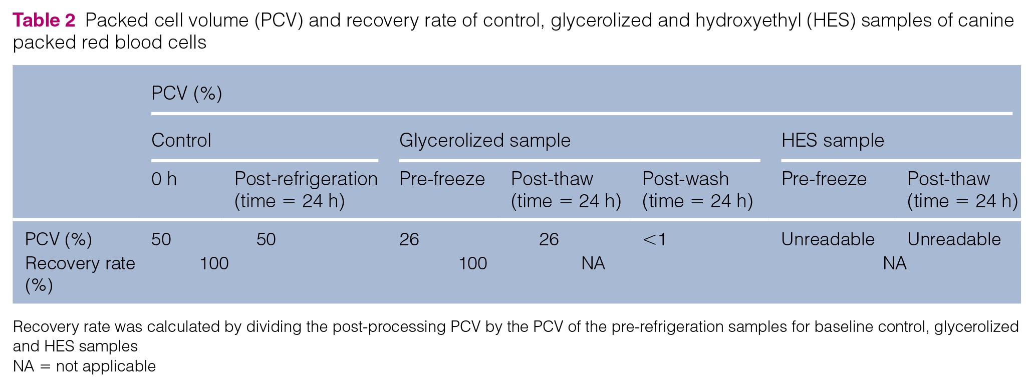
Recovery rate was calculated by dividing the post-processing PCV by the PCV of the pre-refrigeration samples for baseline control, glycerolized and HES samples
NA = not applicable
Erythrocyte morphology
The 24 h refrigerated control sample (Figure 6b) showed similar morphology compared with the baseline control (Figure 6a). Sphere-shaped cells were noted using SEM in the canine RBCs when comparing the pre-freezing and post-thaw glycerolized samples (Figures 6c,d) with the baseline control (Figure 6a). As with the feline samples, no significant changes in morphology as compared with baseline (Figure 6a) were noted in the HES-preserved RBCs pre- and post-freezing (Figures 6e,f).

Scanning electron microscopy of canine red blood cell (RBC) samples. (a) RBCs evaluated from the baseline control unit. (b) RBCs after refrigeration for 24 h. (c) RBCs mixed with 20% glycerol immediately before freezing. (d) RBCs mixed with 20% glycerol immediately after the washing procedure. Note the spherically shaped RBCs in (c) and (d) vs the echinocytes in (a) and (b). (e) RBCs mixed with 12.5% hydroxyethyl starch (HES) immediately before freezing. (f) RBCs mixed with 12.5% HES immediately after thawing. Echinocytes were noted in (e) and (f) but with clumping noted similar to (a). Magnification was × 5000 for all panels
Discussion
In this study, cryopreservation and recovery of feline (and canine) RBCs with glycerol was unsuccessful with a manual mixing and thawing process. Initially, this was surprising as cryopreservation and recovery of RBCs has been performed in human medicine successfully with various automated and manual protocols using glycerol as a cryoprotectant.6–12 Canine cryopreservation techniques have also been successfully described in three previous reports using modifications of both automated and manual human protocols.19–22
However, our findings are consistent with a recent study 23 that evaluated glycerol and HES as cryoprotectants for both canine and feline pRBCs. This study reported that neither HES nor glycerol were able to cryopreserve and recover feline RBCs successfully. 23 Using a different protocol than previous canine studies, including a single-step introduction of glycerol rather than a staged addition and a short exposure time to liquid nitrogen (<30 mins), the study showed severe hemolysis in both canine and feline pRBCs using both cryoprotectants. 23 It is possible that the single step of glycerol introduction and rapid freezing, along with almost immediate thawing post-freezing, might have caused significant osmotic injury to the cells by not allowing establishment of equilibrium between manipulations. Although none of the HES concentrations (6.25%, 10%, 17.5% and 20%) used in the study successfully preserved RBCs with good recovery rates, 23 17.5% HES had the best recovery rate based on post-thaw hemoglobin values in canine and feline samples. However, the lowest hemolysis rates were 16.10% and 17.15%, respectively, which were still higher than the <1% hemolysis rate considered acceptable in human medicine.
The largest issue associated with glycerol cryopreservation in our study was the inability to recover cryopreserved feline or canine RBCs. This led to concerns about the manual protocol employed in this study. The protocol was derived from previous successful studies in both humans and dogs.9–13,15–22 In developing the protocol used in this study, we found that the materials and methods sections of the various publications had limited details related to each protocol, especially the manual techniques involved.9–13,15–22 For instance, while Contreras et al described in detail the manual thawing and washing protocol for cryopreservation of canine pRBCs with glycerol, their protocol did not describe the method of agitation used when adding glycerol to the RBCs. 19 In addition, while Contreras et al used an automated RBC processor for the washing steps, Kim et al did not detail the washing steps in their protocol. 20 Therefore, details of our laboratory technique, such as exact incubation periods, methods of adding/mixing the cryoprotectant, and centrifuge times, are a result of combining the information from several publications.9,10,19
In addition, when details are provided about the protocols used in the literature, variation exists. For example, while Kim et al adopted their freezing and deglycerolization protocol from Contreras et al,19,20 the equilibration time for deglycerolization was extended from 2.5 mins to 5 mins. The use of different cryostorage containers may also affect the cooling rate and survival of RBCs. 16 It is possible that small variations in technique may be crucial for successful cryopreservation using glycerol and may have contributed both to our inability to recover RBCs and to the morphological changes noted in the RBCs.
The use of a manual technique, besides being labor intensive and technically demanding, 6 also increases RBC loss during container transfers and may have contributed to our low recovery rate in both canine and feline RBCs after the washing process (‘washing out the cells’). 24 Pallotta et al reported that deglycerolization using a manual technique was responsible for the loss of approximately 15% of human RBCs. 18 A manual technique for glycerolization and deglycerolization was also employed in a study of cryopreservation of avian red blood cells. 25 The avian study reported a recovery rate of 0% after the washing steps. The authors speculated that the low recovery rate could be due to small details in their protocol, including the addition rate of each aliquot of glycerol, time allowed for equilibration of glycerol with the RBCs, freezing rate of the samples or the deglycerolization process. 25
Another issue with glycerol cryopreservation encountered in our study was the unexpected agglutination during the glycerol wash steps in the feline samples. To rule out agglutination caused by removal of the anticoagulant during the wash steps, we added back anticoagulant to unit #4 both before the first washing steps (aliquot 2) and after the second wash (aliquot 3). Although agglutination was not noted in aliquot 3, there was still visible hemolysis during the last wash and a poor recovery rate was noted.
Agglutination has not been reported as a complication in the human pRBC cryopreservation literature. However, Contreras et al did discuss the possibility of agglutination of canine RBCs and recommended washing the cells with saline immediately after collection from the donor to prevent rouleaux formation and facilitate the addition of glycerol. 19 The absence of this saline washing step in our study could be one of the causes of the agglutination of RBCs. However, our canine glycerolized sample using the same protocol as used for cats did not agglutinate.
There is also a possibility that what was occurring in the sample was not agglutination, but rather aggregation of the RBCs. Aggregation occurs when globular proteins or fibrinogen creates bridges between RBCs; it is essentially the same process as rouleaux formation. 26 As we detail below, feline RBCs do have differences in their RBC membranes vs other species that might make the RBCs more likely to aggregate when stressed by the cryopreservation and thawing process.
The presence of hemolysis is supported by the visible reddish color change in the hematocrit tubes from the glycerolized feline samples pre- and post-freezing. Further support of hemolysis is evidenced by the presence of spherocytes on SEM, suggesting that hemolysis could have occurred. Spherocytes are commonly observed in hemolyzed samples; because they have greater osmotic fragility than normal RBCs, they have a greater tendency to lyse. Pallotta et al noted only small numbers of sphere-shaped cells in glycerolized human RBCs, 18 while Kim et al reported hemispherical shaped canine RBCs rather than spherocytes. 20 Neither of these studies reported significant hemolysis.
Species variations in RBCs, including membrane structure, membrane components and rates of biochemical reactions, are well documented and might affect membrane permeability and osmotic tolerance under cryopreservation.27,28 This, in turn, may be linked with a greater tendency for feline RBCs to hemolyze and possibly to aggregate together. For instance, the water permeability coefficient of RBCs varies significantly from human and dog to cat, with cats having a higher permeability than dogs and humans. 26 This may suggest that the permeability of feline RBCs for glycerol may also be different than in dogs, perhaps leading to greater fragility. In addition, biochemical components of red blood cells, such as ATP and 2,3-diphosphoglycerate (DPG) can affect the stability of the RBC membrane.29,30 Domestic cats have naturally low ATP (as a result of lower adenosine uptake) 31 and 2,3-DPG levels vs dogs and humans, again potentially leading to greater fragility of feline RBCs. 32 Further depletion of ATP and 2,3-DPG concentrations during storage can lead to microscopically sized vesicle formation within the RBC membrane, contributing to a greater tendency for hemolysis. 33
Therefore, protocols developed in one species may require adjustment in concentrations of or equilibration time for glycerol to take into account these differences in membrane permeability while still allowing proper penetration of glycerol into the RBCs. However, as hemolysis was noted macroscopically in the hematocrit tubes and a significant amount of spherocytosis was also noted in the glycerolized canine RBCs in our study, it is possible that the hemolysis seen in this study was at least partially related to technique rather than simply species differences.
Another possible reason for our inability to cryopreserve feline and canine RBCs in glycerol might be related to the blood used in the study. In all the human and canine studies,10–13,15–21 pRBC units were prepared in-house instead of being purchased commercially. One of the goals of the study was to mimic a situation where blood was purchased from a commercial blood bank and cryopreserved in a clinic to allow for extended storage. Previous human studies have shown that pRBCs stored at 4°C for up to 6 days can be glycerolized and frozen with acceptable recovery rates and minimal hemolysis.6–9 However, another study concluded that, despite careful packaging, human blood products shipped overnight by commercial carriers are exposed to variations in ambient temperature, which might affect cell recovery and viability. 24 As none of the previous studies in dogs or humans used commercially prepared and transported blood for cryopreservation, it is possible that shipping conditions related to the blood may have affected our ability to preserve and recover the cells successfully in glycerol.
In the present study, it was difficult to make conclusions about the efficacy of HES. On one hand, we noted minimal morphological changes in feline RBCs exposed to 12.5% HES during SEM analysis. This is comparable to the findings by Kim et al, 20 who also noted fewer deformed canine RBCs using 12.5% HES vs 7.5% and 17.5% HES, suggesting that 12.5% HES offered adequate protection for canine RBCs from freezing-related stresses. However, we were unable to obtain any information about the presence or absence of hemolysis or the recovery rate of RBCs in the HES samples owing to the high viscosity of the samples.
Although none of the literature directly reported the high viscosity of HES samples, various studies discussed centrifuging post-thawing HES samples in dogs for 35 mins at 12,000 rpm prior to analysis.13,20,23 Perhaps if we had tried additional centrifuge time in our feline HES samples and then planned to compare the supernatant (free) hemoglobin content with the total hemoglobin content rather than PCV, we would have an idea of the degree of hemolysis of feline RBCs preserved in HES.15,17,20 Further testing, such as hemoglobin comparisons, will be needed to evaluate the viability of HES-preserved feline RBCs.
Conclusions
Although human and canine RBCs have been successfully preserved in liquid nitrogen using different cryoprotectants, the manual technique for cryopreservation using glycerol described in this study was not able to recover feline RBCs successfully. Further studies evaluating HES as well as newer cryoprotectants, such as polyvinylalcohol or apatite nanoparticles,34,35 for cryopreservation of feline RBCs are also possible avenues of future discovery.
Footnotes
Acknowledgements
We thank Dr Joanne B Messick who provided insight and expertise that greatly assisted the research, Dr Abhijit Mukhopadhya for technical assistance, and Dr Christopher J Gilpin and Laurie Mueller for scanning electron microscopy support.
Conflict of interest
The authors declared no potential conflicts of interest with respect to the research, authorship, and/or publication of this article.
Funding
This work was supported by the Winn Feline Foundation [grant number W17-030]. The contents of this publication are solely the responsibility of the authors and do not necessarily represent the views of Winn.