
Abstract
Many types of phytochemicals have been found to be present in oil palm leaf and could potentially be used as functional ingredients for skincare product. However, as of today, there is no published report on hazard identification and safety assessment of oil palm (Elaeis guineensis) leaf extract (OPLE), particularly on skin and eye irritation. In this study, potential hazard of OPLE on skin and eye irritation was evaluated as an initial step to the safety assessment of OPLE. In vitro cell viability study of OPLE on normal human dermal fibroblasts showed that OPLE was nontoxic to the cells with percentage viability more than 90% after 24 and 48 hours of incubation. Skin irritation potential of OPLE was evaluated using in vitro SkinEthic reconstructed human epidermis (RHE) model (Organization for Economic Cooperation and Development [OECD] Test Guideline 439, 2015), while eye irritation potential was evaluated using in vitro SkinEthic Human corneal epithelium (HCE) model (OECD test guideline 492, 2017). Hazard identification results showed that OPLE at 1%, 5%, and 10% (wt/wt) was classified as nonirritant to the skin and eye where mean tissue viabilities of SkinEthic RHE and SkinEthic HCE were more than 50% and 60%, respectively. Therefore, we recommend a further safety assessment, such as human patch testing, to confirm the nonirritant of OPLE.
Keywords
Introduction
The palm oil industry in Malaysia has grown tremendously where various applications of oil palm biomass have generated enormous income to the economy. For each kilogram of palm oil extracted, there are roughly another 4 kg of dry biomass generated, of which the major contribution was derived from oil palm fronds and oil palm trunk inside the plantation estates, followed by empty fruit bunches, palm kernel shell, palm pressed fiber, and palm oil mill effluent. 1 Oil palm biomass has significant potential in many applications. The biomass can be converted into organic fertilizer, 2 animal feedstock, 3,4 and value-added products for wood industry. 5,6
Numerous studies have reported the presence of phytochemical components in oil palm (Elaeis guineensis) leaf extract (OPLE). The OPLE reported to contain oil soluble antioxidants, such as α-tocopherol and β-tocopherol. 7 Besides oil soluble antioxidants, water soluble antioxidants were also present in OPLE where the leaves are rich in flavonoids 8 and phenolic acids. 8 -11 The total polyphenols content in OPLE was reported to be higher than green tea extract. 9,12 The OPLE comprises (−)-catechin gallate, ferulic acid, and phenolic acids, such as gallic acid and protocatechuic acid. 13,14
A recent study by Yusof et al 11 suggested that OPLE can potentially be used as a functional active ingredient for topical application where they demonstrated that OPLE has antimicrobial activity, antityrosinase activity, and protection against ultraviolet (UV) radiation.
The higher demand of natural plant ingredients in personal care products has raised safety issues and required novel approaches to their safety evaluation. 15 Critical safety testing approaches need to be employed with the purpose of limiting the risks to consumer, whenever new personal care or cosmetic products are about to be launched for public use, or when a new active substance is to be used as a medicinal product in human. 16
Cell culture assays are used to assess the biocompatibility of a material or an extract through the use of isolated cells in vitro. These techniques are useful in evaluating the toxicity or irritancy potential of materials and chemicals. They provide an excellent way to screen materials prior to in vivo safety and efficacy tests. 17
Skin irritation refers to the production of reversible damage to the skin following the application of a test chemical for up to 4 hours as defined by the United Nations (UN) Globally Harmonized System (GHS) of Classification and Labelling of Chemicals. 18 Effectively from March 11, 2009, European Union Cosmetics Directive has banned the skin and eye irritation testing for cosmetic ingredients on animals. Therefore, several in vitro test systems have been proposed, such as the use of reconstructed human epidermis (RHE) models. 16 SkinEthic RHE is one of the validated test models for the determination of skin irritation according to the Organization for Economic Cooperation and Development (OECD) test guideline 439. This in vitro test system closely mimics the biochemical and physiological properties of the upper parts of the human skin, that is, the epidermis. 19
Eye irritation refers to the production of tissue damage in the eye, following application of a test chemical to the anterior surface of the eye, which are fully reversible within 21 days of application according to UN GHS. 20 One of the validated test method suggested in the OECD test guideline 492 was using SkinEthic human corneal epithelium (HCE). 21 The tissue constructs are produced using human immortalized corneal epithelial mucosa cells 22 that are structurally very similar to the corneal mucosa of the human eye.
As of today, there is no published report addressing the hazard identification and safety assessment of OPLE, particularly on skin and eye irritation. A tiered approach using in vitro test method for skin irritation assessment of cosmetics was proposed using hazard identification as an initial decision approach followed by safety assessment. 23 Hence, the hazard identification of OPLE through cytotoxicity effect against normal human dermal fibroblasts (NHDF) cell was described. Then, the skin and eye irritation potential of OPLE was further evaluated through in vitro reconstructed human model. Data obtained in these hazard identification studies will be useful in considering OPLE as a potential functional ingredient in topical application.
Materials and Methods
Chemical and Biological Material
Ethanol and isopropanol were purchased from Friedemann Schmidt, Parkwood, Western Australia. Folin-Ciocalteu phenol reagent, gallic acid, sodium nitrite, aluminium chloride hexahydrate, (−)-catechin gallate, 2,2-diphenyl-1-picrylhydrazyl (DPPH), and 6-hyroxy-2,5,7,8-tetramethylchroman-2-carboxylic acid (TROLOX) were purchased from Sigma-Aldrich (Steinheim, Germany). The remaining chemicals were supplied by different companies as follows: sodium carbonate anhydrous (R&M Chemicals, Essex, United Kingdom), sodium hydroxide (Merck KGaA, Darmstadt, Germany), sodium dodecyl sulfate (SDS) ≥99.0% (Sigma-Aldrich, Steinheim, Germany), methyl acetate 99% (Acros Organics, Morris Plains, New Jersey), and glycerine (Croda, Seraya Avenue, Singapore). The NHDF (CC-2511) were purchased from Lonza (Walkersville, Maryland). Dulbecco’s modified Eagle medium (DMEM), fetal bovine serum, penicillin streptomycin, 0.25% trypsin ethylenediaminetetraacetic acid, phosphate buffer solution (PBS), and 3-(4,5-dimethylthiazol-2-yl)-2,5-diphenyltetrazolium bromide (MTT) assay were purchased from Life Technologies, Grand Island, Nebraska. SkinEthic RHE model, SkinEthic HCE model, and SkinEthic culture maintenance medium were purchased from EpiSkin SA (Lyon, France).
Preparation of OPLE
Fresh oil palm (E guineensis) leaves from mature trees, aged 6 years, were obtained from Malaysian Palm Oil Board, Bangi, Selangor, Malaysia. The leaves collected were washed with tap water and chopped coarsely and left to dry in the oven at 40°C for 24 hours. The dried leaves were ground to powder using a mechanical blender, and 20.0 g of the leaf powder was extracted with absolute ethanol at a 1:10 (wt/vol) under soxhlet extractor for 2 hours at 78.0°C. The extracts were then filtered through filter paper Whatman no 1 (Merck KGaA, Darmstadt, Germany) and vacuum dried in a rotary evaporator (Premium 7, Heidolph Instruments GmbH, Schwabach, Germany) at 40°C, until the solvent was completely removed or one-tenth its volume to yield dark green waxy material. The crude OPLE was used for determination of phytochemicals compound, antioxidant activity, and cytotoxicity study. The crude OPLE was further prepared in 1%, 5%, and 10% (wt/wt) in glycerine as a carrier for hazard identification: in vitro skin and eye irritation assessment.
Total Phenolic and Flavonoid Contents
Total phenolic content in the crude ethanolic extract OPLE was determined using the Folin-Ciocalteu method. 24 In brief, 0.1 mL OPLE (1:10, wt/vol ethanol) was added with 0.5 mL Folin-Ciocalteu reagent, followed by 7 mL distilled water. The mixture was allowed to stand at room temperature in the dark for 5 minutes. Then, 1.5 mL sodium carbonate (15% Na2CO3/H2O, pH 11.2) solution was added and the mixture was left at room temperature for another 2 hours. The UV absorbance at 765 nm was measured using microplate reader spectrophotometer (Synergy H1, BioTek, Winooski, VT). Samples were measured in 3 replicates. Gallic acid was used as reference. Standard curve of gallic acid solution (0, 10, 20, 40, 60, 80, and 100 ppm) was prepared using the similar procedure. The results were expressed as milligram gallic acid equivalent (GAE) per gram of extract (Equation 1).
The total flavonoid content of crude extract was determined by the aluminium chloride colorimetric method by Liu et al, 25 with slight modification. Briefly, 0.5 mL extract (1 mg/mL ethanol) was diluted with 1.25 mL distilled water, followed by 0.15 mL 5% sodium nitrite (NaNO2) solution. The mixture was allowed to stand for 6 minutes at room temperature. Then, 0.3 mL 10% aluminium chloride hexahydrate (AlCI3.6H2O) solution was added, and the mixture was allowed to stand for another 5 minutes. Then, 1 mL of 1 M sodium hydroxide (NaOH) was added, and the total was made up to 5 mL with distilled water. The solution was vortexed and 360 µL of solution was transferred to 96-well plates, and UV absorbance at 510 nm was recorded using microplate reader spectrophotometer (Synergy H1, BioTek). Samples were measured in 3 replicates. Standard curve of catechin gallate (0, 10, 20, 40, 60, 80, and 100 μg/mL) was prepared using the similar procedure. The results were expressed as milligram catechin gallate equivalent per gram of extract (Equation 1).
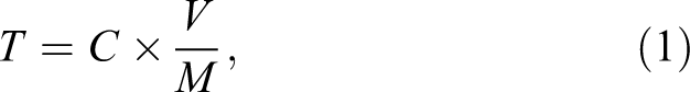
where T is the total phenolic/flavonoid content in mg/g of the extract as GAE, C is the concentration of gallic acid/catechin gallate established from the calibration curve in mg/mL, V is the volume of the extract solution in mL, and M is the weight of the extracts in grams.
Antioxidant Activity Assay
The DPPH free radical scavenging activity of OPLE was determined according to the method described by Blois, 26 with slight modifications. Briefly, 4.2 mg of DPPH powder was dissolved in 50 mL of methanol (0.2 mmol/L). Then, 50 µL OPLE at different concentrations of 62.5, 125, 250, and 500 µg/mL in methanol was prepared and reacted with 195 µL of DPPH methanolic solution in 96-well microtiter plate. After 60 minutes of incubation, the absorbance values of the samples were read at 540 nm using a microplate reader (Synergy H1, BioTek). The TROLOX was used as the standard. The absorbance of the DPPH and methanol was used as a negative control (NC). The analysis was done in triplicate to confirm the reproducibility of the data. The antioxidant activity, expressed as the percentage of DPPH radical scavenging, was calculated using Equation 2.
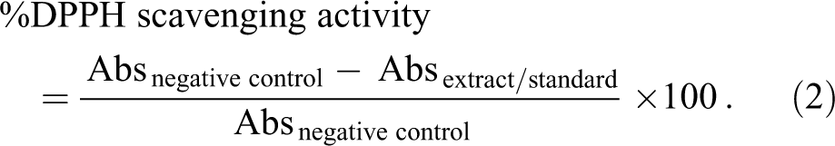
The half maximal inhibitory concentration (IC50) of DPPH assay represents the concentration of the tested sample needed to reduce the DPPH by 50%, where the value obtained from linear regression graph.
Assessment of Direct Test Substance Reduction of MTT
The ability of each test substance to directly reduce the MTT assay was assessed. 19,20 Approximately 16 µL and 30 µL of test substance for SkinEthic RHE and SkinEthic HCE tissues, respectively, were added to 300 µL of MTT solution (1 mg/mL in PBS), and the mixture was incubated in the dark at 37°C for approximately 3 hours ± 15 minutes. If the MTT solution color turned blue or purple, the test substance was presumed to have reduced the MTT. It is then necessary to evaluate the part of optical density (OD) due to the nonspecific reduction of the MTT (ie, using killed epidermis). 27,28
In Vitro Cytotoxicity Assay on Human Dermal Fibroblasts Cell
Cytotoxicity assay was used for determination of the treatment concentration that does not have a toxic effect on normal cells. In this assay, NHDF cells were used as a normal cell modeling. The NHDF cells were grown in complete growth medium (CGM) containing DMEM, supplemented with 10% fetal bovine serum and antibiotics (penicillin 100 U/mL and streptomycin 0.1 mg/mL). The cultures were maintained at 37°C ± 2°C in 5% CO2 atmosphere using a CO2 incubator (INC80, Memmert, Germany). The cells in confluence of 80% to 90% were trypsinized, centrifuged for 3 minutes at 1,200 rpm, and transferred to 96-well plates. The DMEM with fetal bovine serum was used to neutralize the trypsin. The cell density used for cytotoxicity was 2 × 10⁵ cells/mL. The 96-well plate was incubated for 24 hours for complete cell adhesion to the plate.
The stock solution of OPLE with concentration of 10 mg/mL in dimethyl sulfoxide (DMSO) was prepared, followed by working stock of OPLE in CGM with concentration of 100 μg/mL. In brief, 100 μL of CGM was added into each well of 96-well plate. Then, 100 μL of OPLE (100 μg/mL in CGM) were added and serial dilution of 50, 25, 12.5, 6.25, 3.13, and 1.56 μg/mL was performed on the 96-well plate. The assay was performed in 2 plates: one for 24 hours and another for 48 hours of treatment. The plates were incubated at 37°C ± 2°C in 5% CO2 atmosphere for 24 and 48 hours. After incubation period, the solution was removed, and the plates were gently washed with PBS. Cell viability was measured by quantitative colorimetric assay using MTT as described by Carmichael et al. 29 In brief, 20 µL of MTT (5 mg/mL in PBS) was added to each well. The microplates were incubated at 37°C ± 2°C for 4 hours, protected from light, to allow the growing of formazan violet crystals. 30 The solubilization of formazan crystals was carried out by adding 100 µL of DMSO solution into each well. The absorbance was read at 570 nm in microplate reader (Synergy H1, Winooski, Vermont).
The OD value of the control (untreated) cells was taken as 100% viability. The percentage of dead and live cells was calculated in regard to the NC and represents the cytotoxicity of each treatment (Equation 3).
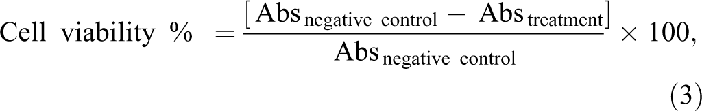
where Abs is an absorbance. The morphology of NHDF cells from initial, after 24, and 48 hours of treatment with concentration of OPLE at 100 µg/mL, was observed under inverted microscope (CKX41, Olympus, Japan) at ×20 magnification.
Reconstructed Human Epidermis Model (SkinEthic RHE)
The SkinEthic RHE model consists of normal, human-derived keratinocytes, which have been cultured to form a fully differentiated 3-dimensional epidermis cultured for 17 days on an inert polycarbonate filter at the air–liquid interface in a chemically defined medium. 31,32 The reconstructed 0.5 cm2 epidermis was received on day 17 and maintained for 2 hours in a SkinEthic growth culture medium (GCM) at 37°Cin 5% CO2. 27
The SDS in 5% (wt/vol) aqueous solution was used as reference irritant (positive control [PC]) and PBS served as NC, in each series of experiments. The treatments for each test substance were conducted in triplicate tissues.
The RHE tissues were topically exposed to 16 ± 0.5 µL, that is, 32 µL/cm2 of 1%, 5%, and 10% OPLE in glycerine for 42 minutes at room temperature. Then, the RHE tissues were rinsed 25 times with 1 mL each time of sterile PBS. Treated tissues were further incubated for 42 hours at 3°C in 5% CO2 with 2 mL of SkinEthic GCM. After the incubation period, the RHE tissues were assessed for tissue viability. 27
Reconstructed HCE Model (SkinEthic HCE)
The SkinEthic HCE model was purchased from EpiSkin, Lyon, France. The HCE model was composed of immortalized human corneal epithelial cells cultured in a chemically defined medium and seeded on a synthetic membrane at the air–liquid interface. The tissue structure obtained was a multilayered epithelium resembling the in vivo epithelium representing about 5 to 7 cell layers and a surface area of 0.5 cm2.
The SkinEthic HCE tissues were topically exposed to 30 µL of 1%, 5%, and 10% OPLE (wt/wt) in glycerine for 30 ± 2 minutes at 37°C at 5% CO2 in a humidified incubator (standard culture conditions). Two tissues were used per test substance (NC, PC, or test sample). After 30 minutes of treatment, tissues were rinsed at least 2 times with 10 mL PBS to remove the residual test substance from the tissue surface. After rinsing, the tissues were immersed into 1.5 mL fresh maintenance medium (750 µL underneath and 750 µL topically) for an incubation period of 30 ± 2 minutes in standard culture conditions. After the incubation period, duplicate tissues were assessed for tissue viability. 28
Tissue Viability Assessment
The MTT test was used to measure the viability of living cells via dehydrogenase enzyme activity. 30 The ring of MTT, yellow, was cleaved by dehydrogenase, yielding blue-purple formazan crystals, which were insoluble in culture medium. An intense purple color was observed when the tissue was healthy, while the culture remained white when necrosis occurred. 33
Each SkinEthic RHE and HCE tissue was transferred to a new well containing 300 µL of freshly prepared MTT (1 mg/mL) solution for an incubation period of 3 hours ± 15 minutes under standard culture conditions. Then, the tissue inserts were rinsed with 300 µL PBS and transferred into new plate containing 1.5 mL of isopropanol per well (750 µL underneath and 750 µL topically) for either 4 hours at room temperature or overnight at 4°C to extract the reduced MTT (formazan crystals) out of the tissues. Then, 200 µL aliquots of formazan solution extracts were transferred to a 96-well plate for OD measurement at 570 nm using a microplate reader (Synergy H1, Winooski). Isopropanol was used as a blank. The percentage viability of each of the treated cultures was calculated from the percentage MTT conversion in the treated cultures relative to the corresponding NCs (100% viability). Results were expressed as percentage viability relative to NC as shown in Equation 4.

Prediction Model
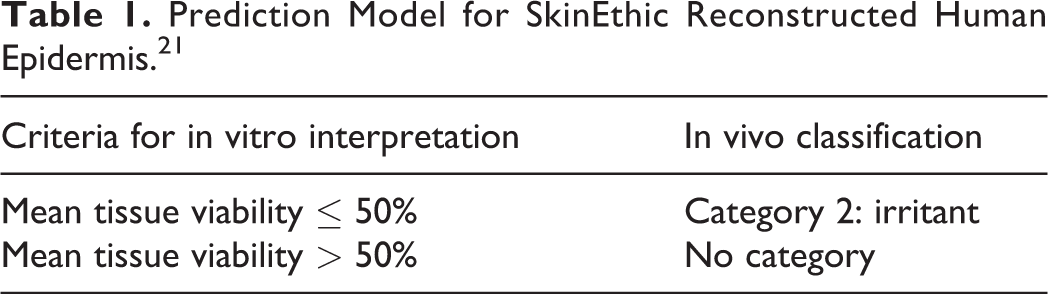
Irritation potential of test substance is determined according to the UN GHS 18,20 and European Commission Regulation no 1272/2008 on classification, labeling, and packaging of chemicals and mixtures (EU CLP) 34 as category 2: irritant or “no category.” In the present study, the irritancy potential of test substance is predicted by mean tissue viability of tissues exposed to the test substance. The test substance is considered to be irritant to skin (category 2) if the mean relative viability after 42 minutes exposure and 42 hours postincubation is ≤50% of the NC. The prediction model is defined in SkinEthic RHE test procedure 21 and described in Table 1.
Prediction Model for SkinEthic Reconstructed Human Epidermis. 21
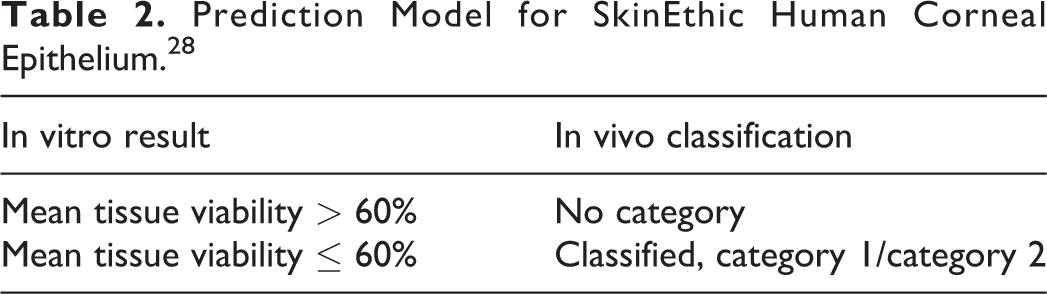
A test chemical is predicted as ocular irritant, according to the UN GHS of Classification and Labelling of Chemicals, 18,20 and as implemented in the EU CLP, if the mean relative tissue viability (%) of 2 tissues exposed to the test chemical is ≤60%. The prediction model is defined in SkinEthic HCE test procedure 28 and described in Table 2.
Prediction Model for SkinEthic Human Corneal Epithelium. 28
Statistical Analysis
The statistical data were expressed as the mean (standard deviation) and performed using 1-way analysis of variance with Student t test. Statistical analyses were conducted using the Statistical Package for Social Science (SPSS) version 17.0 (SPSS Inc, Chicago, Illinois). A P value <0.05 was considered to be significant.
Results
Total Phenolic and Flavonoid Contents
The total phenolic and flavonoid contents of OPLE are listed in Table 3.
Total Phenolic and Flavonoid Contents of OPLE.
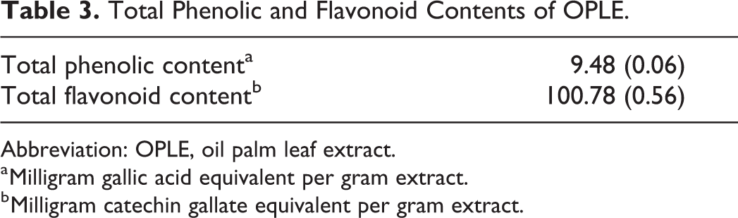
Abbreviation: OPLE, oil palm leaf extract.
a Milligram gallic acid equivalent per gram extract.
b Milligram catechin gallate equivalent per gram extract.
Antioxidant Activity Assay
The OPLE at 500 µg/mL shows higher antioxidant activity, 94.36% (0.29%) with an IC50 value of 247.00 (0.58) µg/mL (Table 4).
Antioxidant Activity of OPLE on Scavenging Effect Using DPPH Assay.
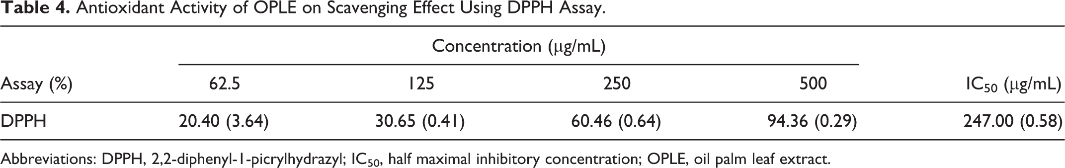
Abbreviations: DPPH, 2,2-diphenyl-1-picrylhydrazyl; IC50, half maximal inhibitory concentration; OPLE, oil palm leaf extract.
In Vitro Cytotoxicity Assay
The viability of NHDF cells with different concentrations of OPLE, measured by MTT assay, was ranged between 90.50% and 106.61% after 24 hours of incubation. Further incubation after 48 hours recorded a mean viability (%) from 98.79% to 101.48% (Figure 1). Increasing the OPLE concentrations up to 100 µg/mL or longer incubation time to 48 hours did not affect the cell viability; thus, no cytotoxic effect was found at the chosen experimental conditions. Furthermore, analysis of variance and Student t test did not detect any significant differences between the OPLE concentrations or different incubation time with P value more than 0.05 (Figure 1).
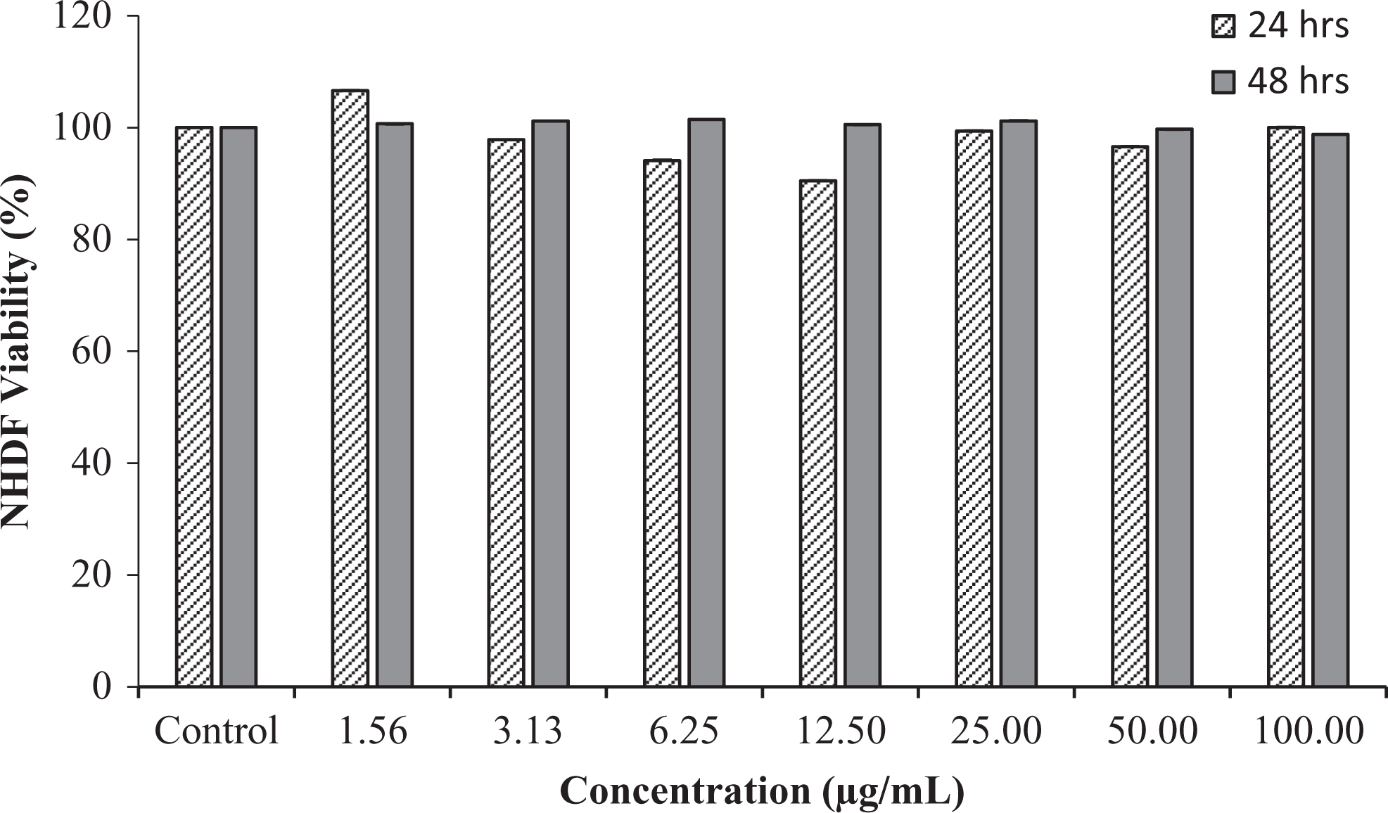
Cell viability (%) of normal human dermal fibroblasts determined by 3-(4,5-dimethylthiazol-2-yl)-2,5-diphenyltetrazolium bromide (MTT) assay after incubation with different concentrations of oil palm leaf extracts at 24 and 48 hours. Each bar represents mean (standard deviation) triplicate samples.
Fibroblast cells are often described morphologically as elongated, spindle-shaped cells that are characterized by high proliferative and migration potential. 31 The morphology of NHDF cells from initial, after 24, and 48 hours of incubation at highest concentration of OPLE (100 µg/mL) was captured using inverted microscope (CKX41, Olympus, Japan) at ×20 magnification and is depicted in Figure 2. It was observed that NHDF cells remain viable and more confluent after the incubation period (Figure 2B and 2C). These results suggest that OPLE is not toxic to dermal fibroblasts cell.
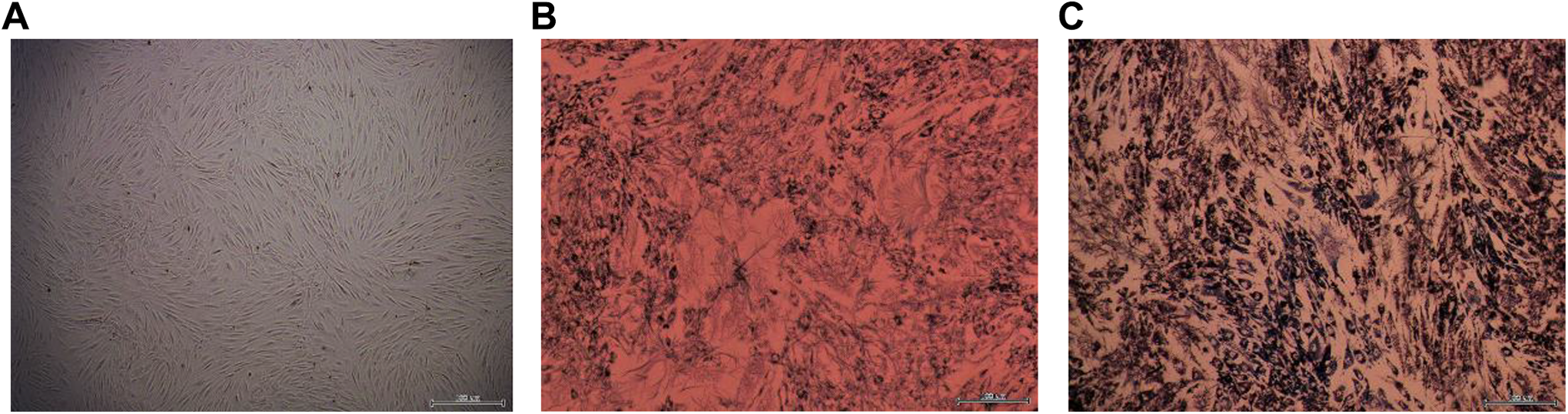
Morphology of normal human dermal fibroblasts cell (A) initial, (B) after 24 hours, and (C) after 48 hours of treatment with oil palm leaf extract at 100 µg/mL at ×20 magnification.
In Vitro Skin Irritation Assessment Using SkinEthic RHE Model
In vitro skin irritation assessment of OPLE at 1%, 5%, and 10% (wt/wt) in glycerine was evaluated using SkinEthic RHE model. 19 Concurrent NC and PC were used in each run to demonstrate that viability (with the NC), barrier function, and resulting tissue sensitivity (with the PC) of the tissues are within a defined historical acceptance range. The PC was a 5% aqueous SDS, and the NC was PBS, as suggested by the test protocol. 21
The ability of test substance to reduce MTT was assessed, and none of the test substances were shown to directly reduce MTT after incubation in the dark at 37°C for approximately 3 hours ± 15 minutes. As such, it was not necessary to include freeze-killed tissue control.
Figure 3A depicts the NC ODs for 3 RHE tissues. All tissues showed NC values with mean of 2.160 (0.029), 2.239 (0.026), and 2.403 (0.017), respectively. Therefore, the NC data from this study met the acceptance criteria where mean OD value of the tissues is ≥1.2 at 570 nm. 21
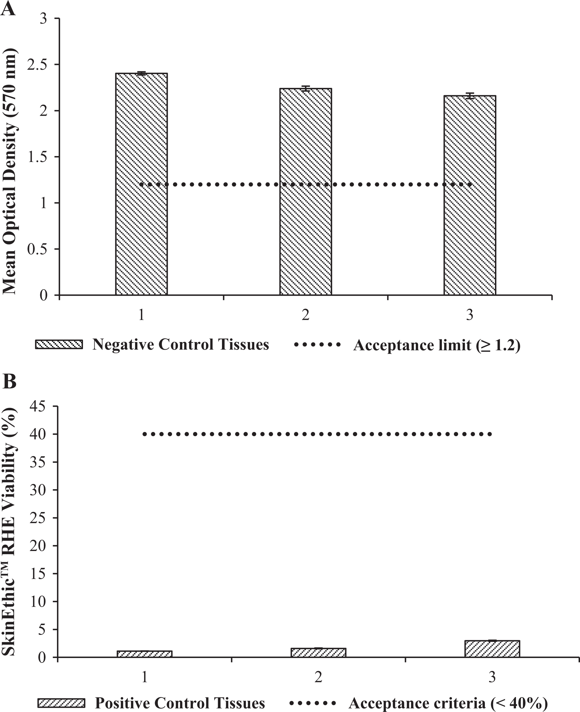
(A) Negative control optical densities (ODs) and (B) positive control (PC) values expressed as viability (%) relative to the negative control for skin irritation using reconstructed human epidermis model.
The mean viability of tissues treated with PC, expressed as percentage of the NC, is below the acceptance limit of 40% (Figure 3B) as described in the test method. 21
Figure 4 depicts the results of MTT assay on RHE skin model and its percentage viability when treated with PBS, 5% SDS, and 1%, 5%, and 10% OPLE in glycerine. The results of PC and OPLE at 1%, 5%, and 10% in glycerine were compared to NC, which was set at 100% viability. The RHE tissues exposed to PC (5% SDS) had significant % viability decreased from 1.12% to 2.98% with a mean value of 1.90% (0.96%). These data were expected as SDS is a known irritant to the skin. 35
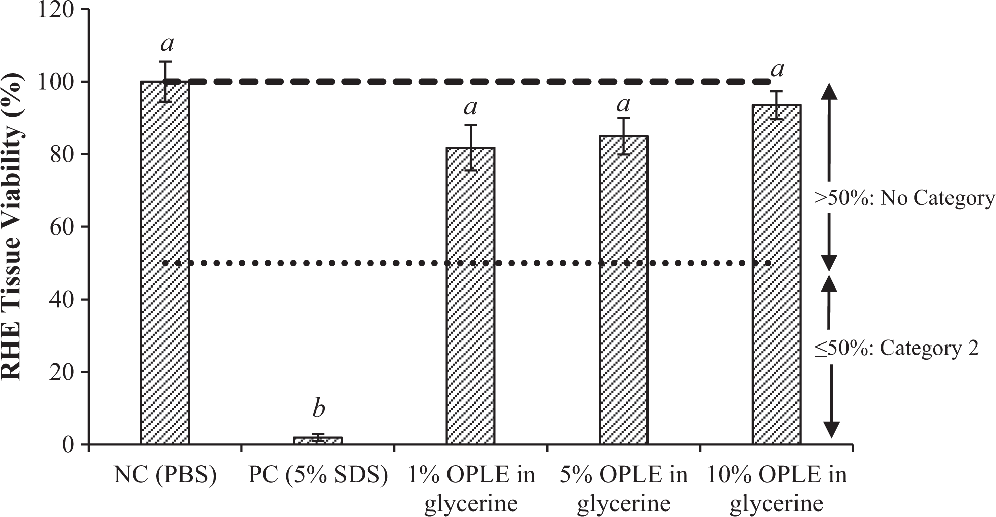
Mean SkinEthic reconstructed human epidermis tissue viability (%) of oil palm leaf extract at 1%, 5%, and 10% in glycerine as compared to positive and negative controls. Values in the same column followed by different letters indicate significant different (P < 0.05).
Tissues treated with 1%, 5%, and 10% OPLE (wt/wt) in glycerine recorded a mean viability of 81.74% (6.30%), 84.97% (5.07%), and 93.49% (3.82%), respectively (Figure 4). The mean viability was more than 50%, and therefore OPLE at 1%, 5%, and 10% (wt/wt) in glycerine was classified as nonirritant to the skin. These data will be used to support the nonirritant of OPLE on the skin and therefore can be further used as a functional active ingredient in topical application.
In Vitro Eye Irritation Assessment Using SkinEthic HCE Model
Eye irritation assessment of OPLE at 1%, 5%, and 10% (wt/wt) in glycerine was performed using SkinEthic HCE model. The ability of test substance to reduce MTT was assessed, and none of the test substances were shown to directly reduce MTT after incubation in the dark at 37°C for approximately 3 hours ± 15 minutes. As such, it was not necessary to include freeze-killed tissue controls.
Figure 5A depicts the NC ODs for 3 treated HCE tissues. All NC values were within the acceptance limit of OD 1.4 to 2.5, according to the eye irritation test method for liquid test substances. 28 The mean viability of tissues treated with PC, expressed as percentage of the NC, was below the acceptance limit of 30% (Figure 5B) as described in the test method. 28
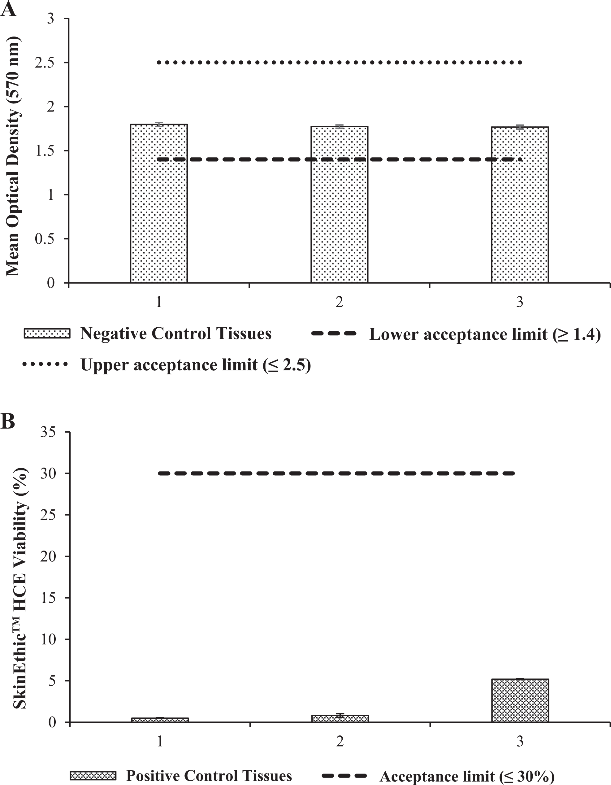
Negative control optical densities (A) and positive control values expressed as viability (%) relative to the negative control (B) for eye irritation using human corneal epithelium (HCE) model.
The OPLE at 1%, 5%, and 10% (wt/wt) in glycerine recorded a mean viability higher than 60% as shown in Figure 6 and therefore classified as nonirritant test substance. These data were important as it verified that OPLE at maximum concentration 10% (wt/wt) in glycerine is safe to be used as an active ingredient in topical product application.
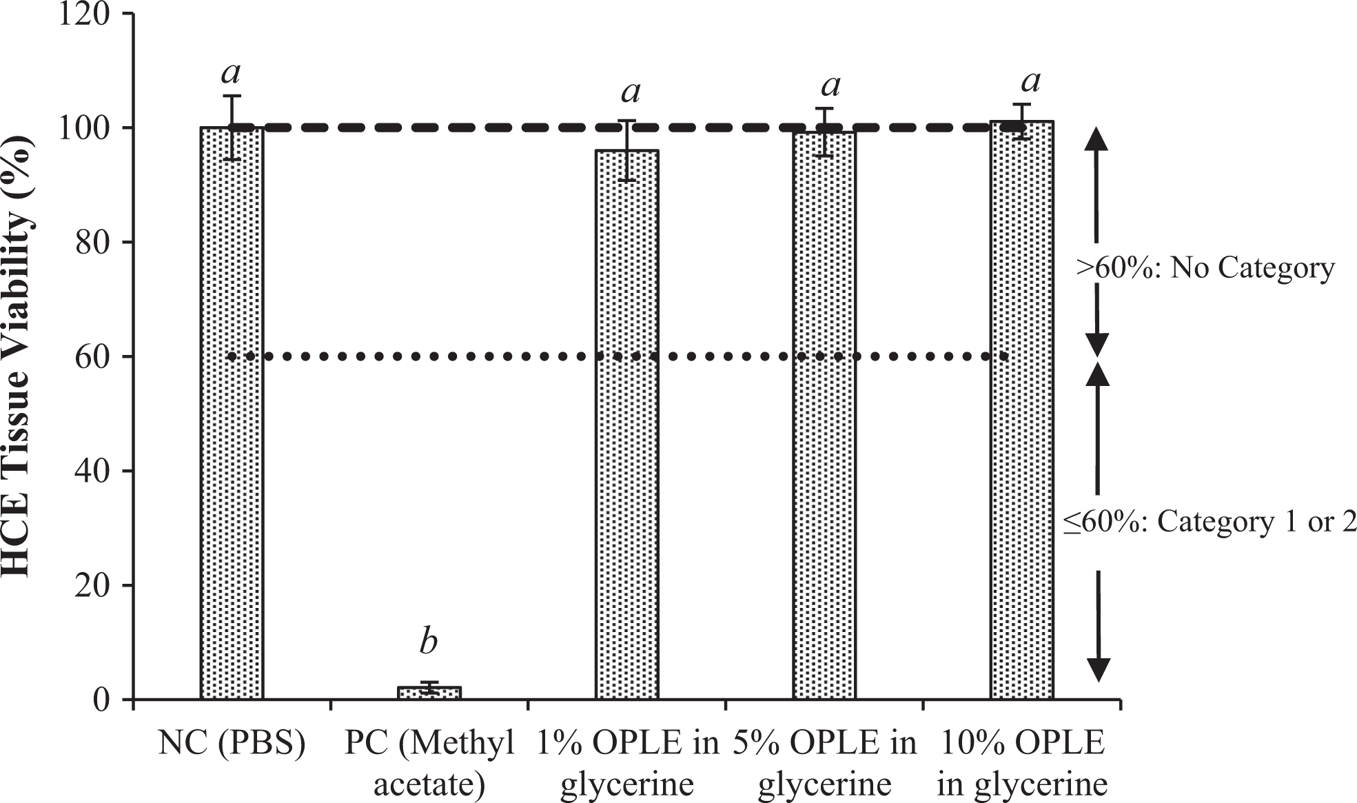
Mean human corneal epithelium (HCE) tissue viability (%) incubated with oil palm leaf extract at 1%, 5%, and 10% in glycerine as compared to positive and negative controls. Values in the same column followed by different letters indicate significant different (P < 0.05).
Discussion and Conclusion
The importance of phenolic antioxidants has remarkably increased in the last decade due to the high capacity to scavenge free radicals. 36 Phenolic-rich plants could be used, for example, for prevention of skin harmful effects of UV radiation. 37 Total phenolic content of OPLE recorded in this study was 9.48 (0.06) mg GAE/g extract, and it was close to value reported by Ng and Choo, 8 with 10.2 mg GAE/g extract. This finding suggested that gallic acid is one of the phenolic compounds that may present in OPLE. This result was supported by a patent filed in 2009, 14 where the extract enriched with (−)-catechin gallate, ferulic acid, and phenolic acids, such as gallic acid and protocatechuic acid. Catechin, epicatechin, and epigallocatechin that normally present in green tea were also present in OPLE, as studied by Jaffri et al. 13 Previous study conducted by Ng et al 10 reported that total flavonoid content of methanolic OPLE was 257.00 (3.055) mg quercetin equivalent per gram dry extract. The data might be higher from this study due to the more polar solvent used which is methanol and different standard used in calculation which is quercetin. The data on antioxidant activities of OPLE in this study were adjacent with antioxidant activities reported by Ng et al, 10 with values ranged from 56% to 93% when determined by the DPPH assay. However, the calculated IC50 value was much lower than values reported by Vijayarathna and Sasidharan 38 with IC50 814 µg/mL and Ng et al 10 with IC50 646 µg/mL. Lower IC50 value indicates higher antioxidant activity. The OPLE was also reported to have antimicrobial activities, 11,39 tyrosinase inhibition for skin whitening purpose, and protection against UV radiation. 11 Thus, OPLE potentially can be used as active ingredient for topical application. The extract was also shown to have wound healing properties. 40 Previous study conducted by Vijayarathna and Sasidharan 38 reported that methanol extract of OPLE was able to inhibit the proliferation of breast cancer cell line, MCF-7 at 15 µg/mL and therefore possible to be developed as promising anticancer drugs. Due to these valuable potentials of OPLE mainly for topical application, this study is focused on the hazard identification of OPLE through MTT assay on NHDF cell and reconstructed human model. Dermal fibroblasts play a vital role in maintaining skin function. They not only synthesize and secrete extracellular matrix molecules but also produce a complex mixture of bioactives factors, which both contribute to immune regulation and wound healing. 41 Treatment of crude OPLE at 100, 50, 25, 12.5, 6.25, 3.13, and 1.56 µg/mL for 24 and 48 hours of incubation against NHDF cells showed that OPLE did not affect or reduce the cell viability. These findings were supported by the morphology’s observation under microscope where NHDF cells remain intact and more confluence after 24 and 48 hours of incubation. The hazard identification of OPLE was further tested through in vitro skin and eye irritation tests using RHE and HCE models. Both tests were originally developed to predict the human skin and eye irritation potential of raw chemicals and final cosmetic products. The hazard identification results on skin and eye irritation potential of OPLE at 1%, 5%, and 10% (wt/wt) in glycerine through in vitro RHE and corneal epithelium models showed that OPLE was nonirritant with tissue viabilities more than 50% and 60%, respectively. However, further safety assessment using different steps, such as human patch testing on case-by-case basis, in the context of its formulation with other ingredients need to be considered in order to specify the skin irritancy of OPLE.
Footnotes
Acknowledgments
The authors wish to thank the Director General of MPOB for the approval to publish this article. The authors also gratefully acknowledged the members of the Consumer Product Development Unit, MPOB, for their technical support.
Author Contribution
Yusof Nor Zuliana substantially contributed to conception or design, contributed to acquisition, analysis, or interpretation of data, and drafted the manuscript; Abd Gani contributed to conception and design, contributed to acquisition, and critically revised the manuscript. Azizul Hasan contributed to conception and design, contributed to acquisition, and critically revised the manuscript; Idris critically revised the manuscript. All authors gave final approval and agree to be accountable for all aspects of work ensuring integrity and accuracy.
Declaration of Conflicting Interests
The author(s) declared no potential conflicts of interest with respect to the research, authorship, and/or publication of this article.
Funding
The author(s) received the following financial support for the research, authorship, and/or publication of this article: The work was supported by Malaysian Palm Oil Board.