
Abstract
Local reactions are the most frequent adverse event associated with vaccines. Adjuvants are major constituents of many vaccines and they are frequently involved in these reactions, associated with their irritating effect and the stimulation of local inflammation. The hen’s egg test on chorioallantoic membrane (HET-CAM) is an alternative toxicological method widely used to determine ocular irritation potential, but very few studies have demonstrated the utility of this method for assessing the irritant properties of vaccine adjuvants. In this work, known/experimental adjuvants were evaluated by both HET-CAM and an in vivo local toxicity study in mice to compare irritation scores to determine whether there was a correlation (Pearson test). Based on these data (r = 0.9034; P < 0.0001), the HET-CAM assay can be used as an alternate method for the prediction of the local toxicity potential of adjuvant candidates to be used in vaccines.
Introduction
Adjuvants are widely used as vaccine constituents. They include a large number of diverse chemical entities, many of which were developed empirically and without the knowledge of their specific mechanisms of action. 1 However, the severity of side effects induced by adjuvants hinders their incorporation into vaccines for human use. 2,3 Local reactions are the more frequent side effects causing different symptoms that include local tenderness, swelling, painful abscesses, sterile cysts, and nodules. 4 The mechanisms of these reactions are diverse depending on the formulation, but undoubtedly one of the more important is the early direct irritation of the tissue in the site of administration, inducing a strong inflammatory response. 5,6
According to the regulatory guidelines that address the reduction of laboratory animal use, the assessment of local tolerance of adjuvants can be performed as a parameter in repeat dose studies. 7 However, in such studies, it is difficult to assume the existence of a primary irritant effect. Local inflammation and other reactions detected at the end of the repeat dose studies can be due to other causes, not associated with the direct disruption of the tissue integrity caused by the adjuvant. 6 In vitro methods for the detection of adjuvant cytotoxicity can offer valuable and direct information for a more complete evaluation of toxicity from the data obtained.
The hen’s egg test on chorioallantoic membrane (HET-CAM) is an alternative in vitro method developed by Lüepke, 8 modified by Spielmann 9 and Steiling et al, 10 to classify eye irritants and that substitutes the classic Draize test in rabbits. 11 In Europe, the HET-CAM has been accepted for this purpose. 12 Other uses of HET-CAM include studies of tissue engineering, angiogenesis/antiangiogenesis, 13 drug delivery, 14 and inflammation. 15 Versatility, possibility for using different types of substances, including those insoluble in water or solids, speed, and simplicity of the method, makes the HET-CAM assay an interesting/feasible alternative for evaluating the potential irritant properties of adjuvants. This assay takes into account the wide spectrum of formulations, the potential for mucosal use, and addresses local reactions, which are the primary toxicological concerns. 6,16 The objective of this study is to evaluate the utility of HET-CAM for detecting the irritation properties of several known and new adjuvant formulation. This method may be used for irritation screening as part of weight-of-evidence assessments or in tiered approaches to assess local tolerance of vaccine adjuvants.
Materials and Methods
Adjuvants
The following adjuvants were used in this study: Freund complete adjuvant (FCA); Freund incomplete adjuvant (FIA); saponin; aluminum hydroxide (Al-gel); Montanide IMS 1313 VG; Montanide ESSAI IMS D 12802 VG PR; Adjuvant Finlay Proteoliposome 1 (AFPL1); Adjuvant Finlay Cochleate 1 (AFCo1); Adjuvant Finlay Cochleate 3 (AFCo3); and clinoptilolite (Cliptox; Table 1). All the formulations were prepared according to the manufacturer’s instructions using phosphate-buffered saline (PBS) as a vehicle without antigens immediately before the experiments under sterility conditions.
Details of the Adjuvant Formulations Used in This Study.
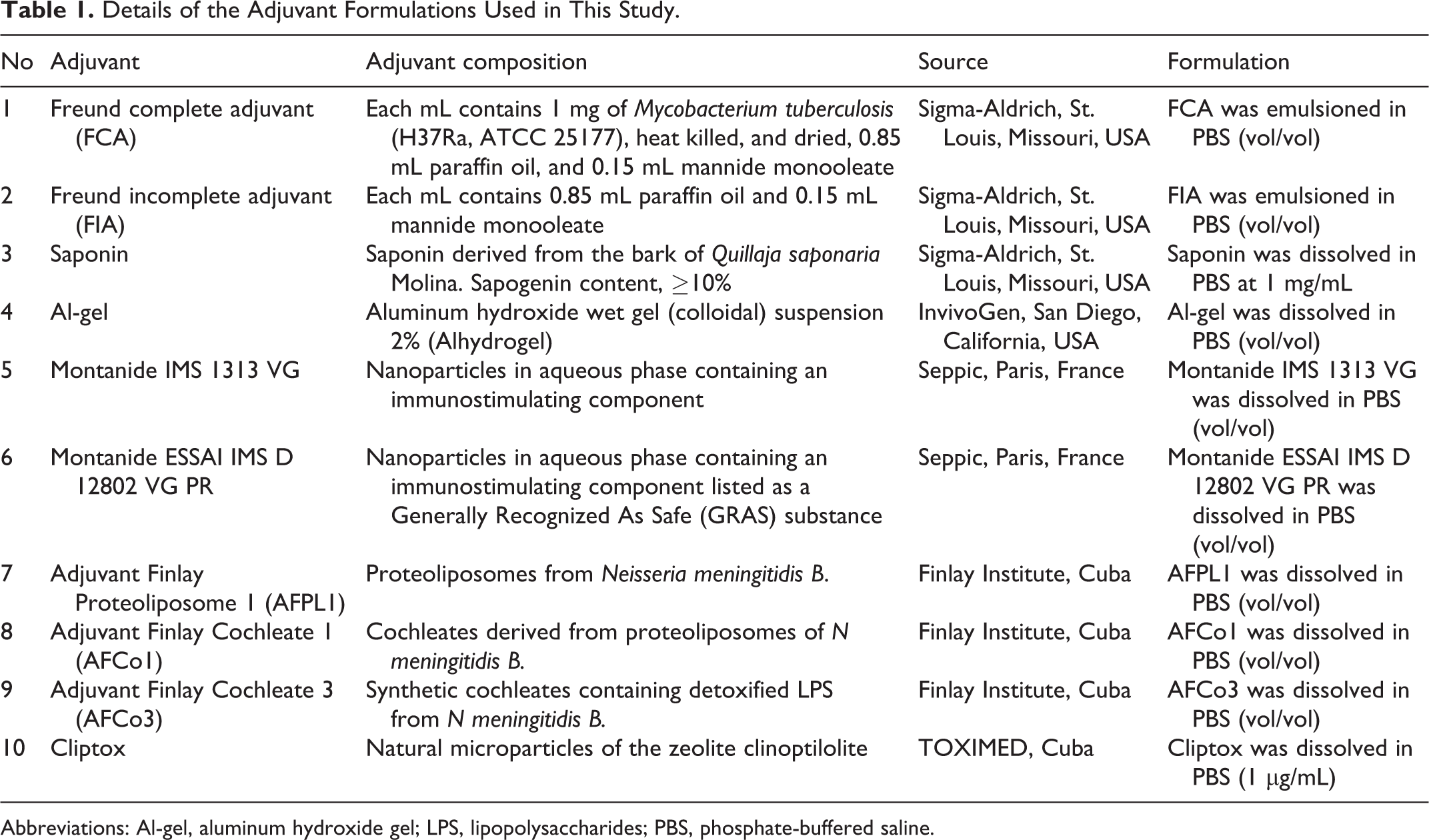
Abbreviations: Al-gel, aluminum hydroxide gel; LPS, lipopolysaccharides; PBS, phosphate-buffered saline.
Hen’s Egg Test on Chorioallantoic Membrane
Hen’s egg test on chorioallantoic membrane was performed according to the standard protocol 47 INVITTOX 9 for nontransparent or solid substances. Briefly, 9-day-old fertilized eggs from white Leghorn chickens were incubated for 24 hours at 37.5°C and 55% relative humidity. The egg shell was opened at the side of the air chamber, and the egg white membrane was removed while avoiding any damage to the fine blood vessels. Two eggs were used in pretest for the determination of the irritation score (IS) for the positive controls (internal standard), and 3 eggs were used to test each adjuvant formulation in the main study. For pretest, 0.1 N NaOH and 1% sodium dodecyl sulfate (SDS) in distilled water, well known for its irritation properties, were used as a benchmark (positive controls) for the categorization of the irritation potential and calculating the IS. The time taken in seconds for the appearance of hemorrhage (H), lysis (L), or coagulation (C) was noted and the overall ISs were calculated according to the following formula: (IS = 301 − H × 5/300 + 301 − L × 7/300 + 301 − C × 9/300). The model is considered optimal if IS of 0.1 N NaOH is 15 ± 3 and 1% SDS is 10 ± 2.
For the main study, 300 mL of the adjuvant formulations was applied to the chorioallantoic membrane (CAM). After 5 minutes of contact, the membrane was rinsed with 5 mL of isotonic NaCl solution and the severity of each of the reactions (N = 3) was recorded according to the following criteria: 0 = no reactions, 1 = slight/mild, 2 = moderate, and 3 = severe (Table 2). If any reaction of degree 3 was observed, it was repeated with 3 new eggs, rinsing after 1 minute. The final result was the highest degree obtained for any of the 3 reactions’ by-product, and the mean score of the 3 eggs was determined. At the end of each assay, the embryos were killed quickly by placing the eggs into a freezer at −20°C.
Semi-Quantitative Scoring for HET-CAM and In Vivo Local Reactions.
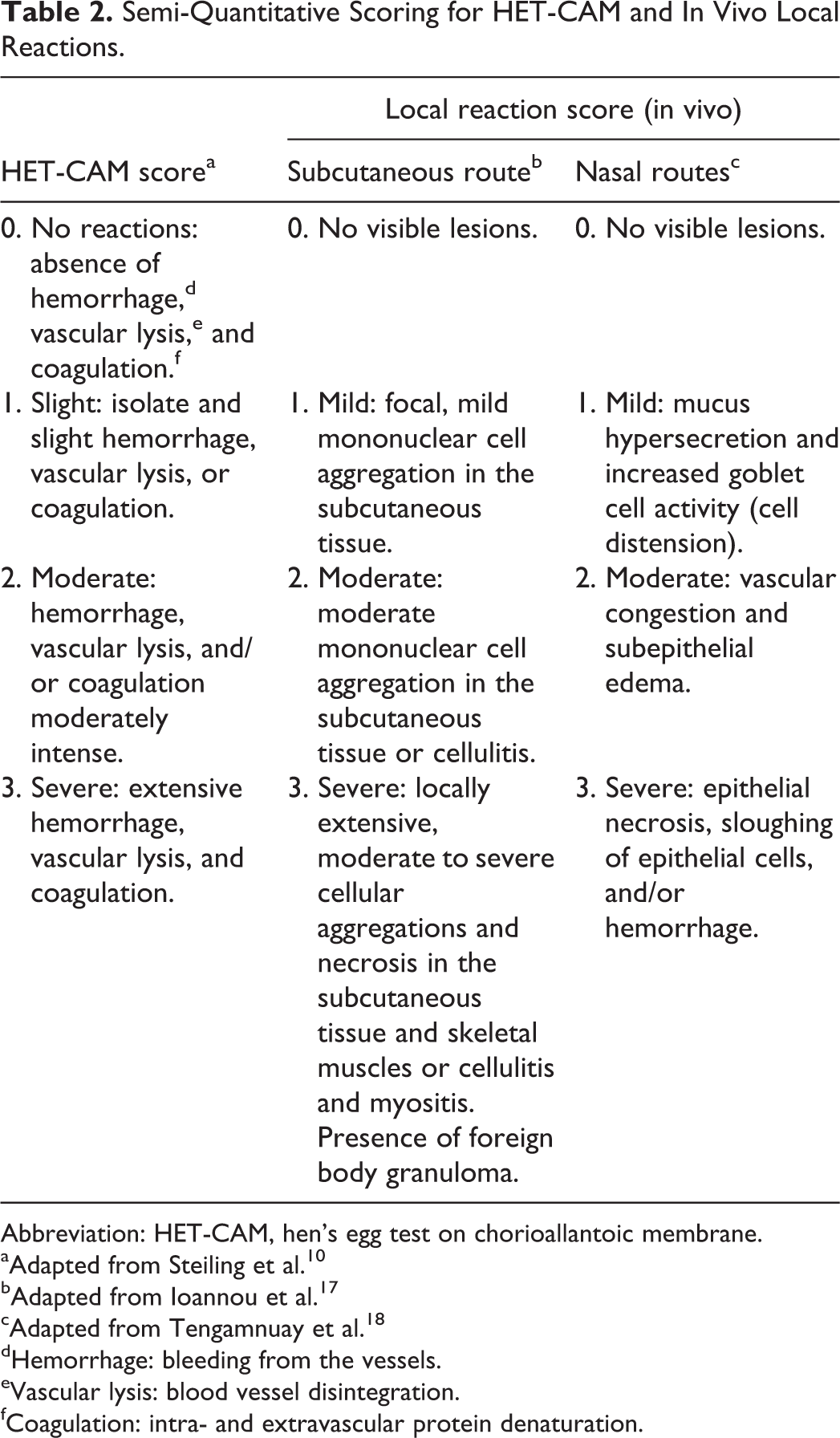
Abbreviation: HET-CAM, hen’s egg test on chorioallantoic membrane.
aAdapted from Steiling et al. 10
bAdapted from Ioannou et al. 17
cAdapted from Tengamnuay et al. 18
dHemorrhage: bleeding from the vessels.
eVascular lysis: blood vessel disintegration.
fCoagulation: intra- and extravascular protein denaturation.
Local Tolerance Assessment In Vivo
Female Balb/c mice (20 g body weight) were purchased from National Center for the Production of Laboratory Animals (CENPALAB, Cuba). All mice received humane care according to the guidelines of the National Institutes of Health. At day 0, groups of 5 mice were subcutaneously (SC) inoculated in the back with 100 µL of each adjuvant formulated with PBS solution (1 group of mice per adjuvant formulation, including a control group with PBS alone). The same dose was used as a booster on day 7 following initial administration. For the group inoculated with FCA, the booster was FIA. Due to the potential alternative use of AFPL1, AFCo1, AFCo3, and Cliptox in vaccine formulations by an intranasal (IN) route, they were also evaluated through this route. Ten microliters of each adjuvant formulation or PBS was administered to the nostril to the respective mice using an Eppendorf pipette. In the case of AFPL1, AFCo1, and AFCo3, boosters were administrated on 5th and 10th day after the first dose, according to the manufacturer’s instructions, and Cliptox was applied on days 0 and 7.
Two weeks after the last administration, mice were anaesthetized by intraperitoneal administration of 0.1 mL of ketamine (50 mg/mL) and then they were euthanized in a CO2 chamber. The necropsy included macroscopic and microscopic examination of injection sites using standardized methods for SC 17 and IN administration. 18,19 A histological score from 0 to 3+ was assigned based on the tissue reactions to the various vaccine adjuvants, as described in Table 2. All experiments (in vitro and in vivo) were analyzed and approved by the research ethics committee of Toxicology and Biomedicine Center Medical Science University.
Statistical Analysis
Statistical analysis was performed by GraphPad Prism version 6.01 software. The strength and direction of the linear relationship between HET-CAM and local tolerance in mice for the tested adjuvants were expressed according to the Pearson correlation coefficient (r) for a significance level of 99%.
Results
Hen’s Egg Test on Chorioallantoic Membrane Test
The positive internal controls, 0.1 N NaOH and 1% SDS, exhibited various end points (H, L, and C) at different times. The mean IS for 0.1 N NaOH and 1% SDS was 17.80 ± 0.2 and 11.80 ± 0.1, respectively, which is indicative of a valid model. The evaluation of the 10 adjuvants revealed that FCA, saponin, FIA, and Al-gel all showed an IS above 1.3, whereas the remaining 8 adjuvants showed only a slight or nonirritating effect (Table 3). Figure 1 depicts a characteristic image of severe irritation with Hs, L, and intravascular C on the CAM.
Irritation Score of Adjuvant Formulations Evaluated by HET-CAM and Local Tolerance in Mice.a
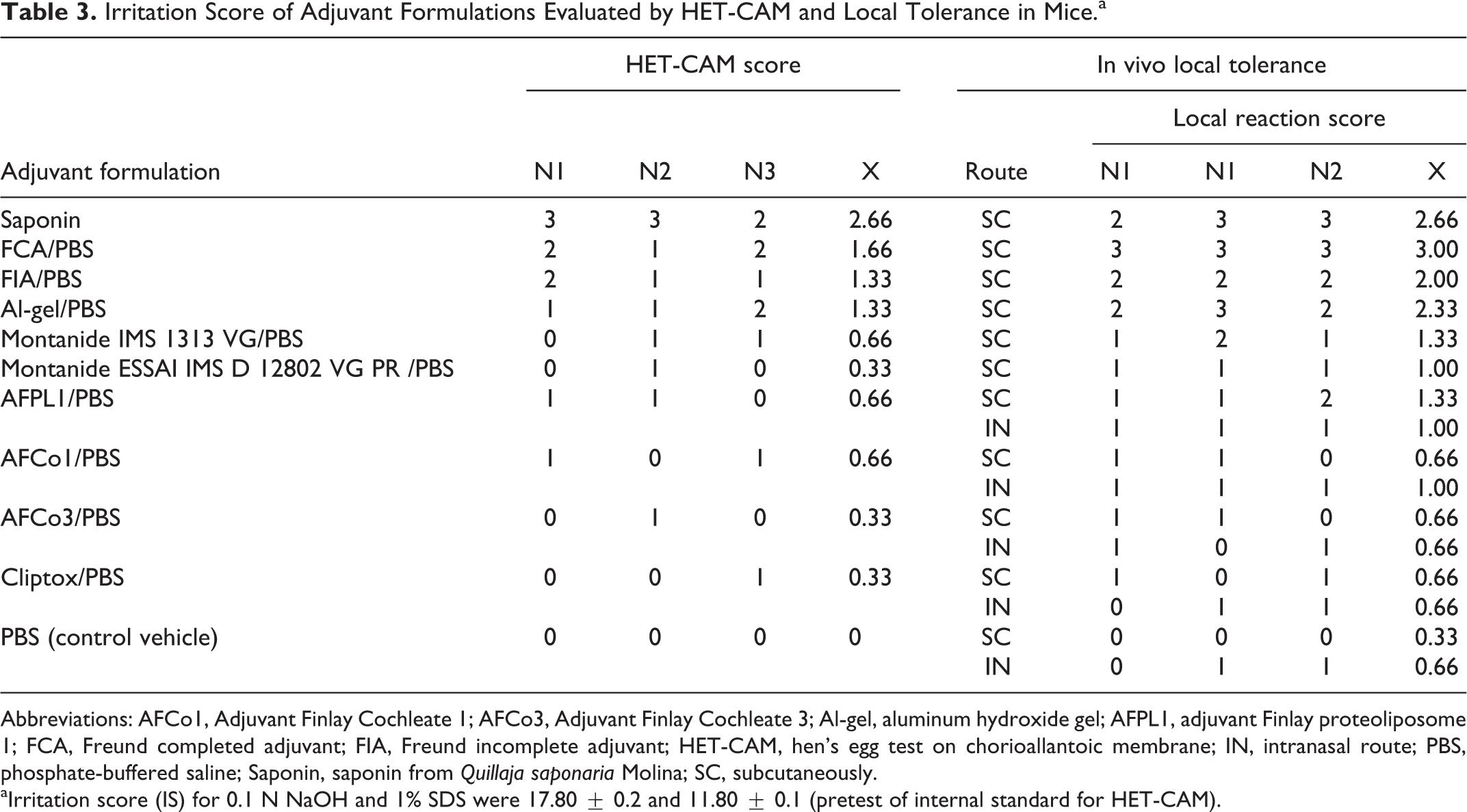
Abbreviations: AFCo1, Adjuvant Finlay Cochleate 1; AFCo3, Adjuvant Finlay Cochleate 3; Al-gel, aluminum hydroxide gel; AFPL1, adjuvant Finlay proteoliposome 1; FCA, Freund completed adjuvant; FIA, Freund incomplete adjuvant; HET-CAM, hen’s egg test on chorioallantoic membrane; IN, intranasal route; PBS, phosphate-buffered saline; Saponin, saponin from Quillaja saponaria Molina; SC, subcutaneously.
aIrritation score (IS) for 0.1 N NaOH and 1% SDS were 17.80 ± 0.2 and 11.80 ± 0.1 (pretest of internal standard for HET-CAM).
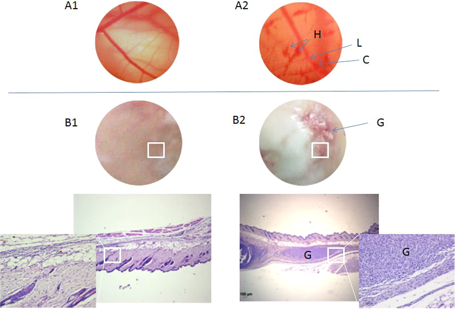
Images of HET-CAM (upper): A1, CAM normal; A2, severe irritation (H: hemorrhage; L: lysis; C: coagulation). Subcutaneous tissue of Balb/c mice (lower): B1, normal; B2, severe local inflammation with granuloma (G). HET-CAM indicates hen’s egg test on chorioallantoic membrane.
Local Tolerance Study
After repeated SC administration, FCA, saponin, FIA, or Al-gel induced different grades of local inflammatory reactions from moderate to severe intensity. The predominant lesion caused by these adjuvants was the foreign body granuloma integrated by lymphocytes, macrophages, fibroblasts, mast cells, and multinucleated cells (Figure 1). These adjuvants were not applied by IN route due to their reported irritant properties. 4 The other adjuvants induced moderate to mild reactions at the inoculation site.
As shown in Figure 2, those adjuvants with severe or moderate irritant effects in HET-CAM assay also produced moderate to severe local reactions in mice. In contrast, those adjuvants with low irritant effects by the HET-CAM assay induced a comparable response in the in vivo study. This association was expressed in a significant correlation value of r = 0.9034, (P < 0.001) between both studies.
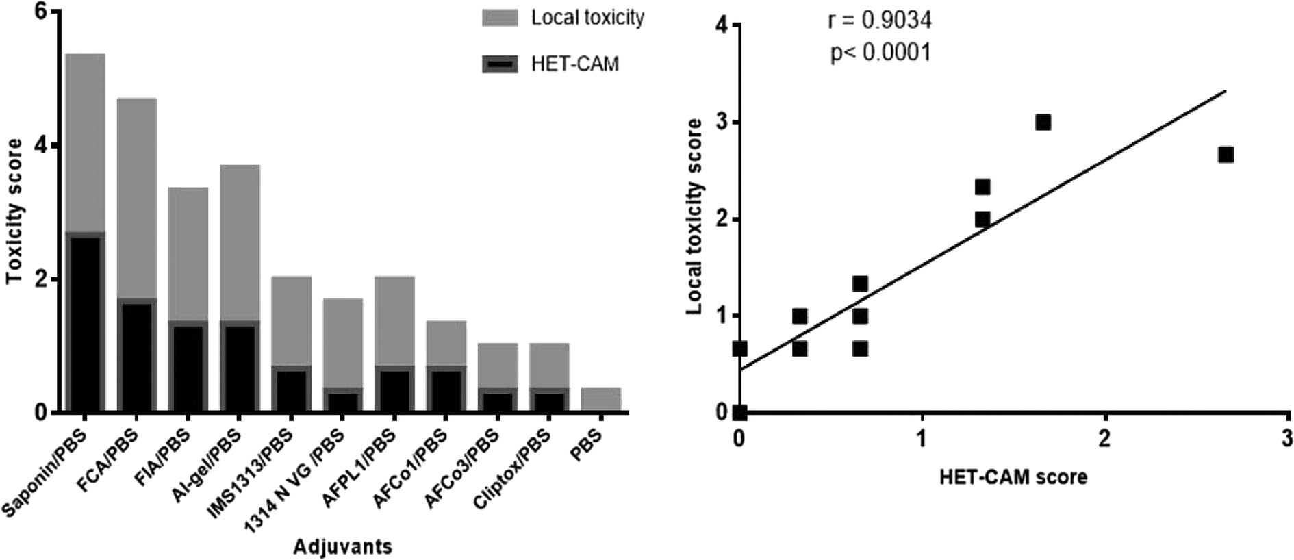
Correlation between score irritation of HET-CAM and in vivo local reaction for 10 adjuvants (P < 0.0001). HET-CAM indicates hen’s egg test on chorioallantoic membrane.
Discussion
Local vaccine toxicity may reflect direct chemical irritation due to a nonphysiological pH, osmolarity, salt concentrations, or direct cell toxicity. Adjuvants are among the most important vaccine constituents responsible for these reactions. Such local irritant effects are typically associated with immediate and severe injection site pain, followed by an inflammatory response triggered by the tissue damage. 6,20 Given the vital importance of adjuvants to modern vaccines, there is an urgent need to develop better in vivo and in vitro models for adjuvant’s safety assessment. 7,20 –24
Often researchers use in vitro tests such as hemolysis 25 –27 or cytolysis in cell culture 26,28 for the assessment of the irritation properties of adjuvanted vaccines, but a wide variety of adjuvants and their different chemical–physical properties, including hydrophobicity, make it difficult to use some of these tests. In addition, with these in vitro models, only specific end points are measured.
This work evaluated the HET-CAM assay as a tool to assess the irritant potential of adjuvants relative to that observed in vivo. The CAM is a highly vascular embryonic membrane formed by the fusion of the allantoic wall with the chorion. 29 As a highly vascular and stratified tissue, it responds to injury in a similar manner as mucosal and subcutaneous tissue. One of the advantages of the HET-CAM is the possibility for using different kinds of formulations, including insoluble or solid substances. 9 This method could be a valuable tool for evaluating the irritation potential for a wide variety of adjuvant formulations, including emulsion, gels, and particles.
Several known and experimental adjuvants were used in this study, formulated with antigen-free PBS, in order to assess their intrinsic irritation potential. Freund adjuvants, saponin, and Al-gel were included as positive controls based on their established toxicological profiles. 4
It should be noted that although there was a significant correlation between the 2 methods, the scores for the HET-CAM assay were consistently lower than the in vivo score. This difference can be due to the fact that HET-CAM detects the immediate, local damage caused by the substance directly in contact with the tissue, whereas the in vivo local tolerance test evaluates the inflammatory reaction induced at the site of inoculation.
The first tissue reactions observed in the CAM after adjuvant application are bleeding from the vessels, intra- and extravascular protein denaturation, and blood vessel disintegration. These reactions can be observed almost immediately after adjuvant application. In contrast, the local reactions observed in laboratory animals after adjuvant inoculation are somewhat delayed. The inflammatory events are presumably a consequence of the initial irritation and other additional causes such as the depot effect caused by emulsions and gels and the presence of immunostimulatory components. 6
In this study, adjuvants that are known irritants such as emulsions, saponins, and aluminum hydroxide showed higher toxicity scores in both. 4,30,31 They can cause tissue damage at the site of injection, which causes the release of danger signals such as damage-associated molecular pattern molecules (DAMPs). Damage-associated molecular pattern molecules are host molecules (eg, DNA, RNA, heat shock proteins, hyaluronic acid, serum amyloid A protein, uric acid, and other intracellular molecules) that can stimulate an early migration of inflammatory cells to the inoculation site, producing clinical manifestations that range from acute mild inflammation to severe tissue necrosis and fibrosis. 6,31
The cytotoxicity of the Freund adjuvants is due to the presence of short-chain hydrocarbons in the mineral oil with detergent-like effects, dissolving the lipid bilayer of the cell membrane, thus causing cell lysis. 4,5 In the case of saponins from Quillaja saponaria Molina, they are amphipathic molecules of glycosides that interact with cell membranes leading to cell lysis. This property have been widely evaluated in hemolysis assay and other in vitro cytotoxicity tests. 25,26 On the other hand, aluminum-based adjuvants are able to cause cell death and the subsequent release of host cell DNA and uric acid, which acts as a potent endogenous immunostimulatory signal mediating its adjuvant activity. 31
Until recently, it was thought that the potency of adjuvants was directly related to the toxicity. 4,32 Interestingly, in this study, all those adjuvants that did not induce irritation were strong immunopotentiators. 33 –37 This seems to contradict the danger model that suggests that tissue disruption is necessary to achieve a good adjuvant effect and immune stimulation induced by the released DAMPs. According to this model, the immune system does not distinguish between self and nonself but rather between things that might cause damage and things that will not. 38
A new generation of adjuvants include agonist of receptors involved in innate immune activation named pattern recognition receptors. These new adjuvants are typically better characterized molecules that can stimulate the immune response with minimal local toxicity. 39,40 This study evaluated some of these new adjuvants such as Montanide IMS 1313 VG, Montanide ESSAI IMS D 12802 VG PR, AFPL1, AFCo1, AFCo3, and Cliptox. According to these results, the HET-CAM assay can be used to screen and evaluate the safety profile of vaccine adjuvant candidates.
Thus, new substances identified as irritants could be excluded from in vivo testing, thereby reducing the number of laboratory animals used for biological testing. This assay could be particularly valuable to evaluate substances specifically designed for mucosal delivery. This is an important factor because products administered to mucosal surfaces should not be irritating. 41 –43
Finally, it is important to highlight that although the focus of this study was the evaluation of the irritation potential of vaccine adjuvants by the HET-CAM assay, antigens may also induce postvaccination local reactions. 44 Thus, the current guidelines recommend the assessment of both the adjuvant alone and in combination with the proposed antigen. 45,46 The evaluation of the adjuvant alone can be important for novel adjuvants that have not been studied previously or will be used in multiple different vaccine formulations. 46
In summary, the HET-CAM assay could be a useful alternative method for evaluating irritation effect of vaccine adjuvants delivered to mucosal and subcutaneous sites. This method is a potential alternative to in vivo toxicological studies, thereby reducing animal use. Further studies are necessary in order to make an extensive validation of HET-CAM assay as an alternative method as part of the toxicity studies of vaccine adjuvants.
Footnotes
Authors’ Note
ABD and OP, contributed to conception and design, acquisition, analysis and discussion of the results, drafted the article, gave final approval, and agreed to be accountable for all aspects of work ensuring integrity and accuracy. GMJ, UPM, and ETN contributed to conception, design, material preparation, implementation of HET-CAM test, and interpretation of results. DPF, BTM, and DTM contributed to conception, design, material preparation, implementation of in vivo local tolerance test, and interpretation of results. JEB contributed to the histological study and interpretation.
Declaration of Conflicting Interests
The author(s) declared no potential conflicts of interest with respect to the research, authorship, and/or publication of this article.
Funding
The author(s) received no financial support for the research, authorship, and/or publication of this article.