
Abstract
BMX-001, a manganese porphyrin that has anti-inflammatory, antioxidant, and antitumor properties, is being developed as a potential therapeutic for high-grade glioma (HGG) and head and neck (H&N) cancer. An IND has been opened for BMX-001 in the treatment of HGG (NCT02655601) and another is in preparation for H&N. The safety of BMX-001 has been evaluated in a battery of nonclinical Good Laboratory Practice (GLP)-compliant studies. Systemic toxicity has been evaluated using the intended cGMP product administered subcutaneously for periods of up to 5 weeks in both the mouse and the monkey and included toxicokinetic evaluations to characterize systemic exposure and tissue distribution and clearance of BMX-001. In additional GLP studies, BMX-001 was not irritating to the skin or eye and caused no changes in cardiac rate or rhythm or blood pressure. Mixed results for genotoxicity were seen with the weight of evidence indicating that BMX-001 poses no genotoxic risk in humans. In systemic mouse and monkey studies, loading/maintenance dose no observed adverse effect levels were 12/2 mg/kg/dose and 6/2 mg/kg/dose, respectively, with maintenance doses administered every 3 days after the initial loading dose. Systemic data were used to determine a Food and Drug Administration-approved safe starting dose for the initial clinical study in patients with HGG. BMX-001 was detected in analyzed tissues, including the brain, persisting well past the short plasma clearance period. The highest levels of BMX-001 were seen in the liver and kidneys, with amounts in these tissues returning to close to undetectable levels after a 2-week cessation of dosing.
Introduction
It is well established that induction of oxidative stress in the surrounding normal tissue after radiation therapy (RT) places a major limitation on its use in the treatment of cancers. 1 –4 A variety of approaches have been investigated to mitigate oxidative stress postirradiation, including the free radical scavenger, amifostine. 5 –7 Although this agent is approved for this use, repeated daily use is limited by side effects. 5,7
Over the past 20 years, a class of manganese (Mn) porphyrin-based compounds has been developed 8 –10 that are potent anti-inflammatory agents and catalytically inactivate a range of reactive oxygen species, including peroxynitrite and superoxide anion, but also exhibit other anti-inflammatory properties.
Porphyrins are made up of 4 pyrrole rings interconnected by methane bridges. These molecules often form complexes with metals by incorporating them into the nucleus of the porphyrin structure, with the metal being situated between the interiorly located vertices of the pyrrole rings. Cationic ortho Mn(III) N-substituted pyridylporphyrins are known to be potent antioxidants and have been demonstrated as effective in ameliorating both inflammation and injury in a large number of animal models of human disease. 8,9 The safety of metalloporphyrins has been evaluated previously with the most common and best studied being heme, an iron-protoporphyrin complex. 11,12 The toxic effects of heme are typically associated with excessive amounts of free heme, resulting in the generation of reactive oxygen species. Target organs of heme toxicity include the kidney, liver, central nervous system, and heart. More recently, we have published safety data for MnTE-2-PyP, a first-generation Mn porphyrin. 13
Manganese porphyrin-based compounds have the unique function of protecting normal tissues while also augmenting tumoricidal activity in patients undergoing radiation and chemotherapy. 8,9,19,20 In addition to inactivation of reactive oxygen species, these drugs inhibit transcriptional activity of stress-induced transcription pathways either by eliminating reactive oxygen species that activate stress responses or by indirectly preventing activation. 8,9,18 Transcription factors known to be inhibited by this class of metalloporphyrin include hypoxia inducible factor-1 alpha (HIF-1), 14,15 nuclear factor κB (NF-κB), 16 specificity protein-1 (SP-1), 17 and activator protein-1 (AP-1). 8
A recently developed compound, BMX-001, is among the most highly potent metalloporphyrins that have been evaluated thus far for safety and efficacy. The chemical name of BMX-001 is Mn(III) meso-tetrakis(N-n-butoxyethylpyridinium-2-yl)porphyrin (Figure 1), abbreviated as Mn-TnBuOE-2-PyP5+. 10
BMX-001 catalytically reacts with a range of reactive oxygen species (eg, superoxide and peroxynitrite). 9,18 BMX-001 blocks signaling by proinflammatory transcription factors, NF-κB and HIF-1α. 8,9,18 The 4 hydrophobic side chains contain an ether linkage that results in disruption of micellar properties yet in preservation of the lipophilicity of this compound and thus facilitate its accumulation in intracellular domains including both mitochondria and nucleus. Multiple studies in animal models have demonstrated that BMX-001 is highly effective in reducing oxidative stress induced by radiation. 9
Weitzel et al 19 studied radioprotection of the brain white matter following treatment with BMX-001. Drug administration at 1.5 mg/kg twice daily (BID) for 1 month followed by 0.5 mg/kg once daily for 1 month provided long-term neuroprotection of mice given 8 Gy of whole brain irradiation. Irradiated mice treated with BMX-001 showed myelin preservation in the corpus callosum and protection against deficits in motor proficiency as assessed by both rotarod tests and running wheel tests compared with saline-treated mice. BMX-001 was shown to distribute to the brain and to have a long tissue half-life. One month after the last treatment with BMX-001, the concentrations of the drug in mouse brain were reported to be 25 to 30 nmol/L.
Ashcraft et al 20 evaluated the protective effects of BMX-001 in a mouse model of head and neck irradiation. The mice received BMX-001 at a dose of 1.5 mg/kg BID subcutaneously and doses of RT to the oral cavity and neck at 11, 13, or 15 Gy. BMX-001 was found to protect normal tissues by reducing RT-mediated mucositis, xerostomia, and fibrosis. A dose-modifying factor of 0.77 was found for protection against xerostomia.
BMX-001 is being developed clinically as a treatment (adjunctive to radiotherapy) for head and neck cancer and high-grade gliomas (HGGs) such as glioblastoma. This drug candidate has undergone a full battery of Good Laboratory Practice (GLP)-compliant nonclinical safety studies and is the subject of a just initiated US clinical trial for HGG (NCT02655601).
Materials and Methods
All of the nonclinical in vivo studies conducted in monkeys, mice, rabbits, and guinea pigs were performed at Calvert Labs (Scott Township, Pennsylvania) and ILS (Hillsborough, North Carolina). For systemic toxicity studies, the cynomolgus monkey and the mouse were chosen based on severe signs of intolerance in rats and dogs in previous studies with BMX-001 and other members of the class of compounds. 13 The genotoxicity studies were conducted at BioReliance (Rockville, Maryland), except for the comet assay, which was conducted by ILS (Research Triangle Park, North Carolina). Consistent with the intended clinical route, nonclinical systemic toxicity studies were conducted by the subcutaneous route. The dosing rationale for these nonclinical studies, including the use of loading and maintenance doses, is based on pharmacokinetic data for BMX-001 and other compounds in this class. Similarly, species-specific lengths of recovery (3 weeks in mice vs 2 weeks in monkeys) were based on previous data indicating a shorter plasma half-life in mice. All studies were conducted in compliance with GLPs (except for the mouse and monkey maximum tolerated dose [MTD] studies) and ICH guidelines. 21,22 Housing and room conditions were consistent with Association for Assessment and Accreditation of Laboratory Animal Care standards unless stated otherwise.
Manufacturing and Characterization of BMX-001
BMX-001 (CASRN 1379783-91-1, molecular weight 1,253.54; Figure 1) was manufactured, tested, and released under cGMP by Albany Molecular Research, Inc (Albany, New York) and supplied as a dark brown to purple solid with 93.5% purity. BMX-001 final drug substance was stored under a blanket of nitrogen in an amber glass bottle with closure. The bottle was placed inside a tie-wrapped 3-mL low-density polyethylene (LDPE) bag containing a desiccant. This bag was sealed in a second tie-wrapped LDPE bag, which was sealed inside a high-density polyethylene pail. The pail was stored at room temperature and protected from light.
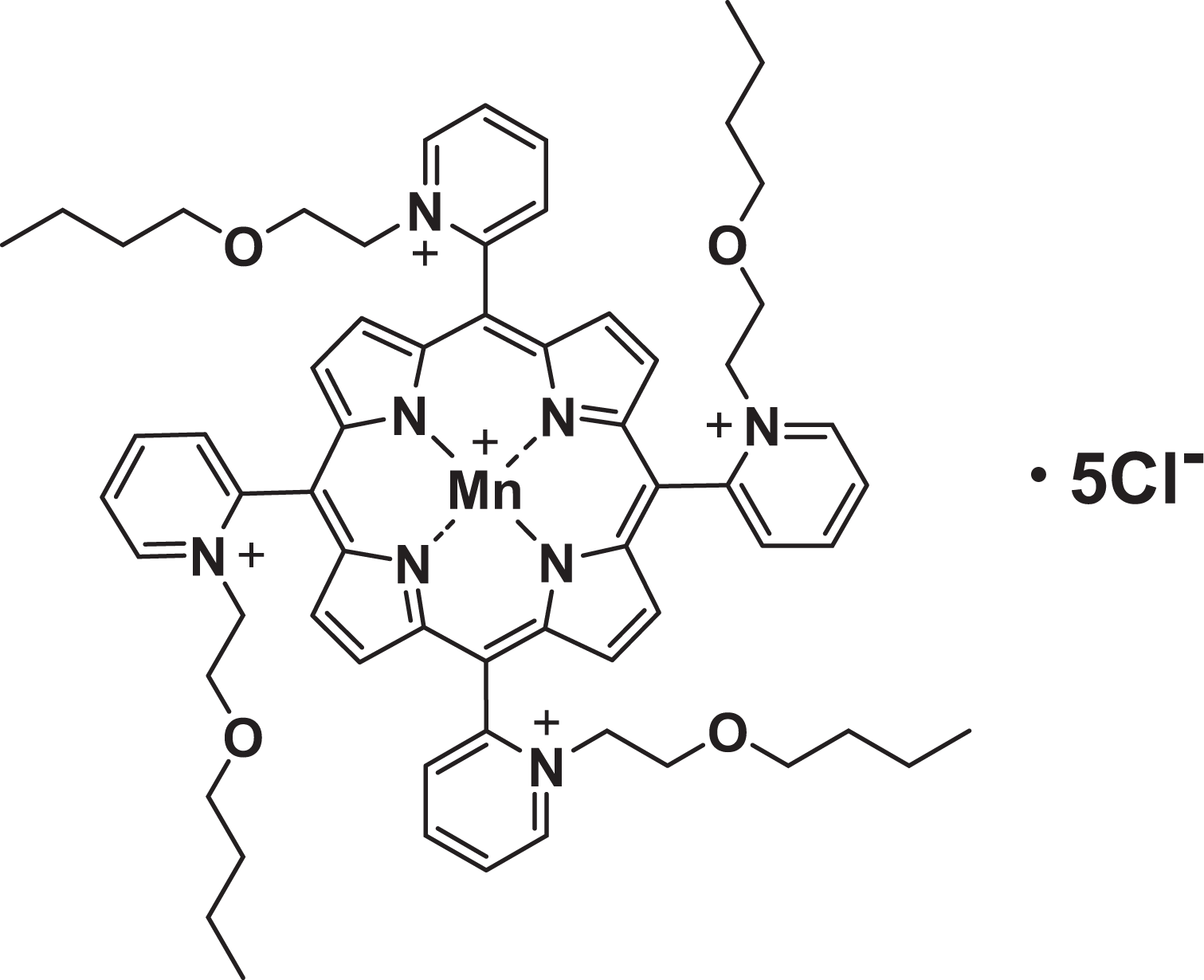
Structure for BMX-001 (MnTnBuOE-2-PyP5+, Mn(III) meso-tetrakis(N-n-butoxyethylpyridinium-2-yl)porphyrin).
Genotoxicity
Bacterial reverse mutation assay
BMX-001 was tested in the bacterial reverse mutation assay using Salmonella typhimurium tester strains TA98, TA100, TA1535, and TA1537 and Escherichia coli tester strain WP2 uvrA in the presence and absence of Aroclor-induced rat liver S9. The assay was performed in 2 phases using the plate incorporation method. In the first phase, the preliminary toxicity assay was used to establish the dose range for the mutagenicity assay. In the second phase, the mutagenicity assay was used to evaluate the mutagenic potential of the test article.
Phosphate-buffered saline (PBS; without magnesium or calcium) was selected as the solvent of choice based on the solubility of the test article and compatibility with the target cells. After sonication at 33.5°C for 15 minutes in the solubility test conducted at BioReliance, the test article formed a clear solution in PBS at a concentration of approximately 10 mg/mL.
In the mutagenicity assay, the maximum dose tested was 5,000 μg per plate; this dose was achieved using a concentration of 10 mg/mL and a 500 μL plating aliquot. The dose levels tested were 6.7, 10, 33, 67, 100, 333, 667, 1,000, 3,333, and 5,000 μg per plate.
Data sets for tester strains TA1535 and TA1537 were judged positive if the increase in mean revertants at the peak of the dose response was ≥3.0 times the mean vehicle control value. Data sets for tester strains TA98, TA100, and WP2 uvrA were judged positive if the increase in mean revertants at the peak of the dose response was ≥2.0 times the mean vehicle control value.
In vitro mouse lymphoma assay
BMX-001 was tested in the L5178Y/TK+/− mouse lymphoma assay in the absence and presence of Aroclor-induced rat liver S9. The preliminary toxicity assay established the concentration range for the mutagenesis assay. The mutagenesis assay was used to evaluate the mutagenic potential of the test article.
Phosphate-buffered saline (without magnesium or calcium) was selected as the solvent of choice based on information provided by the sponsor, the solubility of the test article, and compatibility with the target cells. After sonication at 33.5°C for 15 minutes, BMX-001 formed a clear solution in PBS at approximately 10 mg/mL.
In vivo micronucleus assay
BMX-001 was evaluated for its clastogenic activity and/or disruption of the mitotic apparatus by detecting micronuclei in polychromatic erythrocyte (PCE) cells in mouse bone marrow. Phosphate-buffered saline without magnesium or calcium was selected as the vehicle. To ensure target organ exposure, BMX-001 or control article formulations were administered at a dose volume of 10 mL/kg by intravenous injection. Cyclophosphamide, the positive control, was administered by oral gavage.
The definitive assay dose levels tested were 0.25, 0.5, and 1.0 mg/kg. Dose levels were selected based on the results of the 28-day mouse systemic toxicity study. Groups 1 and 4 consisted of 10 animals/sex designated for either 24- or 48-hour bone marrow collections, and groups 2, 3, and 5 consisted of 5 animals/sex designated for 24-hour bone marrow collection. Following scheduled euthanasia times, femoral bone marrow was collected; bone marrow slides were prepared and stained with acridine orange. Bone marrow cells (2,000 PCEs/animal) were examined microscopically for the presence of micronuclei (micronucleated PCEs [MnPCEs]), and statistical analysis of data was performed using the Kastenbaum-Bowman tables (binomial distribution, P ≤ 0.05). The ratio of PCEs to total erythrocytes (EC) in the test article groups relative to the vehicle control groups was also evaluated to reflect the test article’s cytotoxicity.
Comet assay
Twenty-five male B6C3F1 mice were allocated equally between 5 designated dose groups. Based upon daily body weights, the animals were administered 5, 10, or 30 mg/kg of BMX-001, the vehicle control, or the positive reference item (ethyl methanesulfonate [EMS]) for 2 consecutive days via subcutaneous injection. Dose levels of BMX-001 were selected based on the results of the 28-day mouse systemic toxicity study. Approximately 3 hours following the final dose administration, the animals were humanely euthanized. Liver tissue was collected for the assessment of DNA damage by the comet assay, in conformance with OECD guidelines. 23
Local Tissue Tolerance
Primary dermal irritation in rabbits
BMX-001 (as a 0.1% solution in saline) was initially applied to a clipped dorsal trunk area on 3 male New Zealand white rabbits. The test article (0.5 mL) was applied to an area (∼5 × 5 cm) on the dorsal trunk of the animals and covered with a gauze patch for a 4-hour (±0.5 hours) exposure period. Observations for dermal irritation were recorded immediately after patch removal, at 30 to 60 minutes, and day 2 through day 4 (24-72 hours after patch removal). Grading of irritation was according to the method of Draize et al. 24
Guinea pig maximization
The test article was evaluated in the Magnusson-Kligman guinea pig maximization model of delayed hypersensitivity. The dose levels used were based upon the identification of nonirritating doses in a dose-range finding study. Based upon the intradermal dose range, the intradermal dose utilized was 0.1% BMX-001 in water. Based upon the topical dose range, the topical induction was dosed at 1.5%. The challenge dose utilized was a 0.1% solution, and the rechallenge dose was a 0.01% solution. The main study was conducted with 8 males and 7 females for the test article group and 5 animals/sex for the vehicle control group.
For the intradermal induction phase (day 1), each guinea pig received intradermal injections (0% or 0.1% BMX-001, 0.1 mL each) between the scapulae. On day 8, BMX-001 (1.5%) was spread over a 2 × 4 cm filter paper (0.3 mL) and applied to the injection site areas of the test group. Blenderm tape was used to occlude the injection area. The dressings were removed following approximately 48 hours of exposure, and the areas were wiped clean with gauze and water. The vehicle group was similarly treated with vehicle.
Two weeks after the topical induction, the hair was removed from the right and left flank regions. On day 25, all test article-treated and vehicle control animals were challenged with occluded patches for approximately 24 hours on the left flank and right flank regions. For the test groups, a 2 × 2 cm filter paper was saturated with 0.2 mL of the test article and applied to the left flank. Another 2 × 2 cm filter paper was saturated with 0.2 mL of the respective vehicle control and applied to the right flank. For the vehicle groups, a 2 × 2 cm filter paper was saturated with the respective test article and applied to the right flanks. Another 2 × 2 cm filter paper was saturated with the respective vehicle control and applied to the left flanks. Approximately 24 hours after dosing, sites were unwrapped and wiped clean with gauze and water. Approximately 21 hours after unwrapping, the sites were depilated with Nair lotion hair remover. Approximately 4 hours later, the sites were graded for elicited skin reactions (24-hour grade). Approximately 23 hours, the sites were graded a second time (48-hour grade).
Eye irritation in rabbits
Prior to dosing, the eyes were anesthetized using 2 drops of tetracaine in each eye. The test article, BMX-001 (dosed at 0.1% in normal saline), was initially instilled into the anesthetized right eye of 1 male New Zealand white rabbit at a dose of 0.1 mL. Upon instillation of the test article, the head was pointed upward and the eyelids were held closed approximately 1 second to limit loss of material. The anesthetized left eye was left untreated. Following the 1-hour scores of the first animal, 2 additional animals were dosed. Both eyes of all 3 animals were examined and scored for ocular irritation according to the method of Draize et al 24 immediately prior to dosing, approximately 1 hour after dosing, and at 24 and 48 hours postdose (Note: The FDA recommendation to utilize the bovine corneal opacity and permeability assay in lieu of the in vivo test was promulgated subsequent to the submission of this study.).
Safety Pharmacology Study
Evaluation of cardiovascular function in conscious telemetered male cynomolgus monkeys
This study evaluated the qualitative analysis of telemetric electrocardiographic (ECG) recordings from 4 male monkeys (previous work had determined that dogs are extremely sensitive to the hypotensive effects of this class of compounds). One minute of continuous ECG tracing was collected at baseline, 15 minutes prior to dosing, and then at 30 minutes, 1 hour, 2 hours, 4 hours, 8 hours, 12 hours, and 22 hours after test article administration. BMX-001 was administered once per dose level via subcutaneous injection on study days 1, 2, 9, 16, and 23 as defined by a 4 × 4 Latin square crossover study design. Dose levels and frequency of dosing were based on the short plasma half-life of BMX-001. Dosing was with vehicle at 0 mg/kg (vehicle control) or with BMX-001 at 1 mg/kg (low dose), 3 mg/kg (low-mid dose), 6 mg/kg (mid-high dose), and 9 mg/kg (high dose). For each trace, the recording was evaluated for rate and rhythm disturbances and changes in the general configuration of the complexes.
Systemic Toxicity
Maximum tolerated dose studies in mice and monkeys
To determine the MTD of BMX-001, non-GLP escalating dose-range finding and 5-day repeat dose studies by intravenous injection were conducted in CD-1 mice and cynomolgus monkeys. In the escalating portion of the study, animals were administered BMX-001 by intravenous slow bolus injection over a period of 3 minutes. After a minimum of approximately 48 hours, the dose escalated or de-escalated until an MTD was determined. Once the MTD was determined, a confirmatory phase was conducted in which animals were administered BMX-001 once daily for 5 consecutive days by intravenous slow bolus injection. Mortality was evaluated BID during treatment. Clinical observations were evaluated once prior to dosing, immediately postdose, and approximately 1 hour postdose and additionally as needed. Body weights were recorded prior to dose administration and prior to termination. A gross necropsy was performed on all animals. In the confirmatory phase, blood was collected for the evaluation of hematology and clinical chemistry, and tissues were harvested at necropsy with selected organs weighed.
A 5-week subcutaneous study in mice with a 3-week recovery period
The purpose of this study was to evaluate the toxicity and toxicokinetics of BMX-001 when administered twice weekly by subcutaneous injection in mice for a minimum of 5 consecutive weeks followed by a 3-week recovery period. Animals were dosed as shown in Table 1. Animals were 7 to 8 weeks old, with males weighing approximately 29 to 40 g and females weighing 21 to 31 g at the outset of the study.
Dosing Regimen for the 5-Week Mouse Study.
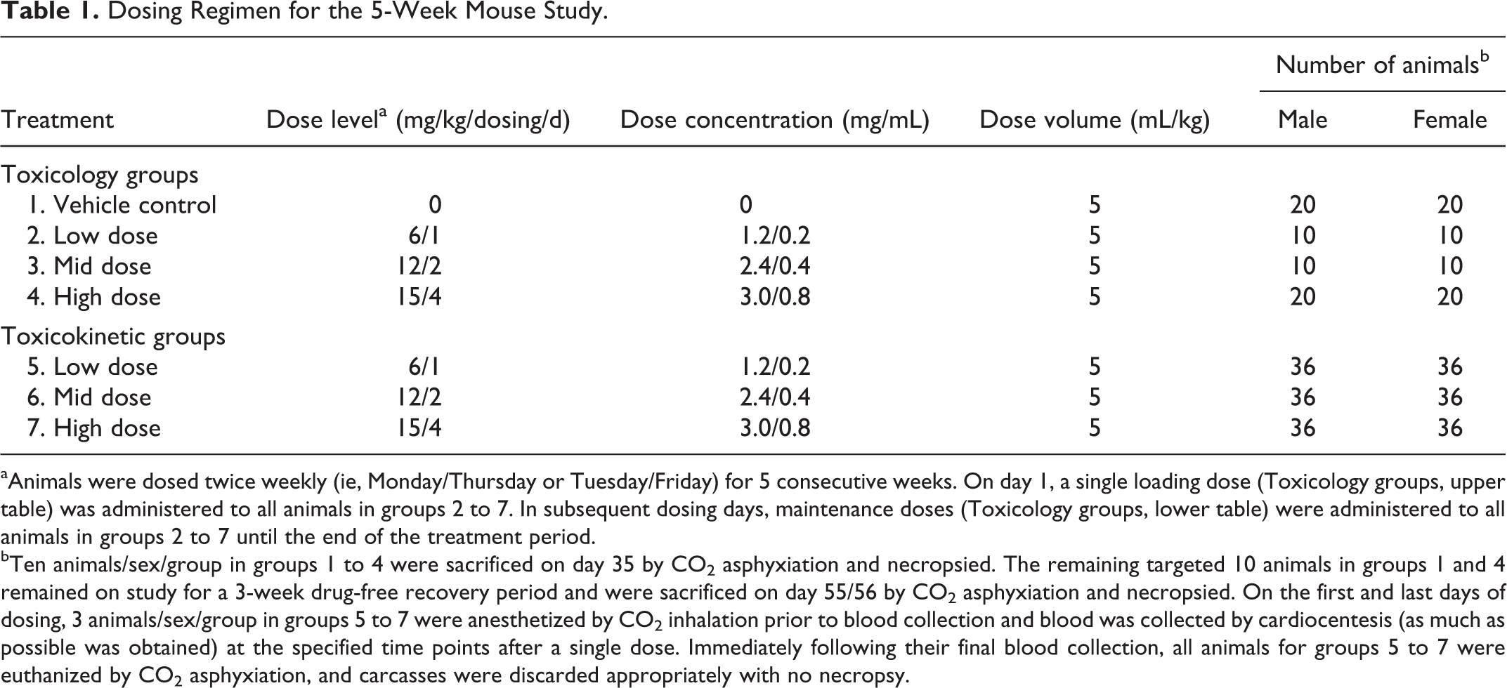
aAnimals were dosed twice weekly (ie, Monday/Thursday or Tuesday/Friday) for 5 consecutive weeks. On day 1, a single loading dose (Toxicology groups, upper table) was administered to all animals in groups 2 to 7. In subsequent dosing days, maintenance doses (Toxicology groups, lower table) were administered to all animals in groups 2 to 7 until the end of the treatment period.
bTen animals/sex/group in groups 1 to 4 were sacrificed on day 35 by CO2 asphyxiation and necropsied. The remaining targeted 10 animals in groups 1 and 4 remained on study for a 3-week drug-free recovery period and were sacrificed on day 55/56 by CO2 asphyxiation and necropsied. On the first and last days of dosing, 3 animals/sex/group in groups 5 to 7 were anesthetized by CO2 inhalation prior to blood collection and blood was collected by cardiocentesis (as much as possible was obtained) at the specified time points after a single dose. Immediately following their final blood collection, all animals for groups 5 to 7 were euthanized by CO2 asphyxiation, and carcasses were discarded appropriately with no necropsy.
Mortality/morbidity was performed BID and once prior to sacrifice. Animals were observed prior to each dose administration and at approximately 1 to 2 hours postdose. Animals were observed once daily on nondosing days. During the 3-week recovery period, animals in the designated recovery groups were observed once daily. Animals in the toxicology groups were also observed once prior to scheduled necropsies on day 35 and day 55/56 (recovery animals). All animals were weighed at the time of randomization, weekly, and prior to termination. Animals in toxicology groups were weighed (fasted) prior to scheduled sacrifice on day 35 and 55/56 (recovery animals). Food consumption was recorded weekly during the dosing and recovery phases of the study.
Ophthalmology examinations were performed prior to treatment initiation and during the final week of the dosing period. Blood for the evaluation of hematology and clinical chemistry parameters was collected on day 35 and day 55/56 (recovery phase). Blood for toxicokinetic evaluation was collected from animals in the toxicokinetic groups on the first and last days of dosing. All surviving animals in the toxicology groups were sacrificed on day 35 and day 55/56 (recovery). Selected tissues were harvested at necropsy, selected organs weighed, and selected tissues from the control and high-dose groups were evaluated microscopically.
A 5-week subcutaneous study in monkeys
The purpose of this study was to evaluate the toxicity and toxicokinetics of BMX-001 when administered a loading dose followed by lower maintenance doses every 3 days by subcutaneous injection to cynomolgus monkeys for a minimum of 5 consecutive weeks followed by a 2-week recovery period. Animals were dosed as shown in Table 2.
Dosing Regimen for the 5-Week Monkey Study.
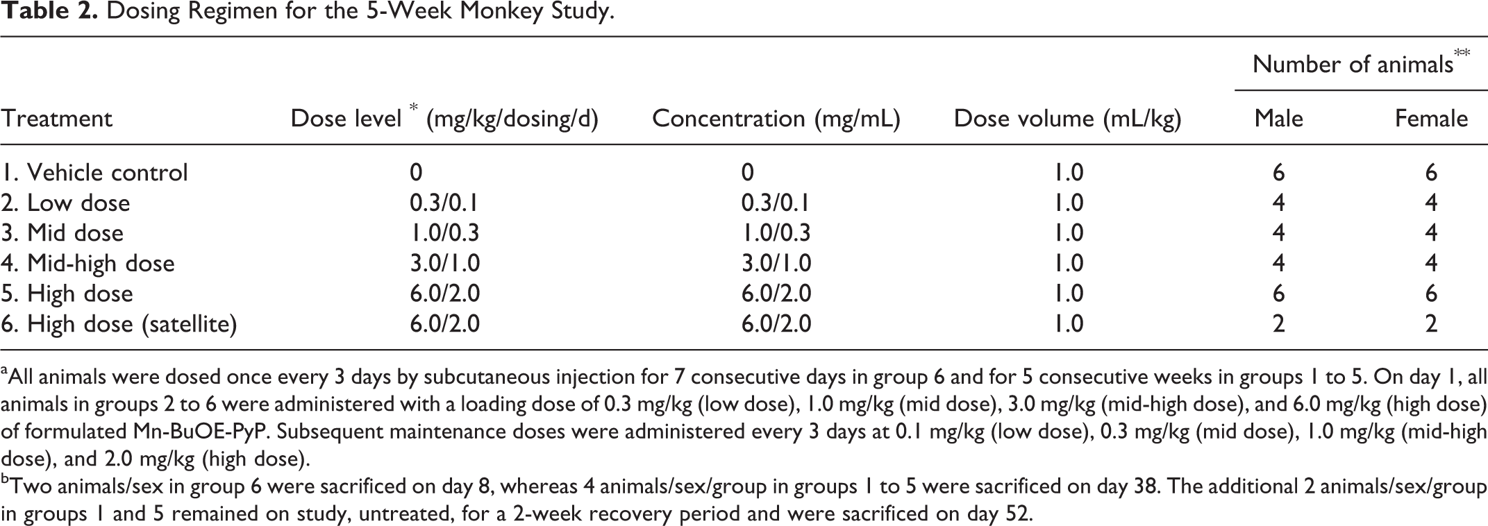
aAll animals were dosed once every 3 days by subcutaneous injection for 7 consecutive days in group 6 and for 5 consecutive weeks in groups 1 to 5. On day 1, all animals in groups 2 to 6 were administered with a loading dose of 0.3 mg/kg (low dose), 1.0 mg/kg (mid dose), 3.0 mg/kg (mid-high dose), and 6.0 mg/kg (high dose) of formulated Mn-BuOE-PyP. Subsequent maintenance doses were administered every 3 days at 0.1 mg/kg (low dose), 0.3 mg/kg (mid dose), 1.0 mg/kg (mid-high dose), and 2.0 mg/kg (high dose).
bTwo animals/sex in group 6 were sacrificed on day 8, whereas 4 animals/sex/group in groups 1 to 5 were sacrificed on day 38. The additional 2 animals/sex/group in groups 1 and 5 remained on study, untreated, for a 2-week recovery period and were sacrificed on day 52.
BMX-001 was prepared into dosing solutions for subcutaneous administration. Fifty-two experimentally naive cynomolgus monkeys (26 males and 26 females), approximately 2 years 7 months to 3 years of age and weighing 2.0 to 3.2 kg for males and females, were used. Mortality and clinical observations were evaluated daily. Body weights were recorded weekly. Food consumption was recorded daily. Ophthalmology examinations were performed prior to treatment initiation and during the last week of treatment.
Electrocardiogram tracings were obtained prior to treatment initiation and prior to terminal sacrifices. Blood for the evaluation of hematology, coagulation, and clinical chemistry parameters was collected from all animals prior to the initiation of treatment, from animals in group 6 on day 8 prior to sacrifice, from all animals on day 38, and from recovery animals on day 52. Blood for toxicokinetic evaluation was collected from group 6 animals at selected time points on days 1 and 7 and from all animals in groups 1 to 5 at selected time points on days 1 and 37. All animals were sacrificed on day 8 (for tissue levels), day 38, or day 52. Selected tissues were harvested at necropsy, selected organs weighed, and tissues from the control and high-dose groups evaluated microscopically. Selected tissues including the brain, liver, kidneys, lungs, mandibular lymph nodes, prostate, rectum, salivary glands, tongue, and urinary bladder were evaluated for levels of BMX-001.
Results
Genotoxicity
Bacterial reverse mutation assay
In the preliminary assay, dose responsive increases in revertant counts (2.8- to 6.5-fold maximum increases) were observed with tester strains TA98, TA100, and WP2 uvrA (data not shown). No precipitate was observed in the treatment medium. Toxicity was observed beginning at 100, 333, or 667 μg per plate, respectively. Based on the results of the toxicity assay, the maximum doses tested in the mutagenicity assay were 1,000 µg per plate with tester strains TA98, TA100, TA1535, and WP2 uvrA and 3,333 μg per plate with tester strain TA1537.
In the bacterial mutagenicity assay, positive mutagenic responses (2.4- to 11.0-fold maximum increases) were observed with tester strains TA100 and WP2 uvrA (Table 3). No positive mutagenic responses were observed with tester strains TA1535 and TA1537. No precipitate was observed in the treatment medium. Due to the presence of numerous intermediately sized colonies that made determination of the true revertants difficult, tester strain TA98 was not evaluated for mutagenicity but was retested. Tester strain TA100 in the presence of S9 and tester strain WP2 uvrA in the absence of S9 were also retested due to variability in the plate counts and a possible dosing error (ie, the 100 µg per plate dose level was more toxic than 333 µg per plate), respectively.
Summary of Results of Mutation Assay for BMX-001.a
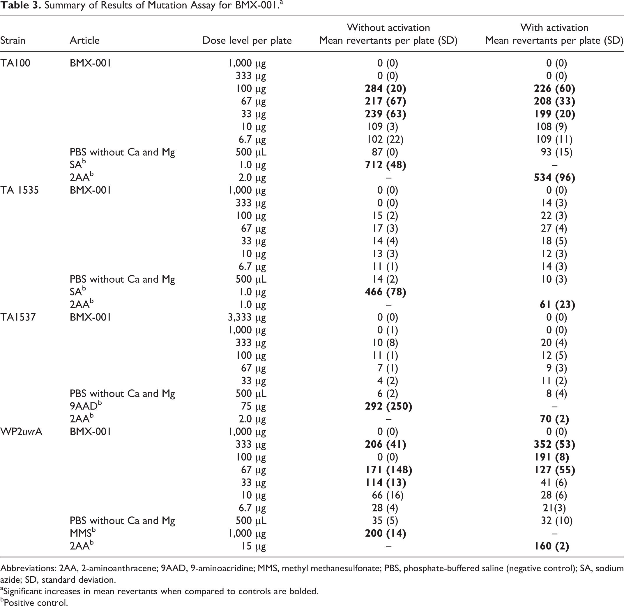
Abbreviations: 2AA, 2-aminoanthracene; 9AAD, 9-aminoacridine; MMS, methyl methanesulfonate; PBS, phosphate-buffered saline (negative control); SA, sodium azide; SD, standard deviation.
aSignificant increases in mean revertants when compared to controls are bolded.
bPositive control.
In the retest of the mutagenicity assay, positive mutagenic responses (2.9- to 9.1-fold maximum increases) were observed with tester strain TA98 in the presence and absence of S9, TA100 in the presence of S9, and WP2 uvrA in the absence of S9 (Table 4). Under the conditions of this study, BMX-001 was concluded to be positive with tester strains TA98, TA100, and WP2 uvrA in the bacterial reverse mutation assay.
Retest of Mutagenicity Assay for BMX-001.a
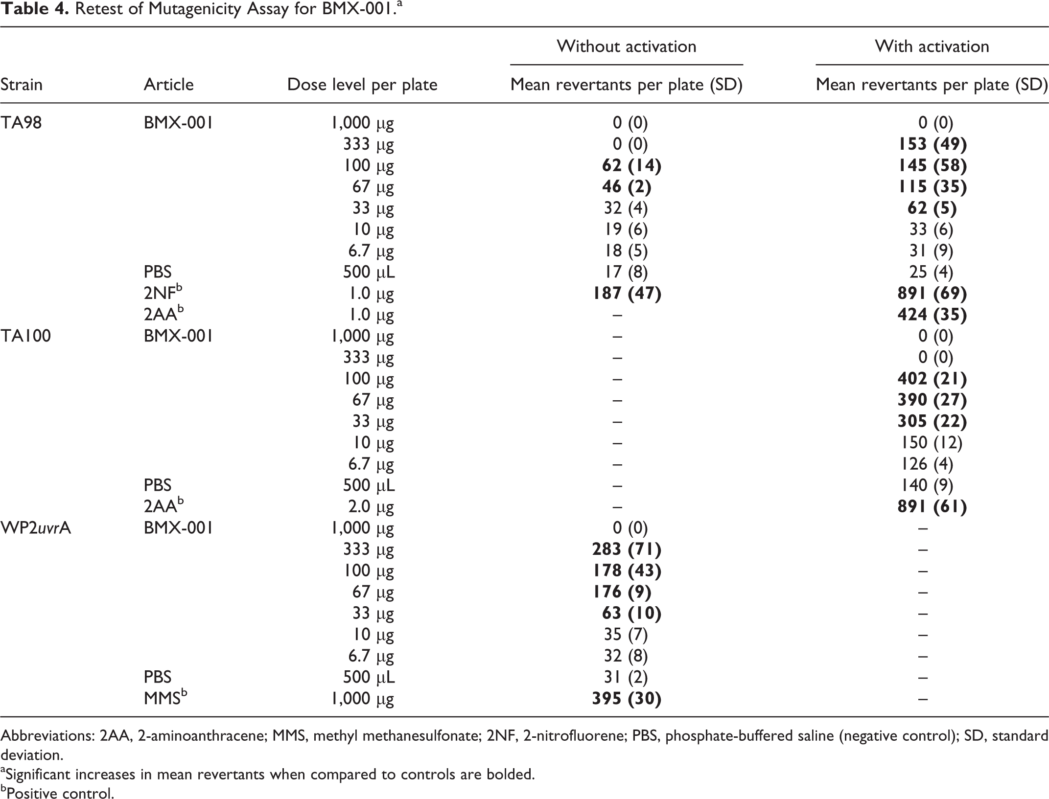
Abbreviations: 2AA, 2-aminoanthracene; MMS, methyl methanesulfonate; 2NF, 2-nitrofluorene; PBS, phosphate-buffered saline (negative control); SD, standard deviation.
aSignificant increases in mean revertants when compared to controls are bolded.
bPositive control.
In vitro mouse lymphoma assay
In the preliminary toxicity assay, the maximum concentration in the treatment medium was 500 μg/mL (the limit dose for the assay). No visible precipitate was observed to be present in the treatment medium at the beginning or end of the treatment. Selection of concentrations for the mutagenesis assay was based on observations of reduction in suspension growth relative to the solvent control. Suspension growth relative to the solvent control at 500 μg/mL was 42% for the nonactivated cultures with a 4-hour exposure, 8% for the S9-activated cultures with a 4-hour exposure, and 0% for the nonactivated cultures with a 24-hour exposure.
Based on the results of the preliminary toxicity assay, the concentrations treated in the initial mutagenesis assay (Table 5) ranged from 25 to 500 μg/mL for the nonactivated and S9-activated cultures with a 4-hour exposure. No visible precipitate was present in the treatment medium at the beginning or end of the treatment. No cloned cultures in the absence of S9 exhibited induced mutant frequencies (IMFs) ≥90 mutants per 106 clonable cells. Cloned cultures at concentrations ≥100 μg/mL in the presence of S9 exhibited IMFs ≥90 mutants per 106 clonable cells.
Initial L5178Y/TK+/− Mouse Lymphoma Assay (4-Hour Exposure) With and Without Metabolic Activation.
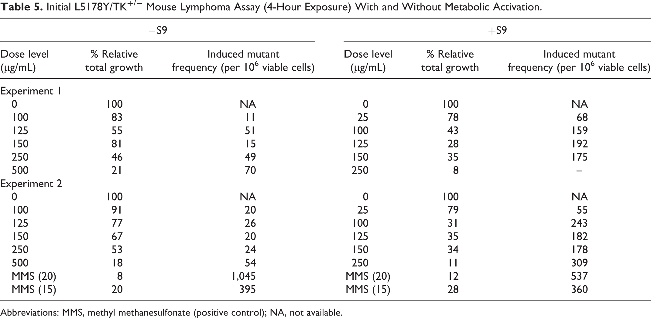
Abbreviations: MMS, methyl methanesulfonate (positive control); NA, not available.
Based on the results of the preliminary toxicity assay, the concentrations treated in the extended mutagenesis assay ranged from 75 to 500 μg/mL for the nonactivated cultures with a 24-hour exposure (Table 6). No visible precipitate was present in the treatment medium at the beginning or end of the treatment. The concentrations chosen for cloning were 75, 100, 150, 200, and 250 μg/mL. One cloned culture at a concentration of 250 μg/mL exhibited an IMF ≥90 mutants per 106 clonable cells; however, the average IMF at that concentration was 76 mutants per 106 clonable cells. There was no concentration-related increase in mutant frequency.
Extended L5178Y/TK+/− Mouse Lymphoma Assay (24-Hour Exposure) Without Metabolic Activation.
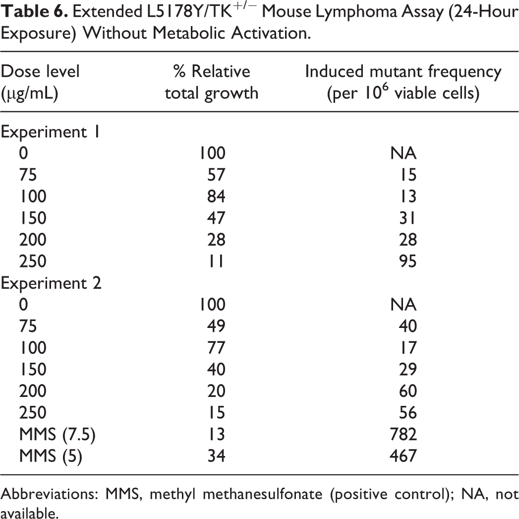
Abbreviations: MMS, methyl methanesulfonate (positive control); NA, not available.
The trifluorothymidine-resistant colonies for the cloned test article-treated cultures with a 4-hour exposure in the presence of S9 and the positive and solvent control cultures for each exposure group were sized according to diameter over a range from approximately 0.2 to 1.1 mm. The data on colony size distributions showed both an increase in the frequency of small (≤0.63 mm) and large colonies (>0.63 mm) when the test article-treated cultures were compared to the solvent control cultures (Figure 2). An increase in the frequency of small colonies is indicative of damage to multiple loci on chromosome 11 in addition to functional loss of the thymidine kinase (TK) locus. An increase in large-colony mutants is indicative of localized damage in the form of a point mutation or small deletion within the TK locus. The colony sizing for the methyl methanesulfonate and 7,12-dimethylbenz[a]anthracene-positive controls yielded the expected increase in small colonies (verifying the adequacy of the methods used to detect small-colony mutants) and large colonies.
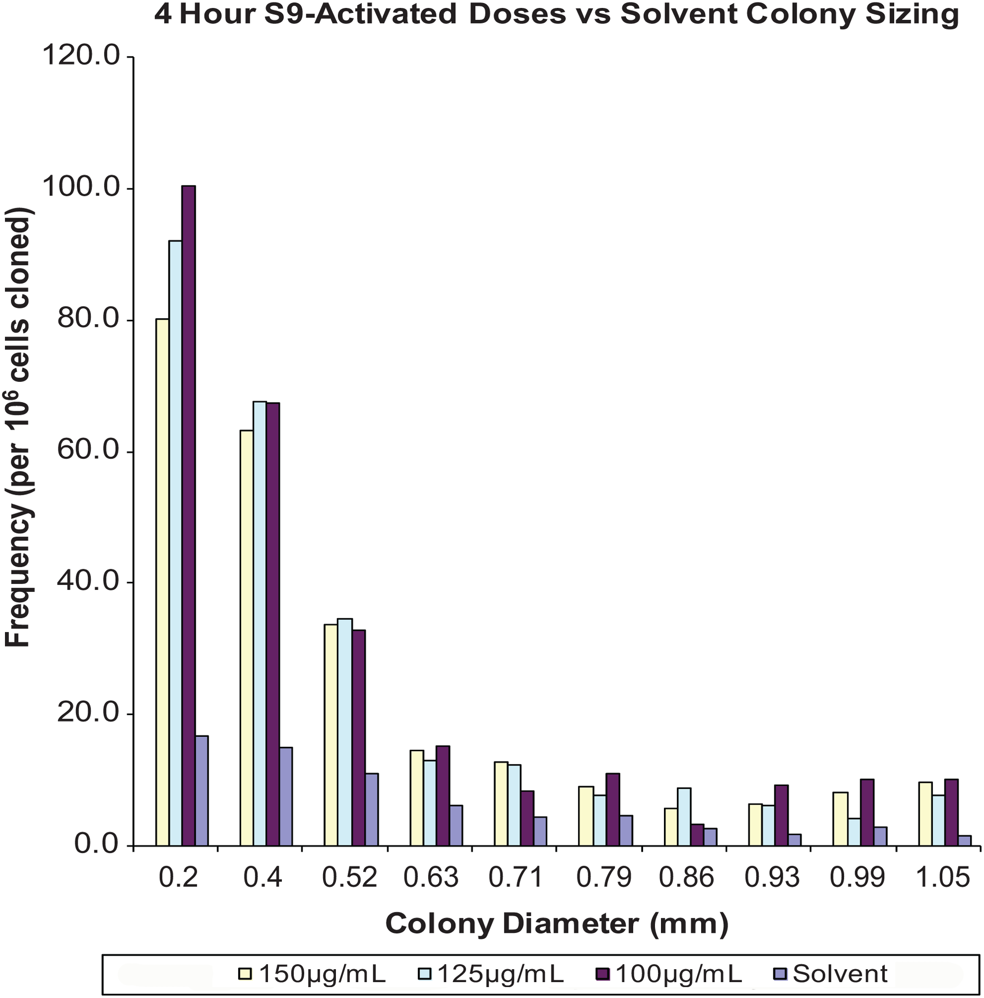
Initial mutagenesis assay with BMX-001 with activation, 4-hour exposure colony size distribution in the presence of metabolic activation (S9-activated doses compared with solvent control).
Under the conditions of this study, BMX-001 was concluded to be negative following 4- and 24-hour exposures in the absence of S9 activation and positive following a 4-hour exposure in the presence of S9 activation in the L5178Y/TK+/− mouse lymphoma assay.
In vivo micronucleus assay in mice
No mortality occurred at any dose level during the study. Clinical signs were limited to piloerection observed at a dose level of 1.0 mg/kg. The results are summarized in Table 7. No appreciable reductions in the PCE–EC ratio in the treated groups were seen when compared to vehicle controls, indicating that the test did not induce cytotoxicity. No statistically significant increase in the incidence of MnPCEs in the test article-treated groups was observed relative to the negative control group. The positive control induced a statistically significant increase in the incidence of MnPCEs. The number of MnPCEs in the vehicle control groups did not exceed the historical control range. Under the conditions of this study, the administration of BMX-001 at doses up to 1.0 mg/kg was concluded to be negative in the micronucleus assay.
Summary of Bone Marrow Micronucleus Assay in Mice.
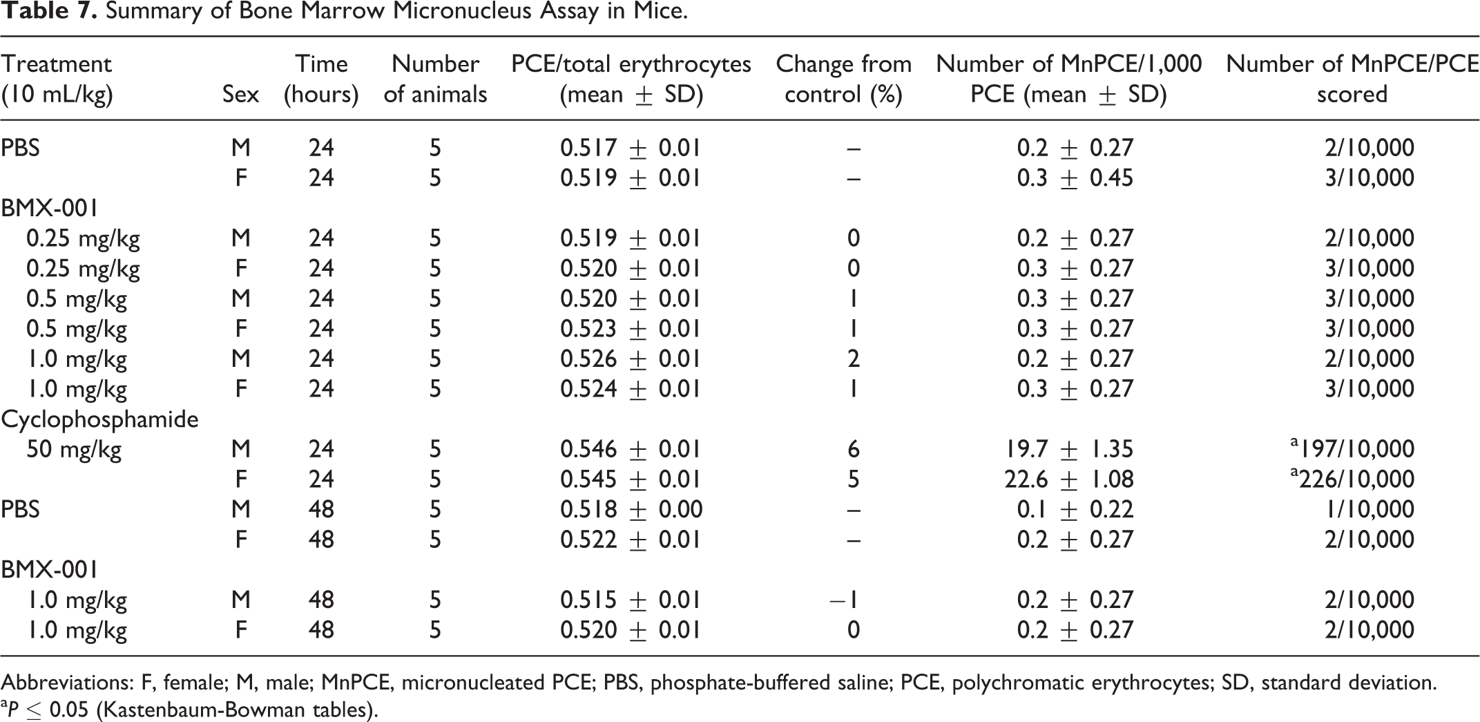
Abbreviations: F, female; M, male; MnPCE, micronucleated PCE; PBS, phosphate-buffered saline; PCE, polychromatic erythrocytes; SD, standard deviation.
a P ≤ 0.05 (Kastenbaum-Bowman tables).
Comet assay
Based on the results in the man genotoxicity study battery of 3 studies, and in accordance with ICH S2(R1), the comet assay was undertaken to clarify potential human risks. There were no changes observed in final body weight or body weight gain in animals administered BMX-001. Following dose administration on days 1 and 2, animals administered the high dose (30 mg/kg) were noted to exhibit decreased movement or lethargy. There was no increase in DNA damage in the livers of male mice administered BMX-001 when compared to the concurrent control group (Table 8). Increases in DNA damage were observed in animals administered with the positive control article, EMS. BMX-001 was concluded to be negative for genotoxicity in the comet assay.
Summary of Comet Assay Results in Male Mice.
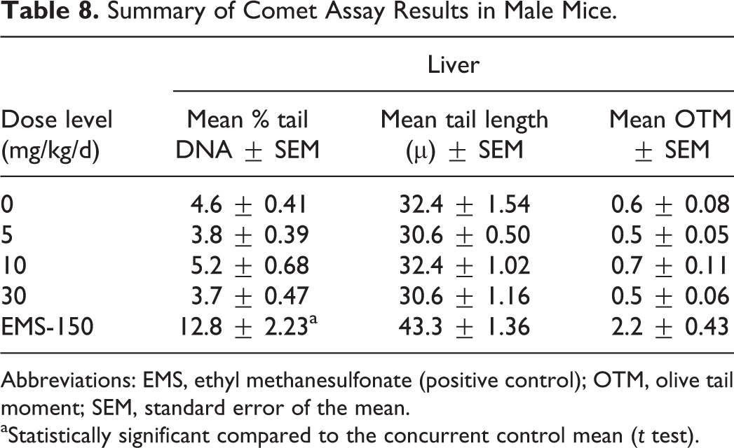
Abbreviations: EMS, ethyl methanesulfonate (positive control); OTM, olive tail moment; SEM, standard error of the mean.
aStatistically significant compared to the concurrent control mean (t test).
Local Tissue Tolerance
Dermal irritation study in rabbits
No mortality or clinical signs were recorded during the course of the study. BMX-001 was found to be nonirritating dermally as no erythema or edema was reported. The primary irritation index was 0.
Guinea pig maximization test
No mortality was observed, and all animals appeared normal throughout the course of the main study. No biologically relevant differences in the final body weights of the animals in the main study occurred, except in a single animal that experienced transient weight loss between day 1 and the final body weight at challenge but was comparable to other animals by challenge and rechallenge.
Based on the scores of the dose-range finding study, a challenge dose of 0.1% was selected. Scores of 0 to 3 were observed in the vehicle control animals and the test article-treated animals at 24 or 48 hours following the challenge. Because some equivocal responses were observed in the initial challenge, a rechallenge was performed on day 30. The test article at 0.01% was administered to a naive site on the right mid flank area of each test article group animal and to a group of 5 naive animals (right mid flank area). The left mid flank areas of both groups received the vehicle (Base PCCA Lipoderm).
Under the conditions of this study and based on the results, an intradermal induction of 0.1% BMX-001 in Base PCCA Lipoderm and a topical induction of BMX-001 at 1.5% followed by a topical challenge at 0.1% to guinea pigs did not elicit a positive response in the test article-treated animals at 24 or 48 hours following the challenge. A rechallenge was performed with the test article at 0.01% in the vehicle. A topical rechallenge at 0.01% to guinea pigs showed a moderate (grade 3) elicitation response at 24 hours decreasing to a mild (grade 2) elicitation response at 48 hours.
Eye irritation in rabbits
There were no treatment-related clinical signs noted. No mortality was observed in any animal dosed during the study. No corneal, iris, or conjunctival irritation was observed in the untreated eye of the rabbit at any time point. No irritation of the cornea was observed in the treated eyes of the rabbits throughout the study. Conjunctival irritation (scores of 1 for redness) was observed at 1 hour in 2 of 3 animals. All irritation was resolved by 24 hours, and the study was terminated following the 72-hour scoring interval. BMX-001 (dosed at 0.1% in saline) was found to be nonirritating to the eye.
Safety Pharmacology Studies
Cardiovascular function in conscious telemetered male cynomolgus monkeys
All monkeys maintained sinus rhythms throughout the study. No consistent, dose-related ECG morphologic changes were detected on any traces. Cardiovascular data collected by telemetry revealed a transient increase in heart rate in all treated monkeys at 30 minutes postdose. The increased heart rate occurred in a dose-dependent manner and was modestly increased at dose levels (6 and 9 mg/kg) above those intended for clinical use (0.067 mg/kg). Subcutaneous administration of BMX-001 at up to 9 mg/kg did not have any toxicologic effects on cardiac rate or rhythm or ECG morphology in monkeys in this study.
Systemic Toxicity
Maximum tolerated dose studies in mice and monkeys
There were 4 treatment-related deaths in mice following intravenous administration of 10 mg/kg BMX-001 during the dose-escalation phase of the study. No other early deaths were seen in either species. No treatment-related effects were seen on body weights, clinical chemistry values, hematology parameters, organ weights, or during gross necropsy. Clinical signs of toxicity were seen in both mice and monkeys. In mice, clinical findings including piloerection, decreased activity, abnormal stance and gait, labored respiration, body twitching, sudden intermittent movement, and abnormal yellow color at the injection sites were noted when BMX-001 was administered once at 2, 3, and 10 mg/kg and daily at 2/1.5 mg/kg/d for 5 consecutive days. High-dose mice were initially dosed at 2 mg/kg/d. However, because of mortality, the dose level was lowered to 1.5 mg/kg/d starting on day 4 for males and day 3 for females. No mortality was seen once doses were lowered to 1.5 mg/kg/d, but severe clinical signs of toxicity were still noted. Monkeys showed clinical signs of toxicity, including decreased activity, salivation, ataxia, and pink skin in the upper lip, cheeks, and nose, when dosed intravenously with BMX-001 once at 1.5, 2.0, and 2.5 mg/kg and daily at 2.0 mg/kg for 5 consecutive days. Clinical signs were dose dependent with all animals returning to normal during the recovery period. Based on these findings, the MTD for BMX-001 was determined to be 1.0 and 2.0 mg/kg/d in mice and monkeys, respectively, based on daily maintenance dosing.
A 5-week subcutaneous study with BMX-001 in mice
One high-dose female dosed on day 1 at 15 mg/kg of BMX-001 was found dead on nondosing day 2. There were no adverse clinical findings observed during the study. Common findings on dosing and nondosing days in treated animals included abnormal coloration of the injection sites (yellow to dark yellow) and amber-colored urine. The number of animals and the frequency of occurrence of abnormal coloration at the injection sites on nondosing days were observed to be directly proportional to dose levels. On dosing days, some animals exhibited mildly decreased activity and partially closed eyes, while a few animals had small amount of rough hair coat over the entire body. During recovery, some animals were observed to exhibit persistent yellow injection sites. However, by days 54 to 56 and days 48 to 56, all males (10 of 10) and all surviving females (9 of 9) appeared normal, respectively.
There were no test article-related effects observed on body weights, food consumption values, ophthalmological findings, or hematology parameters. There were statistically significant changes in some clinical chemistry parameters. At day 35, sodium levels in males administered 2 or 4 mg/kg/dose maintenance doses were statistically higher by 2% and 3.5%, respectively, when compared to controls. At day 55 in females administered 4 mg/kg/dose BMX-001, the alanine aminotransferase level was noted to be 286% higher, whereas the blood urea nitrogen value was 25% lower. The level of alanine aminotransferase was above the upper limit of the reference range, whereas all the other parameters were within historical limits. There were test article-related gross necropsy findings at terminal sacrifice on day 35. The most common finding was the yellow discoloration of the injection sites in mid- (12/2 mg/kg/dose) and high-dose animals (15/4 mg/kg/dose). There were no test article-related effects on absolute or relative organ weights.
Microscopic findings for 1 high-dose (15 mg/kg/dose loading dose) female that was found dead on day 2 included minimal to marked individual lymphocyte necrosis in the thymus, spleen, mandibular, and mesenteric lymph nodes and mild to moderate individual cell necrosis in the bone marrow. Necrosis in lymphoid tissues and bone marrow were attributed to nonspecific agonal stress. The actual cause of death was undetermined. No correlative microscopic findings were identified for test article-related yellow discoloration at the injection site in all 15/4 mg/kg/dose males and females at terminal sacrifice on day 35.
At terminal sacrifice, test article-related microscopic findings were noted in the injection sites, liver, and spleen (Table 9). At the injection site, 15/4 mg/kg/dose animals were observed to have increased acanthosis, dermal fibrosis, and degeneration/regeneration and/or mononuclear cell inflammation of the panniculus muscle. Minimal to marked acanthosis was noted in all 15/4 mg/kg/dose animals but was only minimal in 2 control males. Minimal to mild dermal fibrosis was not noted in controls but was seen in 5 males and 2 females at the 15/4 mg/kg/dose. Within the panniculus muscle, minimal to marked muscle degeneration/regeneration (characterized by shrinkage and loss of myofibers, individualization and swelling of myofibers, centralized myofiber nuclei, and/or basophilic cytoplasm) was seen in all 15/4 mg/kg/dose males and females but was only minimal in 3 control females. Degeneration was often accompanied by mononuclear cell inflammation in the panniculus muscle, ranging from minimal to moderate. These injection site findings were considered nonadverse because they did not impact significantly on the overall health or well-being of the animals on this study. In the liver, scattered Kupffer cells contained minimal amounts of brown granular pigment. In addition, there was a small but noteworthy average decrease in glycogen-type vacuolation in the 15/4 mg/kg/dose group overall when compared to the controls. The significance of this finding is uncertain; decreased hepatic glycogen stores might occur with decreased food consumption, increased metabolic state, or a longer period of fasting before necropsy. A subtle increase in incidence and severity of extramedullary hematopoiesis in the spleen was also noted in high-dose animals. The reason for this increase was not apparent; no differences were noted in the bone marrow. The findings discussed above were all considered nonadverse as they did not significantly impact the overall health or well-being of the animals on this study.
Select Histopathology Findings From 5-Week Mouse Study Following Day 35 Sacrifice.
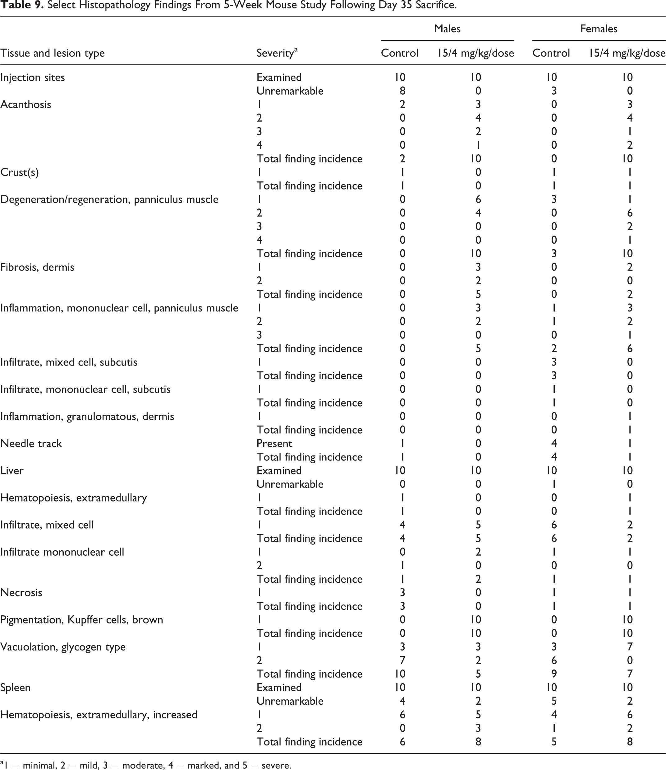
a1 = minimal, 2 = mild, 3 = moderate, 4 = marked, and 5 = severe.
Dose-dependent increases in plasma levels of BMX-001 as characterized by area under the curve and Cmax were seen on days 1 and 34. Levels were lower following the last administration (day 34) when compared to the first administration on day 1. When compared to males, lower levels were seen in females after the first and last administration. Mean plasma half-lives ranged from 0.81 to 2.62 hours for males and from 1.44 to 3.57 hours for females. The calculated TK parameters are shown in Table 10.
Pharmacokinetic Parameters for the Mouse Repeat-Dose Study.
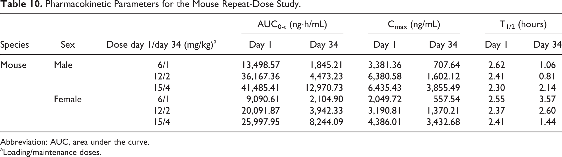
Abbreviation: AUC, area under the curve.
aLoading/maintenance doses.
In conclusion, BMX-001 administered subcutaneously twice weekly in mice for a minimum of 5 consecutive weeks starting with a single loading dose on day 1 followed by maintenance doses on subsequent dosing days caused nonadverse clinical signs in animals given ≥1 mg/kg/dose of BMX-001. The death of 1 high-dose (15 mg/kg/dose loading dose) female on day 2 may be attributed to biological variation. Based on the findings discussed above, including mortality and increased alanine aminotransferase in 15/4 mg/kg/dose females, this study identified a no observed adverse effect level (NOAEL) in mice of 12 mg/kg/dose for the loading dose and 2 mg/kg/dose (0.57 mg/kg/d) for the maintenance dose.
A 5-week subcutaneous study with BMX-001 in monkeys
There were no unscheduled deaths that occurred during the study. All animals survived until their terminal sacrifice on day 8 (group 6 satellite), day 38 (groups 1-5), or day 52 (groups 1 and 5 recovery). There were clinical findings during the 5-week treatment period that were test article related. On day 1, groups 5 and 6 monkeys given 6 mg/kg of BMX-001 (high loading dose) exhibited decreased activity, dilated pupils, and partially closed eyes. Also noted on day 1 and following administration of subsequent maintenance doses were amber-colored urine and/or yellow discoloration of the injection sites. The yellow discoloration of the injection sites persisted throughout the study (including through recovery) but had no histologic correlate other than tissue pigmentation.
There were no test article-related effects on mean body weights. However, there were test article-related effects on mean food consumption values that were observed during the study. On several occasions, lower food consumption values with statistical significance were noted in groups 4 (3/1 mg/kg/dose) and 5 (6/2 mg/kg/dose) animals when compared to controls. There were no test article-related ophthalmological and electrocardiology findings. There were no test article-related effects on hematology values, EC morphology, coagulation, and clinical chemistry parameters on days 8, 38, and 52.
At days 38 and 52 necropsy, green mandibular and tracheobronchial lymph nodes, green discolored subcutis at the injection site, and green and/or prominent mesenteric lymph nodes were noted in groups 4 (3/1 mg/kg/dose) and 5 (6/2 mg/kg/dose) animals. At day 38 necropsy, the mean absolute uterus weight and its relative ratios with mean body weight values and brain weight values were lower with statistical significance in 6/2 mg/kg/dose females when compared to controls but returned to normal by day 52 (data not shown).
Microscopic evaluation indicated that test article-related minimal to mild brown pigment accumulation was noted in multiple tissues at day 38 and interpreted as injected test material pigment (Table 11
Select Findings From a 5-Week Monkey Study Following Day 38 Sacrifice.
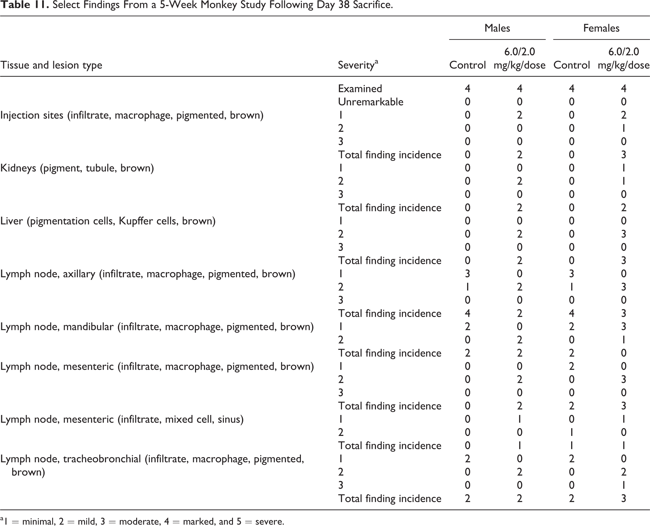
a1 = minimal, 2 = mild, 3 = moderate, 4 = marked, and 5 = severe.
Extensive plasma and tissue toxicokinetic analyses were performed for all monkeys that were exposed to BMX-001 following a single subcutaneous injection of a loading dose of BMX-001 at 0.3, 1, 3, and 6 mg/kg (day 1) and following the subcutaneous injection of a maintenance dose of BMX-001 at 0.1, 0.3, 1, and 2 mg/kg (day 37; Table 12). Absorption of BMX-001 following a subcutaneous injection was fairly rapid as supported by a Tmax time of 0.5 hours (the first blood sampling time) in the majority of animals. Exposure appeared to be dose proportional to slightly greater than dose proportional across 0.1 to 6 mg/kg doses of BMX-001 administered subcutaneously. Plasma T1/2,e was approximately 9 hours following the highest dose level of 6 mg/kg and approximately 1 hour following the lower dose levels of 0.1 and 0.3 mg/kg. There did not appear to be any gender-related differences with regard to exposure or elimination in cynomolgus male and female monkeys. The exposure and elimination of BMX-001 in animals administered 12 maintenance doses of 0.3 and 1 mg/kg after 37 days were similar to that exposure and elimination in animals administered a loading dose of 0.3 and 1 mg/kg on day 1.
Pharmacokinetic Parameters for the Monkey Repeat-Dose Study.
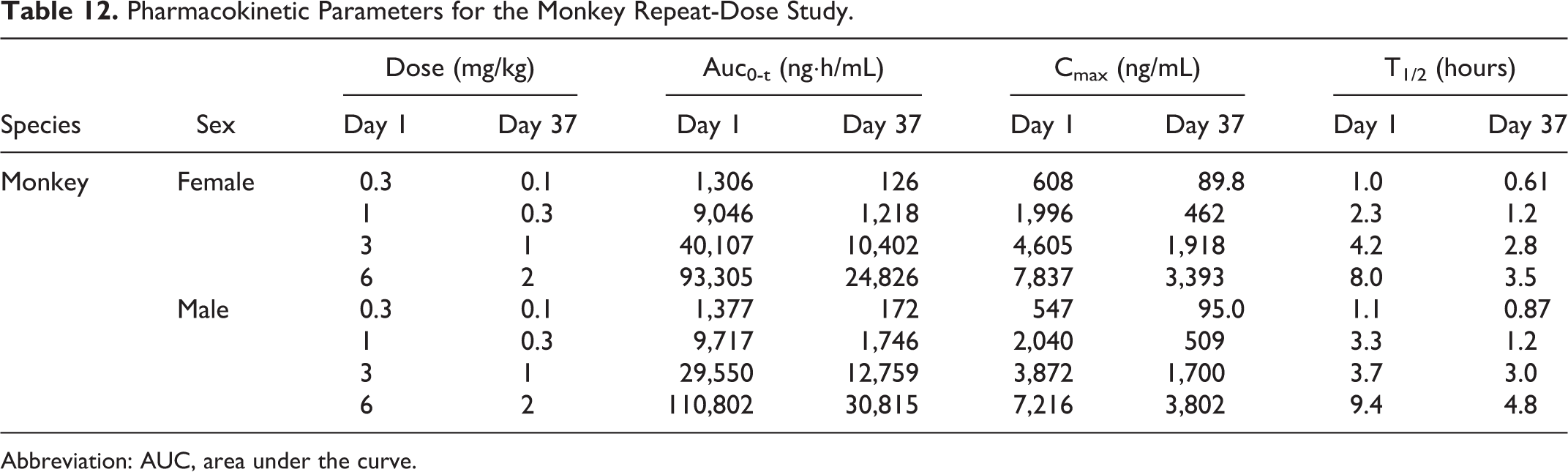
Abbreviation: AUC, area under the curve.
LC-MS/MS analysis detected the presence of BMX-001 in target tissues previously identified in first-generation Mn porphyrins including the brains, livers, kidneys, lungs, mandibular lymph node, prostates, rectums, salivary glands, tongues, and urinary bladders collected at necropsies on days 8, 38, and 52 (Table 13). The increase in levels of BMX-001 in all tissues examined was dose dependent, with the highest levels seen in the livers and kidneys of both males and females. Levels of BMX-001 decreased close to basal levels for most tissues during recovery (day 52), but the levels were still higher in the livers and kidneys as well as the mandibular lymph nodes when compared to all other tissues examined.
Tissue Levels of BMX-001 in the Monkey.
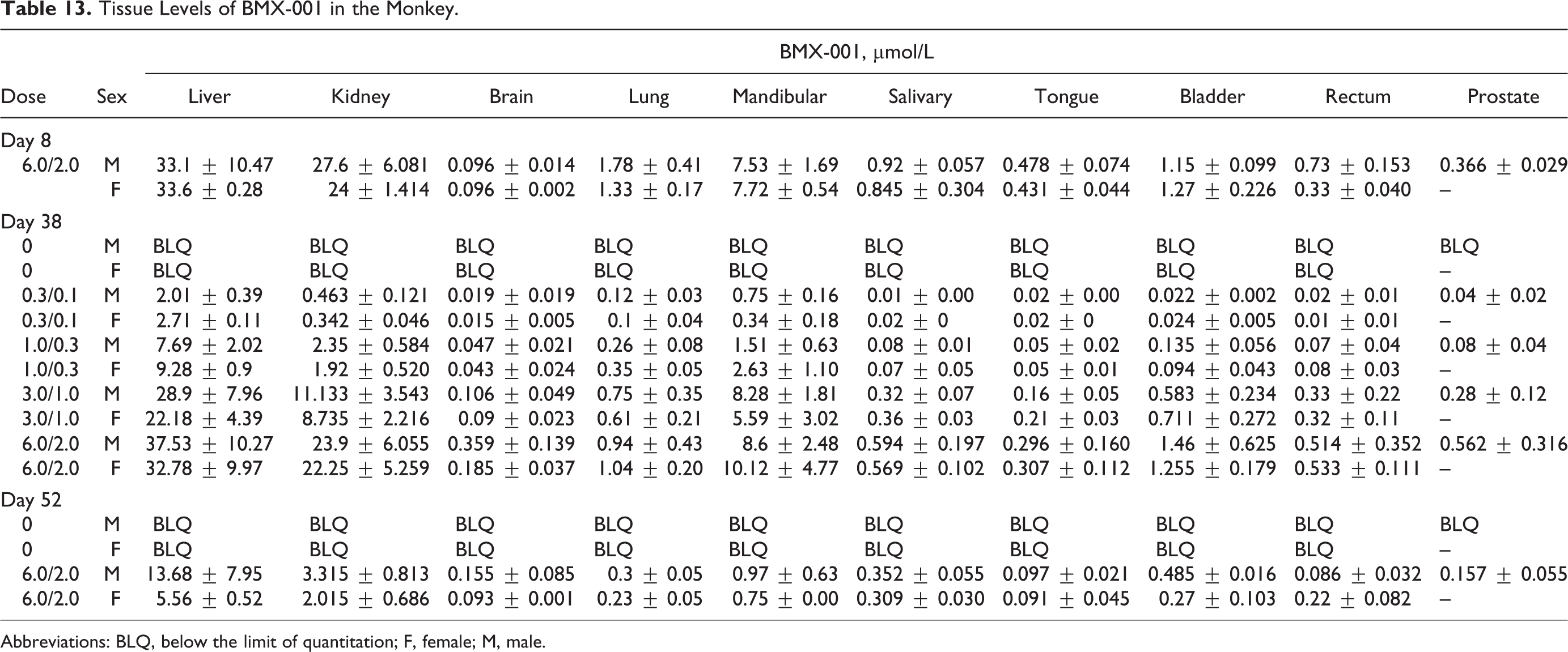
Abbreviations: BLQ, below the limit of quantitation; F, female; M, male.
In conclusion, cynomolgus monkeys given BMX-001 every 3 days by subcutaneous injection for 5 consecutive weeks followed by a 2-week recovery period did not show any clinical signs of intolerance or toxicity. There were no adverse effects observed with regard to the development of any clinical signs, food consumption values, body weights, ophthalmology findings, electrocardiology data, clinical pathology parameters, gross necropsy, and histopathology in any of the dose groups in this study. Based on the absence of adverse effects in the high-dose groups (groups 5 and 6), the NOAEL for monkeys was determined to be 6.0 mg/kg/dose for loading dose and 2.0 mg/kg/dose (0.7 mg/kg/d) for maintenance dose.
Discussion
BMX-001 is a second-generation metalloporphyrin compound that is being developed as a potential therapeutic agent for the treatment of HGG and head and neck cancer. The nonclinical safety for the first-generation compound of this class has been described elsewhere. 13 Here, we have described the nonclinical safety and toxicokinetic profile of BMX-001.
To determine the potential toxicity of BMX-001 prior to human use, a battery of nonclinical studies were performed. BMX-001 was negative for dermal and eye irritation in rabbits and did not alter cardiovascular function in nonhuman primates. When administered intravenously for a period of up to 5 days, BMX-001 was tolerated at doses up to 1.0 and 2.0 mg/kg in mice and nonhuman primates, respectively. When exposure of animals to BMX-001 was extended to 5 weeks using subcutaneous injections, mortality was seen in mice at the highest dose (15 mg/kg/dose loading dose and 4 mg/kg/dose maintenance dose). At loading/maintenance doses of up to 12/2 mg/kg/dose in mice and 6/2 mg/kg/dose in monkeys, treatment-related findings were noted at the injection sites of nonhuman primates and mice and in the livers and spleens of mice. With the exception of the effects seen at the highest dose in mice, none of these were considered to be adverse. The NOAEL for BMX-001 administered every 3 days for 5 weeks was 12 mg/kg/dose loading dose and 2 mg/kg/dose maintenance dose in mice and 6 mg/kg/dose loading dose and 2 mg/kg/dose maintenance dose in nonhuman primates. In the nonhuman primate, BMX-001 is rapidly taken up primarily in the liver, then released for further redistribution into other tissues, and appears to be eliminated via the lymphatic and renal systems.
The genotoxic potential of BMX-001 was initially evaluated in 3 assays described in ICH S2(R1) as the standard test battery for determining the genotoxic potential of pharmaceutical candidates. 22 The results from the initial battery of genotoxic assays were mixed. Two in vitro assays, a bacterial reverse mutation assay and a mouse lymphoma assay, provided positive genotoxicity findings for BMX-001. However, the micronucleus in vivo assay in mice was negative for genotoxicity. To further evaluate the genotoxicity of BMX-001, including potential biological relevance of the positive findings in the in vitro assays, a second in vivo genotoxicity study (comet assay in mice) was conducted. The negative results of the comet assay in mice provided supporting evidence for BMX-001 being negative for genotoxicity in vivo. Although in vitro assays are extremely useful when determining the genotoxicity of a compound, comparative trials have shown conclusively that they can generate false positives. 22 In vivo assays have the advantage of taking into account the absorption, distribution, and excretion of a compound, which are not factors in in vitro tests but are extremely relevant to human exposure. 22 For BMX-001, this appears to be the case, and it is concluded in accordance with ICH S2(R1) 22 and based on the weight of the evidence that BMX-001 does not present a risk of genotoxicity to humans.
Safe starting doses for the proposed clinical studies in patients with HGG and head and neck cancer were based on the 5-week study performed in nonhuman primates as it was the more sensitive species. In the 5-week study, no adverse effects were seen in monkeys administered a loading dose of 6 mg/kg followed by maintenance doses of 2 mg/kg BMX-001 every 3 days. For the clinical study, a safe starting (loading) dose for a 70-kg person can be achieved by dividing the kg dose by a value of 30, which accounts for the extrapolation of monkeys to humans (n = 3) and for differences between individual humans (n = 10). Thus, the safe starting loading dose for BMX-001 would be 6 mg/kg/dose (70 kg body weight)/3(10) = 12 mg/dose loading. Safe consecutive (maintenance) doses would then be 2 mg/kg/dose (70 kg body weight)/3(10) = 4.7 mg/dose.
Footnotes
Author Contributions
S. Gad contributed to conception and design, contributed to acquisition, analysis, and interpretation, and critically revised the manuscript. D. Sullivan contributed to conception and design, contributed to acquisition, analysis, and interpretation, drafted the manuscript, and critically revised the manuscript. I. Spasojevic, C. Mujer, and C. Spainhour contributed to conception and design, contributed to analysis and interpretation, and critically revised the manuscript. J. Crapo contributed to conception and design, contributed to acquisition, and critically revised manuscript. All authors gave final approval and agree to be accountable for all aspects of work ensuring integrity and accuracy.
Declaration of Conflicting Interests
The author(s) declared no potential conflicts of interest with respect to the research, authorship, and/or publication of this article.
Funding
The author(s) disclosed receipt of the following financial support for the research, authorship, and/or publication of this article: This work was supported by BioMimetix JV, LLC.