
Abstract
Insect repellent N,N-diethyl-m-toluamide (DEET) and sunscreen oxybenzone have shown a synergistic percutaneous enhancement when applied concurrently. Both compounds are extensively metabolized in vivo into a series of potentially toxic metabolites: 2 metabolites of DEET, N,N-diethyl-m-hydroxymethylbenzamide (DHMB) and N-ethyl-m-toluamide (ET), and 3 metabolites of oxybenzone, 2,4-dihydroxybenzophenone (DHB), 2,2-dihydroxy-4-methoxybenzophenone (DMB), and 2,3,4-trihydroxybenzophenone (THB). In this study, the metabolites were extensively distributed following intravenous and topical skin administration of DEET and oxybenzone in rats. Combined application enhanced the disposition of all DEET metabolites in the liver but did not consistently affect the distribution of oxybenzone metabolites. The DHMB appeared to be the major metabolite for DEET, while THB and its precursor DHB were the main metabolites for oxybenzone. Repeated once-daily topical application for 30 days led to higher concentrations of DEET metabolites in the liver. Hepatoma cell studies revealed a decrease in cellular proliferation from all metabolites as single and combined treatments, most notably at 72 hours. Increased accumulation of DHMB and ET in the liver together with an ability to reduce cellular proliferation at achievable plasma concentrations indicated that simultaneous exposure to DEET and oxybenzone might have the potential to precipitate adverse effects in a rat animal model.
Introduction
Topical insect repellent and sunscreen products have become an integrated part of summer routines for the general public in developed countries, due primarily to a conscious awareness of the health threats from vector-borne diseases and nonessential sun exposure. These consumer-care products are the most practical, cost effective, and well-accepted choice of defense, and there are a variety of preparations commercially available to consumers including sprays, lotions, aerosols, and cloth wipes. Concurrent skin application of sunscreen preparations along with insect repellents has been prevalent in North America since 1999 when mosquito-transmitted West Nile virus was first detected in the continent.
Numerous chemical repellent and sunscreen ingredients are utilized for commercial civil use; DEET (N,N-diethyl-m-toluamide) and oxybenzone are 2 principal repellent and sunscreen substances, respectively. Their repellency and UV-blockage efficacy have been investigated and documented. 1 ,2 Interactions between active repellent and sunscreen compounds, as well as subsequent percutaneous disposition of these substances after topical skin application, have also been studied and reported. 3 –5
Designated as topical products, repellents and sunscreens remain on the skin surface to achieve their protection efficacy. Percutaneous permeation and systemic absorption of the active ingredients are considered neither desirable nor productive. However, DEET and oxybenzone have been shown to permeate across the stratum corneum into the general circulation after topical skin application. 6,7 In vitro studies have demonstrated permeation synergy between DEET and oxybenzone when both substances were applied simultaneously. 3,4 The dose applied, formulation type, and application method all influenced the rate and extent of permeation, as well as the interaction between the 2 active compounds.
The pharmacokinetics and toxicology of DEET and oxybenzone applied alone have been reported in some previous studies. 8-10 Since concurrent skin application of repellents and sunscreens was rare in the past, studies were only carried out to assess concurrent use of DEET or oxybenzone in association with several specialty chemical substances pertinent to military and/or farming usages. 11,12 In addition, loss of Sun Protection Factor (SPF) of the sunscreens has been reported from mixing DEET-based repellents and commercial sunscreen products. 13,14 With increasing administration of DEET-based repellents together with oxybenzone-containing sunscreens, it has become critical and essential to understand the in vivo disposition profiles of the 2 substances. No studies have indicated whether the documented adverse effects of DEET and oxybenzone resulted from the parent compounds or their subsequent metabolites.
Individual users may apply different doses of repellents and sunscreens at discretion, because there is no medically recommended dosing scheme available, and use conditions are vastly variable. In a 75-kg average adult, for example, a typical 5 mg/kg dose is equivalent to 3.75 mL of 10% DEET liquid spray or 7.5 g of 5% oxybenzone-containing sunscreen lotion. For continuous extensive applications, the topical dosing of DEET and oxybenzone could be much higher, as what was utilized in this study. Previous studies in Sprague-Dawley rats also observed Cmax values of DEET and oxybenzone at 0.5 µg/mL and 2.5 µg/mL, respectively. 15,16 Hence, the objective of this study was to determine the potential metabolite disposition of high-dose DEET and oxybenzone following intravenous and topical skin administration in a rat model. We also performed in vitro hepatoma cell experiments with metabolite concentrations between 0.1 and 10 µg/mL to assess potential toxicity resulting from enhanced disposition of these metabolites in the rat liver.
Materials and Methods
Materials and Reagents
Sodium Chloride Injection USP (0.9%) was obtained from Astra Zeneca Inc (Mississauga, Ontario, Canada) and used directly for intravenous injection. Anhydrous ethyl alcohol was purchased from Commercial Alcohols Inc (Brampton, Ontario, Canada) and Emulphor (ethoxylated castor oil) was purchased from Nihon Emulsion Co Ltd (Tokyo, Japan). Pure DEET and oxybenzone standards were obtained from Fluka Chemika GmbH (Buchs, Switzerland) and Riedel-de Haën GmbH (Seelze, Germany), respectively. Various formulations were prepared for the animal experiments without further purification.
For drug extraction and analysis, acetonitrile, methanol, and sodium hydroxide were purchased from Fisher Scientific (Fair Lawn, New Jersey). Glacial acetic acid was obtained from Mallinckrodt Specialty Chemical Company (Paris, Kentucky). Ammonium acetate was obtained from Aldrich Chemical Co, Inc (Milwaukee, Wisconsin). Nitric acid was purchased from LabChem Inc. (Pittsburgh, Pennsylvania). All solvents were of high-performance liquid chromatography (HPLC) grade and other chemicals were of Analytical Reagent grade. Deionized water was obtained from a Milli-Q Pure Water System (Nepean, Ontario, Canada).
For studies of cell viability, HyClone classical liquid media Minimum Essential Medium with Earle Balanced Salts (MEM/EBSS) was purchased from Thermo Fisher Scientific (Nepean, Ontario, Canada). Fetal bovine serum, pyruvate,
Animal Model
The Animal Use Protocol was approved by the University of Manitoba Fort Garry Campus Animal Use Protocol Management and Review Committee, and research was conducted according to the current guidelines published by the Canadian Council for Animal Care (CCAC). Fifty-seven Sprague-Dawley rats were used in the study, which were randomly divided into 8 study groups, that is 2 groups for intravenous administration (n = 6, 24 hours), 3 groups for single topical application (n = 5, 24 hours), and 3 groups for once-daily repeated application (n = 10, 30 days).
Rats (average body weight of 250 g) were obtained from the Central Animal Care Services, University of Manitoba. Animals were checked by a University veterinarian for general health conditions upon arrival at the Department of Zoology Animal Holding Facility. They were housed individually in holding and/or metabolism cages and provided food and water ad libitum. A 5-day quarantine period was provided to the study animals, allowing them to adapt to the environment before the actual experiment commenced.
Intravenous and Topical Study Samples
For intravenous administration, solutions of DEET (0.3%, w/v) and oxybenzone (0.1%, w/v) were prepared using a mixture of Emulphor:ethanol:water (2:5:3, v/v/v). 17 A total of 167 µL of DEET preparation and 500 µL of oxybenzone preparation were injected into the tail vein of the animals at 2 mg/kg DEET (group 1) and 2 mg/kg oxybenzone (group 2). 16
For single, 24-hour topical skin administration, DEET and oxybenzone standards were weighed and dissolved in anhydrous ethanol at a concentration of 120 mg/mL DEET and 48 mg/mL oxybenzone, either individually or in combination. Prior to administration, an area of 25 cm2 (5 × 5 cm) on the back of each study animal was shaved using an electric clipper. The 3 study doses were 100 mg/kg or 4 mg/cm2 DEET (group 3), 40 mg/kg or 1.6 mg/cm2 oxybenzone (group 4), and combined 100 mg/kg DEET/40 mg/kg oxybenzone (group 5). 16 A total of 250 µL of test solution were measured and applied onto the skin surface using a pipette. The solution was carefully spread over the shaven skin area with a disposable pipette tip, and sufficient time for ambient evaporation of the solvent was allowed before the study rats were returned to their holding cages. The application was performed by an animal care technician in order to ensure the consistency of the drug administration and the accuracy of the study results.
For once-daily, 30-day repeated topical skin administration, DEET and oxybenzone standards were weighed and dissolved in 70% ethanol at a concentration of 100 mg/mL DEET and 12.5 mg/mL oxybenzone, either individually or in combination. The test samples were transferred to amber glass bottles and stored in a refrigerator for the duration of the experiment. 18 Prior to skin application, an area of 4 cm2 (2 × 2 cm) on the upper dorsal end of the study animals was shaved using an electric clipper. This area remained hair free by regular shaving in order to facilitate skin applications and observations over the 30-day study period. The upper dorsal surface was selected for dose application because it was extremely difficult for the study animals to reach this area of the body. The 3 study doses were 40 mg/kg or 10 mg/cm2 DEET (group 6), 5 mg/kg or 1.25 mg/cm2 oxybenzone (group 7), and combined 40 mg/kg DEET/5 mg/kg oxybenzone (group 8). 15 Similar application procedures were utilized as those of single skin application, and the weight of the study animals was measured periodically over the study duration so that the study doses were adjusted accordingly.
Animal Study Sample Collection
Blood samples (150 µL) were collected from the saphenous vein of the study animals using Microvette capillary collection tubes (Sarstedt AG & Co, Nümbrecht, Germany) at 30, 60, 90, 120, 150, 180, 240, 360, 480, 600, and 1400 minutes after intravenous injection and 24-hour single skin dosing. A bolus of 10 mL saline was injected subcutaneously after the 120-minute sample collection to compensate for the loss of blood volume. Plasma was separated by centrifugation of the samples at 13000g for 30 minutes and stored in labeled polypropylene tubes at –20°C until drug analysis. For 30-day repeated skin application, a 300-µL blood sample was collected from the saphenous vein of the rats using Microvette capillary collection tubes at 2, 4, 6, and 24 hours after the last dosing on day 30. The plasma was separated and stored in labeled polypropylene tubes at −20°C until drug analysis. 18
For topical skin studies, skin tape stripping was performed to evaluate percutaneous penetration of DEET and oxybenzone into various skin layers as well as the formation of metabolites. Before skin stripping at 24 hours, the application surface was swabbed once using cotton swabs saturated with 400 µL of acetonitrile. Upon complete drying of the skin, D-Squame stripping disks (CuDerm Corporation, Dallas, Texas) were consecutively applied to the site, gently pressed for 10 seconds, and then peeled off. For 24-hour study, 4 stripping disks were used at 7 hours on one particular area of the application site and 12 stripping disks were collected at 24 hours from a different area of the application site. For the 30-day study, 12 stripping disks were used at 24 hours after the last dosing. All tape strips were placed individually in labeled polypropylene tubes and stored at −20°C until drug analysis. 18
Liver, brain, and kidney specimens were also collected after study animals were euthanized at the end of the study. They were rinsed with saline solution to remove blood, air-dried, weighed, and stored in labeled polypropylene tubes at −80°C until drug analysis.
Urine and feces samples were collected from 10 to 24 hours during the single-dose 24-hour topical study and were stored in labeled polypropylene tubes at –20°C until drug analysis. 18
Concentration Measurement
Concentrations of primary metabolites of DEET and oxybenzone, including N,N-diethyl-m-hydroxymethylbenzamide (DHMB), N-ethyl-m-toluamide (ET), 2,4-dihydroxybenzophenone (DHB), 2,2-dihydroxy-4-methoxybenzophenone (DMB), and 2,3,4-trihydroxybenzophenone (THB), were measured using an HPLC assay developed and validated in our laboratory. 18 The method was capable of quantifying the compounds simultaneously using photodiode array detection.
To extract the metabolites from the biological samples, an automatic solid-phase extraction method was developed using a Zymark Rapidtrace SPE Workstation (Caliper Life Sciences, Hopkinton, Massachusetts). 15 Briefly, the separation was completed on a Waters Oasis MAX 3 cc (60 mg) extraction cartridge, by using acetonitrile, 0.03 mol/L ammonium acetate (pH 4.5) and water as preconditioning and washing solvents; 50 µL of plasma and urine samples were used for drug extraction, and 300 µL of methanol as the final elution solvent. The eluent was vortexed for 15 seconds, transferred to an HPLC vial, and 50 µL of the sample injected. Skin tape strips were dissolved in 1.5 mL of acetonitrile and extracted. Liver, kidney, brain, and feces samples were first homogenized in acetonitrile using an electronic homogenizer (Biospec Products, Bartlesville, Oklahoma). A portion of the homogenate was then subjected to solid-phase extraction, and 50 µL of the eluent was injected to the HPLC system for drug measurement.
Cell Culture Assay
Rat hepatoma cell line 1548 was grown in MEM/EBSS supplemented with 100 U/mL penicillin, 100 µg/mL streptomycin, 10% fetal bovine serum, 4 mmol/L
Cells were then trypsinized from 75 cm2 culture flask and seeded at a density of 3000 cells per well in 96-well plates. Cells were allowed to attach to the plates in an incubator containing 5% CO2 at 37°C for 12 hours. Following attachment, the medium in each well was removed and replaced with 100 µL fresh medium containing DHMB, ET, DHB, DMB, and THB at predetermined concentrations of 0.1 µg/mL, 1 µg/mL, and 10 µg/mL. Cells were then incubated in their respective media for 24, 48, and 72 hours. In order to maintain sufficient concentration and exposure time, medium in each well was renewed every 24 hours by adding 50 µL of fresh medium containing the same concentrations of DHMB, ET, DHB, DMB, and THB. To evaluate the effect of test substances on cell proliferation, Cell Proliferation Reagent WST-1 (1:10 dilution) was added into wells after 24, 48, or 72 hours of exposure. Plates were gently shaken for 5 minutes and the cultured cells were incubated in the presence of WST-1 reagent for 2 hours according to manufacturer’s recommendation. The absorbance of each well was measured using an ELx 808 Ultra Microplate Reader (BIO-TEK Instrument, Winooski, Vermont) at 450 nm. Wells with only the medium were utilized as the background control, while wells containing normal medium with the same amount of cells (3000 per well) were used as the no treatment (NT) control. An additional control with 1% methanol was used because all metabolites required 1% methanol to dissolve into medium.
Data Analysis
Amounts of all metabolites in samples were calculated from the average HPLC calibration curve. Plasma concentrations of DHMB, ET, DHB, DMB, and THB were averaged and plotted on concentration versus time graphs. Metabolite concentrations in liver, kidney, and brain were also calculated for single and combined topical skin applications along with intravenous administration. Correction for blood in the tissues was performed by utilizing blood in wet tissue percentages as reported in the literature. 19 Total wet tissue weight was subtracted by the weight of blood in that particular tissue. Blood-free tissue concentrations were compared using the Kruskal-Wallis Test (PC-SAS 8.02, SAS Institute Inc, Cary, North Carolina) followed by a post hoc analysis using Mann-Whitney U Test (PC-SAS 8.02, SAS Institute Inc). Metabolite concentrations in urine and feces were compared between single and combined skin applications using the Mann-Whitney U Test. For hepatocellular data, a 1-way analysis of variance (ANOVA; PC-SAS 8.02, SAS Institute Inc) followed by the Tukey test was conducted on each metabolite to determine the differences among the 8 study groups at each time interval and drug concentration. Area under the curve (AUC) was calculated using the trapezoidal rule method and compared between single and combined applications using the Mann-Whitney U Test. Nonparametric tests were utilized when variance ratios were unequal. Normally distributed data were expressed as mean ± standard error of the mean (SEM), while nonparametric data were represented as median ± standard deviation (SD). Differences were considered statistically significant at P < .05.
Results and Discussion
Metabolism is one of the important functions to remove foreign substances from the body. While a majority of metabolites are considered weakly active or inactive by-products of a parent compound, certain metabolites may indeed either possess pharmacologic properties that correlate with the parent compound and thus augment its effect, or are toxic compounds that result in adverse effects and toxicity. 20 In addition, disposition of a parent compound may be affected by its metabolites due to competition for plasma- and tissue-binding sites during the course of body metabolism. 20 It is therefore critical to understand and elucidate the metabolic pathway for drug compounds, not only to help optimize the therapeutic outcomes but also to minimize potential for side effects and toxicity of the metabolites. 21 Both DEET and oxybenzone are metabolized to numerous compounds in vivo that have not been adequately studied in the past. With a synergistic percutaneous permeation of the 2 substances from a concurrent topical skin application, it is important to characterize the disposition of these metabolites and their relationship to the parent compounds.
Plasma Concentrations
Figure 1 shows the plasma time course of the formation of DEET and oxybenzone metabolites after a single intravenous administration of DEET (2 mg/kg) and oxybenzone (2 mg/kg). The metabolites of DEET, DHMB (formed via the oxidative hydroxylation of the ring methyl in the meta-position), and ET (formed via N-deethylation) were detected in plasma 30 minutes postadministration. Similarly, the metabolites of oxybenzone, DHB (formed via O-dealkylation of the methoxy side chain at para position on the oxybenzone nucleus), and DMB (formed via aromatic hydroxylation at the ortho position on ring B of the oxybenzone nucleus) were also detected in plasma at the same time. This indicated a rapid metabolism of the 2 parent compounds in rats. The peak concentrations of DEET metabolites were observed within 2 hours of intravenous administration; the peak levels of DHMB in plasma were 14% higher (P < .05) than those of ET. Both metabolites were still detectable in plasma at the end of the study. For oxybenzone metabolites, the peak concentrations were observed within 4 hours of the administration; DHB was the predominant metabolite in plasma with a 15% higher AUC (P < .05) than DMB. The two metabolites were still detectable in plasma 24 hours postadministration of the parent compound.
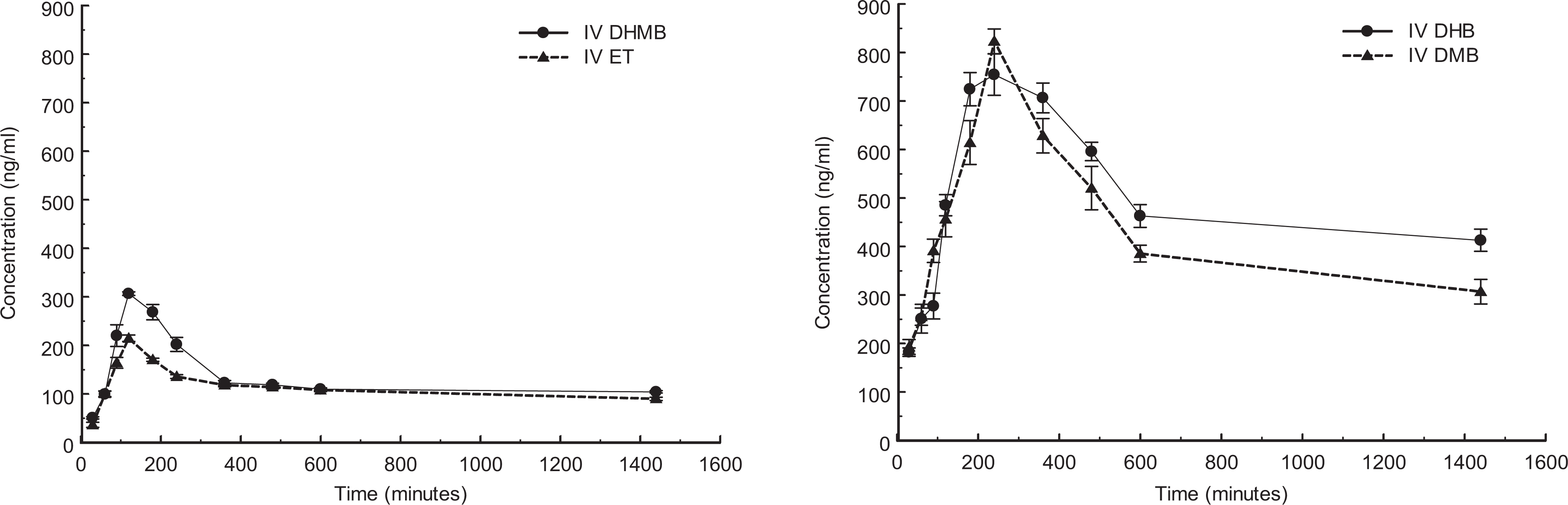
Plasma concentrations of metabolites of DEET (left) and oxybenzone (right) after an intravenous injection of 2 mg/kg DEET and 2 mg/kg oxybenzone (n = 6, mean ± SEM). DEET indicates N,N-diethyl-m-toluamide; SEM, standard error of the mean.
Figure 2 shows the plasma time curves for the appearance of DEET and oxybenzone metabolites in plasma after a single topical skin application. Concurrent application of DEET and oxybenzone resulted in elevated metabolite concentrations for both substances compared to administration of DEET and oxybenzone alone. For DEET metabolites, AUC levels of DHMB and ET were significantly increased by 23% and 17% (P < .05), respectively, following DEET and oxybenzone application, in comparison to that from single skin application. For oxybenzone metabolites, AUC levels of DHB and DMB following DEET and oxybenzone application were significantly increased by 28% and 16% (P < .05), respectively, in comparison to that from single skin application. Metabolite concentrations were still detected in plasma 24 hours postadministration from all application procedures.
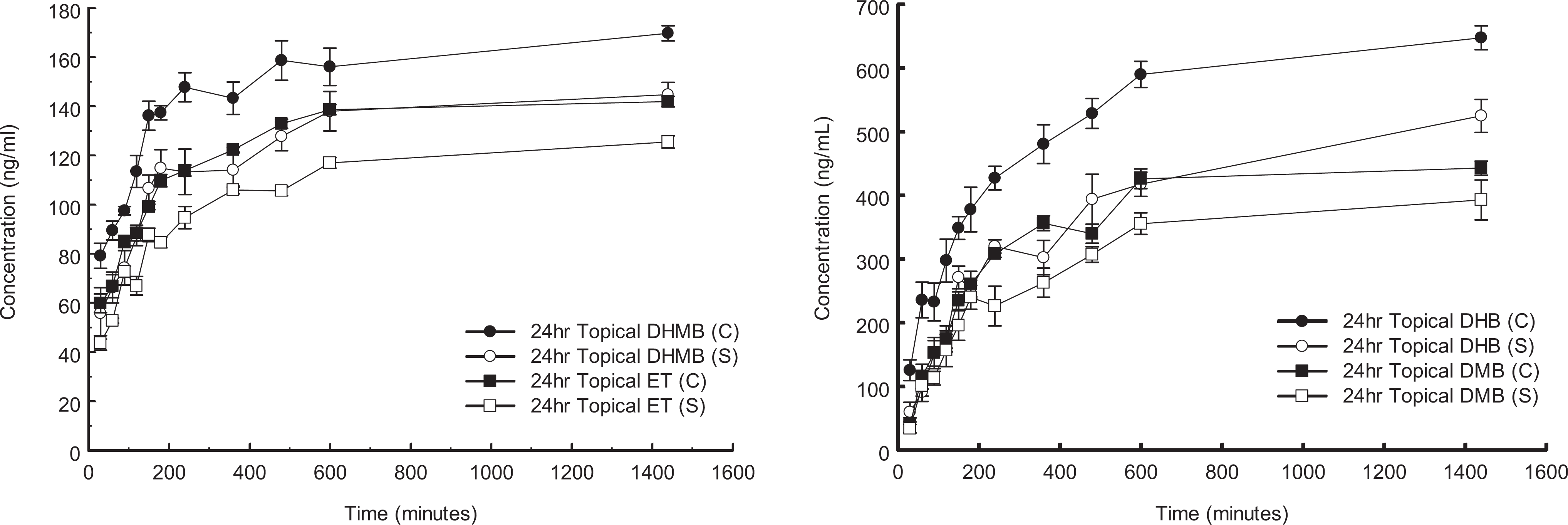
Plasma concentrations of metabolites of DEET (left) and oxybenzone (right) after a 24-hour topical application (n = 5, mean ± SEM). C indicates DEET and oxybenzone combined application; S, DEET or oxybenzone single application.
Figure 3 shows the plasma time curves in the formation of DEET and oxybenzone metabolites after repeated topical skin application once daily for 30 days. Concurrent use of DEET and oxybenzone led to a higher DHMB level (30% increase) than individual administration, but the ET levels were not different whether DEET was applied alone or with oxybenzone. For oxybenzone metabolites, no difference in AUC of the 2 metabolites was observed between single and coapplication with DEET. Like all other studies, these metabolites were still detectable in plasma 24 hours after the skin applications.
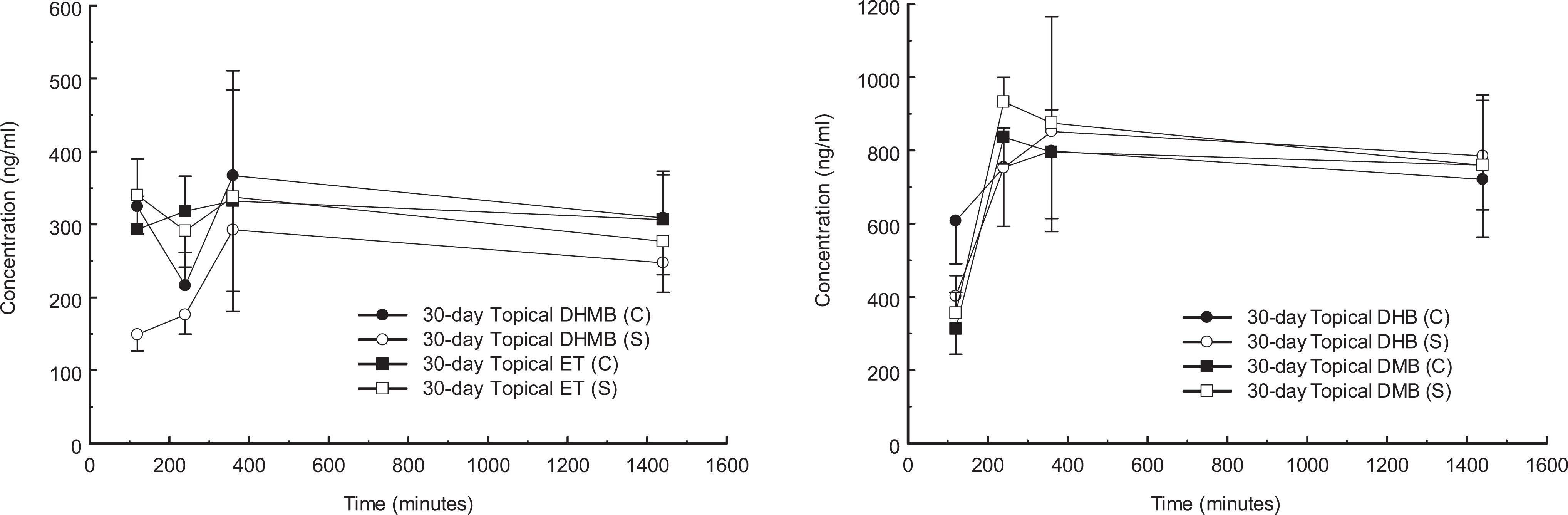
Plasma concentrations of metabolites of DEET (left) and oxybenzone (right) after the last dose of a 30-day repeated topical application (n = 10, mean ± SEM). C indicates DEET and oxybenzone combined application; S, DEET or oxybenzone single application.
It was evident that both DEET and oxybenzone were rapidly metabolized in the liver after intravenous and skin applications. The prompt appearance of metabolites in plasma from skin applications may indicate that metabolism had taken place while the parent compounds were being absorbed across the skin layers. 9 Skin has been proven to be a major organ for various enzyme activities. 22 Nevertheless, no quantitative concentrations of these metabolites were detected in skin tape strips, indicating minimal metabolism of DEET and oxybenzone in the stratum corneum and upper epidermis, which was similar to results reported in a previous study. 4 Several minor metabolites were also recorded in humans from subsequent oxidation, hydroxylation, and glucuronidation reactions (e.g., N,N-diethyl-m-carboxylbenzamide). 23 –25 Additional studies may be beneficial in clarifying the similarities and differences regarding metabolism of DEET and oxybenzone in rats and humans.
The presence of 1 aromatic ring in DEET and 2 aromatic rings in oxybenzone leads to favorable lipophilicity for percutaneous transport; their partition coefficients in octanol and water are 2.0 and 3.8, respectively. 26,27 Topical skin application also tends to supply the general circulation with a continuous, slow delivery of the parent compounds, DEET and oxybenzone, and prolongs their absorption phase, which can subsequently influence in vivo disposition. In comparison, concentrations of the metabolites decreased much more slowly than the parent compounds over time. 15 The long-term impact of these substances may require further investigation. 28
Tissue Dispositions
Table 1 lists the total recovery of DEET and oxybenzone metabolites in liver from the 3 administration methods. Two DEET metabolites, DHMB and ET, were detected in all tissue samples collected, but DHMB appeared to be the dominant metabolite in liver after application of DEET to skin. Coapplication of the 2 parent compounds together seemed to enhance the disposition of DHMB and ET after topical skin application. For single-dosing study, DHMB and ET concentration in liver was significantly increased by 50% and 30%, respectively, when concurrent use was compared to individual use of the compounds. For 30-day repeated-dosing study, DHMB and ET concentration in liver was significantly increased by 22% and 49%, respectively, from concurrent use. In addition, although ET concentration was larger than that of DHMB by a range of 8% to 25% in both single-dosing studies, DHMB concentration exceeded ET by a range of 125% to 175% in a 30-day repeated-dosing study.
Concentration (ng/g) of Metabolites of DEET (DHMB and ET) and Oxybenzone (DHB, DMB, and THB) From Liver Following Intravenous Administration (Group 1/2), Single Skin Application (Groups 3/4/5), and Repeated Skin Application (Groups 6/7/8)a
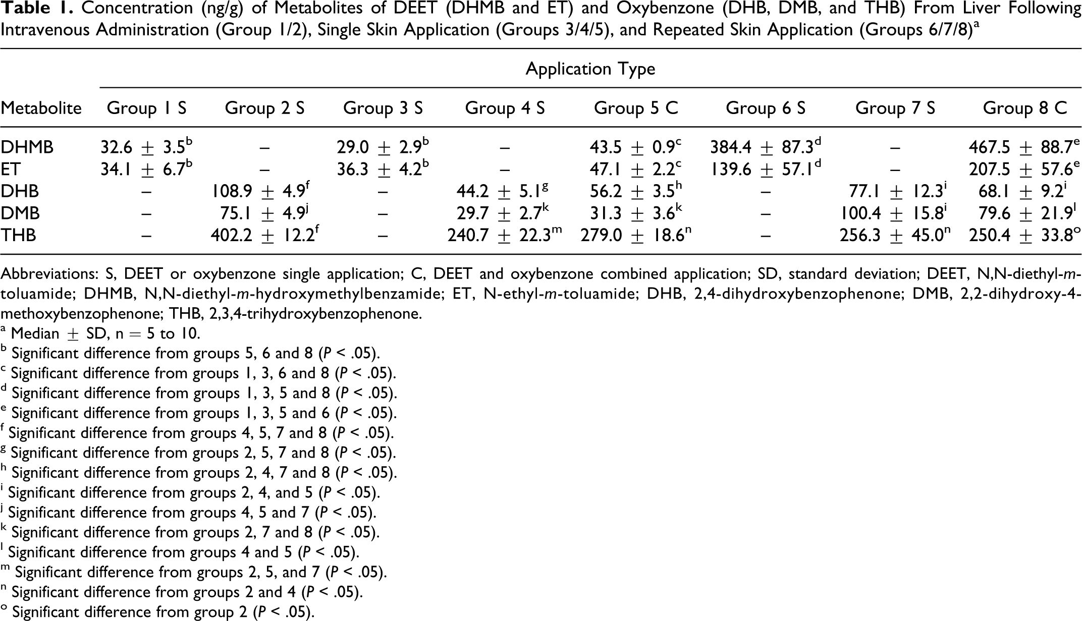
Abbreviations: S, DEET or oxybenzone single application; C, DEET and oxybenzone combined application; SD, standard deviation; DEET, N,N-diethyl-m-toluamide; DHMB, N,N-diethyl-m-hydroxymethylbenzamide; ET, N-ethyl-m-toluamide; DHB, 2,4-dihydroxybenzophenone; DMB, 2,2-dihydroxy-4-methoxybenzophenone; THB, 2,3,4-trihydroxybenzophenone.
a Median ± SD, n = 5 to 10.
b Significant difference from groups 5, 6 and 8 (P < .05).
c Significant difference from groups 1, 3, 6 and 8 (P < .05).
d Significant difference from groups 1, 3, 5 and 8 (P < .05).
e Significant difference from groups 1, 3, 5 and 6 (P < .05).
f Significant difference from groups 4, 5, 7 and 8 (P < .05).
g Significant difference from groups 2, 5, 7 and 8 (P < .05).
h Significant difference from groups 2, 4, 7 and 8 (P < .05).
i Significant difference from groups 2, 4, and 5 (P < .05).
j Significant difference from groups 4, 5 and 7 (P < .05).
k Significant difference from groups 2, 7 and 8 (P < .05).
l Significant difference from groups 4 and 5 (P < .05).
m Significant difference from groups 2, 5, and 7 (P < .05).
n Significant difference from groups 2 and 4 (P < .05).
o Significant difference from group 2 (P < .05).
Three oxybenzone metabolites were detected in the tissue samples collected. For single-dosing study, concurrent skin application of DEET and oxybenzone resulted in increased concentrations of metabolites in comparison to single skin application; that is 27% for DHB, 5% for DMB, and 16% for THB. For 30-day repeated-dosing study, concurrent application of the 2 substances led to a slight decrease in concentration in comparison to single application; that is 12% for DHB, 21% for DMB, and 2% for THB. 2,3,4-Trihydroxybenzophenone is a metabolite formed from secondary aromatic hydroxylation of ring A at the meta-position of DHB. This compound was not detected in plasma samples, but it was the primary oxybenzone metabolite found in the liver with an amount exceeding that of DHB and DMB. 2,3,4-Trihydroxybenzophenone was 269% to 436% more than DHB/DMB after intravenous administration, 396% to 791% more than DHB/DMB after 24-hour single dosing, and 155% to 268% more than DHB/DMB after the 30-day repeated-dosing, respectively.
No comparable tissue disposition data are available from a human study. Nevertheless, it appeared that metabolism of DEET and oxybenzone from dermal administration in rats was qualitatively similar to their metabolism in humans after dermal exposure. 10 ,24 Several case reports have described young girls developing encephalopathy after DEET exposure 29 –33 ; it is unknown whether the parent compounds or metabolites are accountable for DEET toxicity. In addition, the intraindividual variations in metabolism and the correlation between adverse risk and dermal DEET application have not been fully studied. 25,34 The enhanced disposition of DEET metabolites from concurrent skin application raised concerns for these DEET metabolites that may exert pharmacological and/or toxicological effects from prolonged exposure to DEET and oxybenzone. 8,25 Brain and kidney samples collected from this study also contained all DEET metabolites; ET was the predominant metabolite in these organs. However, concurrent application did not lead to significant increases in brain and kidney concentrations. For the 30-day repeated-dosing study, brain concentrations of DHMB and ET were similar after combined use (17.7 ± 3.6 ng/g and 27.5 ± 6.0 ng/g) and single use (16.2 ± 2.9 ng/g and 22.7 ± 7.5 ng/g), respectively. For the single 24-hour topical administration, kidney concentrations of DHMB and ET were also similar after combined use (16.3 ± 4.6 ng/g and 38.8 ± 5.6 ng/g) and single use (14.5 ± 1.9 ng/g and 36.3 ± 3.3 ng/g), respectively. Liver is a prime metabolism site of the body so it was expected that there would be a higher concentration of both parent compound and metabolites in this tissue. 10 The 30-day repeated-dosing study demonstrated increased potential for DHMB and ET accumulation in the liver, as a lower study dosage (vs 24-hour topical application), similar plasma profiles (versus intravenous and 24-hour topical administration), and a less penetrating formulation (70% ethanol for 30-day repeated topical application vs 100% ethanol for 24-hour topical application) led to increased concentrations. Potential for undesirable side effects and drug interactions due to alterations in cytochrome P-450 activity from chronic exposure to repellents in susceptible participants should be further investigated.
Previous studies have indicated that liver contains the highest concentrations of oxybenzone metabolites per gram of tissue, followed by kidney. 9 Higher concentrations of these metabolites in liver was attributed to a high content of cytochrome P-450, which may explain why increased THB amounts were detected in the liver, as THB is derived from DHB through an aromatic hydroxylation reaction mediated by the cytochrome P-450 enzyme system. 9 In addition, oxybenzone was shown to enhance liver and kidney weights in animals after oral and dermal exposure. 35,36 All 3 metabolites were detected in brain and kidney samples, with THB being the predominant metabolite. However, concurrent application did not lead to significant increases in brain and kidney concentrations. For the 30-day repeated-dosing study, brain concentrations of DHB, DMB, and THB were similar after combined use (20.9 ± 2.4 ng/g, 14.5 ± 3.2 ng/g, and 22.1 ± 3.6 ng/g) and single use (23.2 ± 2.0 ng/g, 16.6 ± 1.7 ng/g, and 26.0 ± 4.3 ng/g), respectively. For the single 24-hour topical administration, kidney concentrations of DHB, DMB, and THB were similar after combined use (25.3 ± 4.9 ng/g, 25.6 ± 1.0 ng/g, and 145.8 ± 5.4 ng/g) and single use (28.3 ± 2.9 ng/g, 23.4 ± 3.9 ng/g, and 141.2 ± 6.0 ng/g), respectively. Transport proteins are responsible for transferring endogenous compounds (bile acids) and xenobiotics (toxins) within the body and are therefore an important determinant of metabolite disposition and subsequent excretion. 37 Differences observed in liver between combined and single topical application were considered detrimental from a clinical viewpoint, since oxybenzone and its metabolites enhanced the accumulation and disposition of DEET and its metabolites.
Urine and feces samples were collected from the 24-hour single-dosing study. The DHMB was the major urine metabolite of DEET with combined application (6.3 ± 0.1 µg/mL) significantly exceeding single application (5.9 ± 0.1 µg/mL, P < .05). DHMB and ET were also detected in feces; their concentrations were 1.9 ± 0.4 ng/g (DHMB) and 0.6 ± 0.1 ng/g (ET) in combined use and 1.8 ± 0.5 ng/g (DHMB) and 0.5 ± 0.1 ng/g (ET) in single use, respectively. This suggested that rats utilized both urinary and enterohepatic routes for eliminating DEET from the system as previously reported. 10 Past studies suggested that oxybenzone was excreted via urine and feces following oral administration; DHB was a major metabolite excreted in the urine. 4,38 This study led to similar findings, in which DHB was the predominant metabolite in urine (2.0 ± 0.1 µg/mL for combined use and 1.8 ± 0.1 µg/mL for single use, P < .05) and feces (1.7 ± 0.1 ng/g for combined use and 1.4 ± 0.1 ng/g for single use, P < .05). No THB was detected in the feces. The data suggested that rats utilized both urinary and enterohepatic routes for eliminating oxybenzone and its metabolites.
Cellular Viability
Figure 4 shows the cellular proliferation of rat hepatoma cells after the cultures were exposed to DHMB, ET, DHB, DMB, and THB at concentrations of 0.1 µg/mL, 1 µg/mL, and 10 µg/mL for 48 and 72 hours. Colorimetric assays are used extensively in cell proliferation and cytotoxicity analysis, enzyme analysis, and bacteriological screening. 39 The water-soluble tetrazolium 1 (WST-1) test investigated the metabolic activity of mitochondria as an indication of the vital status of cells. 40 While no significant decrease in cellular proliferation was observed after 24 hour exposure at all concentrations (data not shown), reduced cellular proliferation was noted when the exposure time to all metabolites was increased.
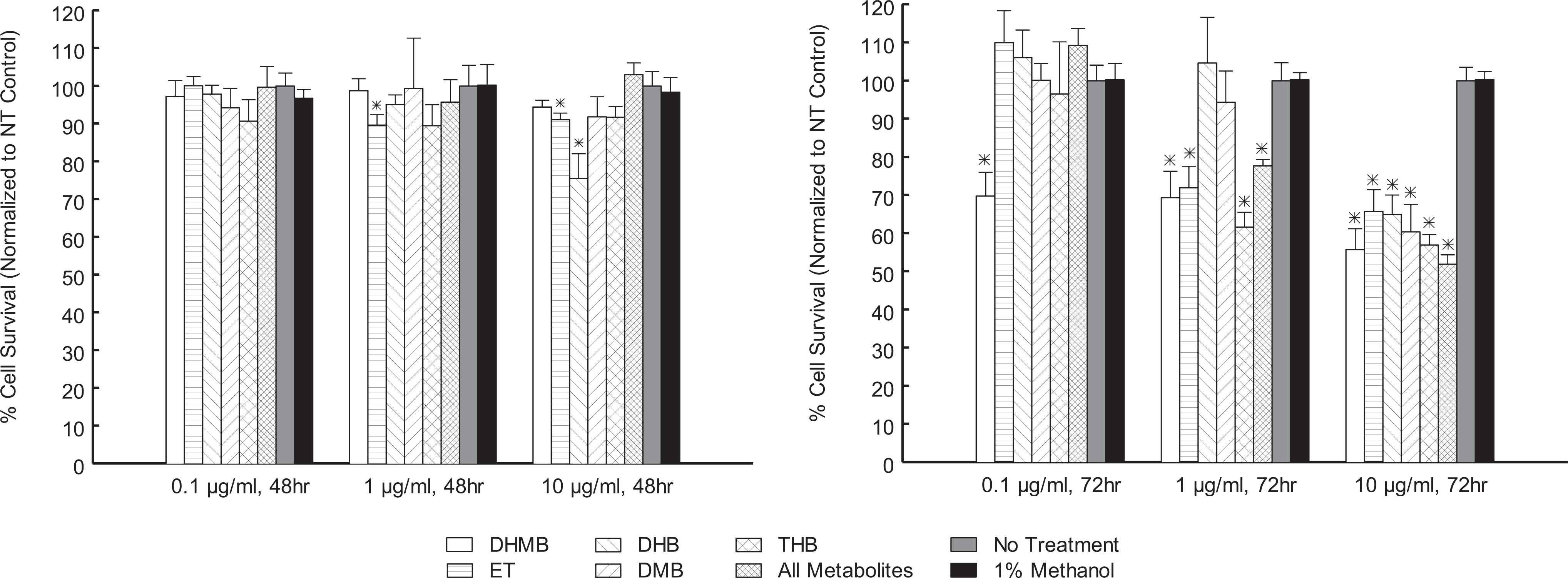
Cellular viability of rat hepatoma cell line 1548 exposed to metabolites of DEET and oxybenzone after repeated dosing every 24 hours at different concentrations and durations (*Significant difference from no treatment control, P < .05, n = 6, mean ± SEM).
For DEET metabolites, DHMB reduced cellular proliferation by 30% to 44% for all 3 treatment concentrations after 72 hours. The ET reduced cellular proliferation by 10% and 9% at 1 and 10 µg/mL, respectively, after a 48-hour exposure; a decrease of 28% and 34%, respectively, after a 72-hour exposure was also observed. For oxybenzone metabolites, DHB reduced cellular proliferation by 25% at 10 µg/mL and 48 hours; a reduction of 35% after 72 hours of exposure was also observed. 2,2-Dihydroxy-4-methoxybenzophenone reduced cellular proliferation by 40% at 10 µg/mL and 72 hours. 2,3,4-Trihydroxybenzophenone also reduced cellular proliferation by 38% and 43% at 1 and 10 µg/mL, respectively after a 72-hour exposure. Combined exposure to all test metabolites reduced cellular proliferation by 22% and 48% at 1 and 10 µg/mL, respectively, after 72 hours. Overall, DEET metabolites affected the proliferation of hepatoma cells more than oxybenzone metabolites, and THB appeared to have the most notable effect on cellular proliferation of all oxybenzone metabolites.
Initiation of programmed cell death (apoptosis) is demonstrated by the release of mitochondrial cytochrome c activity, activation of caspases, elevation of 8-hydroxy-2-deoxyguanosine level, increased levels of 3-nitro-tyrosine, and alterations of p53 gene expression. 41 Previous studies found that concurrent application of DEET and permethrin in rats induced urinary excretion of 3-nitrotyrosine and 8-hydroxy-2′-deoxyguanosine, markers of DNA damage and oxidative stress, and mitochondrial cytochrome c release. 42 Another study using human hepatocytes confirmed that DEET was a mild inducer of adenylate kinase and caspase 3/7, both indicators of apoptotic cell death. 43 Therefore, DEET was capable of inducing apoptotic cell death in hepatocytes in humans and animals. Little information is available for DEET metabolites. Recent investigation indicated significant interindividual variability in CYP450 isoform activity in human microsomes relevant to DEET metabolism. 25 ,34 Individuals with high activity of CYP2B6 and CYP1A2 activity produced the highest DHMB level, whereas individuals with high activity of CYP3A4 or CYP2C19 had the highest ET level. Our study is the first of its kind to observe suppression of cellular proliferation by ET and DHMB after 48 and 72 hours of exposure. Plasma concentrations of DEET metabolites also reached 0.1 µg/mL from in vivo animal studies, which may indicate that cell proliferation could be compromised over extended exposure to the compound.
Cellular proliferation was suppressed after concurrent administration of all metabolites including DHB, DMB, and THB at 1 µg/mL. Plasma concentrations of these metabolites approached this range after intravenous administration and 30-day repeated topical application, which may indicate that in vivo cell proliferation could be affected. No toxicological mechanisms have been clearly defined for oxybenzone or its metabolites. Cases of allergic contact cheilitis, general endocrine disruption, birth weight variations, and even possible carcinogenicity have been reported with oxybenzone use. 44 –48 Although the WST-1 assay measures mitochondrial activity, reduction of WST-1 may also be associated with superoxide, occurring in either the extracellular environment or the plasma membrane. 39 Depletion of NADH or other mechanisms that involve superoxide may potentially interfere with the reduction process of WST-1. Consequently, different cell types that possess variable extracellular superoxide contents could interfere in the reduction capacity of each cell type to different degrees. 39 Hepatocellular experiments conducted in this study did assist in providing a better understanding of the underlying mechanisms causing cellular proliferation deficits from short-term and repeated-dosing to long-term in vitro exposure of these test metabolites, but further analysis and studies with additional cell lines would be beneficial in formulating more conclusions regarding their toxicokinetic effects.
Conclusion
In conclusion, the intravenous and topical application studies demonstrated significant pharmacokinetic differences between administration or application of single and combined preparations for both DEET and oxybenzone. Concurrent topical application of DEET and oxybenzone enhanced the plasma, liver, and urine concentrations of all DEET metabolites in vivo, but this effect was not apparent in the brain, kidney, and feces. Plasma concentrations of all metabolites remained detectable for 24 hours after topical application, which supported the presence of a skin depot by the parent compounds. Increased accumulation in the liver was observed for DHMB and ET after once-daily repeated topical applications for 30 days. A significant decrease in cellular proliferation was observed from all metabolites, occurring as early as 48 hours after exposure for ET and DHB. While combined exposure to DEET and oxybenzone appeared to enhance the distribution of DEET metabolites in the liver, no additional decreases in cellular proliferation related to this synergistic interaction were noted. However, due to the potential for reduced cellular proliferation as a result of exposure to DEET, oxybenzone, and their metabolites, either alone or in combination, consumers must exercise caution when applying these widely available over-the-counter topical skin products. Both DEET and oxybenzone are intended for remaining on the surface of the skin, and thus other studies are in progress to develop new formulations of the insect repellent and sunscreen, aiming at decreasing their overall percutaneous penetration and further evaluating their concurrent toxicological profiles.
Footnotes
Acknowledgment
The authors acknowledge research support from the Canadian Institutes of Health Research (CIHR)/Manitoba Health Research Council (MHRC), grant No. 2005ROP-148547, and Canada Foundation for Innovation (CFI). We also acknowledge technical assistance and animal care by Heather Simpson, Susan Blair and Teri Whittington of University of Manitoba Veterinary Services.
Declaration of Conflicting Interests
The author(s) declared no potential conflicts of interest with respect to the research, authorship, and/or publication of this article.
Funding
The author(s) disclosed receipt of the following financial support for the research, authorship and/or publication of this article: research support from the Canadian Institutes of Health Research (CIHR)/Manitoba Health Research Council (MHRC), grant No. 2005ROP-148547, and Canada Foundation for Innovation (CFI).