
Abstract
Gas-filled microbubbles are used as contrast agents in diagnostic ultrasound imaging. A preclinical, acute toxicity study of 2 surfactant-stabilized ultrasound contrast agents (ST68 and ST44) was conducted. Subjects were 104 Sprague-Dawley rats (experimental doses, 0.1, 0.2, 0.8, and 1.0 mL/kg; control, 1.0 mL/kg saline) that were studied for 14 days after contrast; clinical signs, weight, blood, and urine were evaluated. Histopathology was performed following euthanasia. Of the 40 animals receiving ST44, 4 died prematurely and a dose dependency was demonstrated (P = .011), whereas in the ST68 groups only 1 death occurred (no dose dependency; P = .48). Only the weight of rats injected with ST44 varied significantly (P = .0003). This dependency was also found for 3 of 5 urine parameters and 4 of 36 blood parameters (P < .05). For ST68, only 1 urine parameter showed significance (P < .0001). Giant cell infiltration in the lungs was significantly higher than controls in the ST44 0.1 mL/kg and the ST68 0.8-1.0 mL/kg groups (P < .01). It is concluded that the prudent choice for future nonrodent, toxicology studies and potentially for human clinical trials is ST68 (given the deaths in the ST44 groups).
Keywords
Gas-filled microbubbles can be used as contrast agents in diagnostic ultrasound imaging. 1 Three such contrast agents have been approved by the US Food and Drug Administration (FDA) for use in echocardiography, whereas numerous agents have been permitted for clinical use in both radiology and cardiology in Europe, Asia, and the rest of the Americas. Although many studies have reported that ultrasound contrast agents are safe and efficacious with low incidents of adverse events,2-5 there are still regulatory concerns about the safe use of ultrasound contrast agents in patients with severe cardiac disease. 6 This ongoing discussion highlights the need to establish the safety profile of any ultrasound contrast agent prior to initiating human clinical trials.
Our laboratory has been developing surfactant-stabilized microbubbles as ultrasound contrast agents for more than a decade. 7-9 The chemical composition of these bubbles has been investigated, and it has been shown that bubbles from the nano- to the micro-scale can be prepared. 10,11 The agents are composed of mixed nonionic surfactants of the polysorbate ester series, namely selected Spans and Tweens. One advantage of this system is the variety of combinations of Span and Tween that have been found to form stable microbubbles, allowing flexibility in the choice of the final formulation. 9 In vivo animal studies have demonstrated that these contrast agents can produce up to 28 dB of enhancement over the course of 10 minutes. 12,13 The agents also display excellent nonlinear properties. 14 Intriguingly, it has been shown that a specific surfactant (Tween 80) inhibits the activities of efflux transporters P-glycoprotein and multidrug-resistance-related protein (MRP), both of which are implicated in conferring multidrug resistance (MDR) in tumors. 15 The next generation of contrast agents is being developed with therapeutic as well as diagnostic capabilities, and new agents will carry not only a gas core but also drug within the bubble shell. 16 An ability to prevent MDR would be highly desirable in this next stage of development; especially in the use of ultrasound contrast agents as drug carriers for cancer therapy. 17-19 However, this evidence of membrane activity increases the importance of conducting an initial toxicology study at this stage of the contrast agent development. Hence, a preclinical, acute toxicity study of 2 candidate surfactant-stabilized ultrasound contrast agents (named ST68 and ST44) was conducted in adult rats.
Methods
Preparation of ST 68 and ST 44
Two different ultrasound contrast agents, ST68 and ST44, consisting of surfactant-stabilized microbubbles, were prepared as described by Wheatley. 10 These mixtures of nonionic surfactants can form very stable microbubbles when subject to cavitation in phosphate-buffered saline (PBS). Briefly, the agents were produced from an intimate mixture of 1.50 g of sodium chloride (NaCl) and either Span 60 and Tween 80 (in the case of ST68) or Span 40 and Tween 40 (in the case of ST44). The mixture, containing 1.48 g of Span and 1.00 mL of Tween, was suspended in 50 mL of PBS and crushed in a mortar and pestle before being stirred and heated to a temperature of 50°C and held there for 3 minutes. Then the mixture was cooled to room temperature before autoclaving (Tuttnauer Brinkmann 3850E, Westbury, NY) using a liquid cycle for 12 minutes at 120°C at atmospheric pressure. The cooled solution was purged of dissolved air using a stream of perfluorocarbon (PFC) gas. The purged solution was then continuously probe sonicated at 110 W while in a 250 mL beaker (held in an ice bath), with constant purging using a steady stream of PFC gas (~4 mL/min) for 3 minutes. The ultrasonic liquid processor used for sonication (W 385; Misonix, Farmingdale, NY) was equipped with a tapped 1/2 inch horn. The sonicated mixture separated into 3 phases, and the middle phase, consisting of microbubbles, was removed using a separatory funnel and washed 3 times with 60 mL of PBS.
The number of microbubbles per milliliter of PBS was measured using a Coulter Multisizer Mark II (Coulter Electronics, Luton, Bedfordshire, UK), with a 30 μm aperture, a 100 μL manometer setting, and an aperture current of 1600 μA on diluted samples in Isoton solution. The concentration was adjusted to around 1.1 × 10⁹ bubbles per milliliter (using PBS), and the mean bubble diameter was measured using a Zetasizer Nano ZS Particle Sizer (Malvern Instruments, Malvern, Worcestershire, UK) as approximately 1.2 μm for both agents. Samples were stored under PFC in glass vials capped with a rubber septum and closed with a band of parafilm, at 15°C. Prior to use, the vials were brought to room temperature and gently rotated to evenly distribute the bubbles that had collected at the top of the sample.
Animals
Subjects were 104 Sprague-Dawley rats (Taconic, Germantown, NY), 6 to 8 weeks old, with a body weight range of 219 to 306 g. Prior to study initiation, all rats were quarantined for 7 days and were ensured to be in good health by a veterinarian. The animals were kept in environmental conditions in accordance with established guidelines. 20 The animal studies described here were carried out in an ethical and humane fashion under supervision of a veterinarian, and the university’s animal care and use committee approved all protocols.
Study Design
This was a preclinical, acute toxicity study of the surfactant-stabilized ultrasound contrast agents ST68 and ST44 conducted in adult rats. Five groups of 5 male and 5 female rats were studied per agent (ie, 100 rats in total). Because the highest clinical dose of the ultrasound contrast agents is estimated at 0.1 mL/kg, 10 the study tested dosages of 0.1, 0.2, 0.8, and 1.0 mL/kg with 1.0 mL/kg of saline injected as control. Following acclimatization, the animals were anesthetized and received the test or control articles at an approximate rate of 0.05 mL/s through a single lateral tail vein injection.
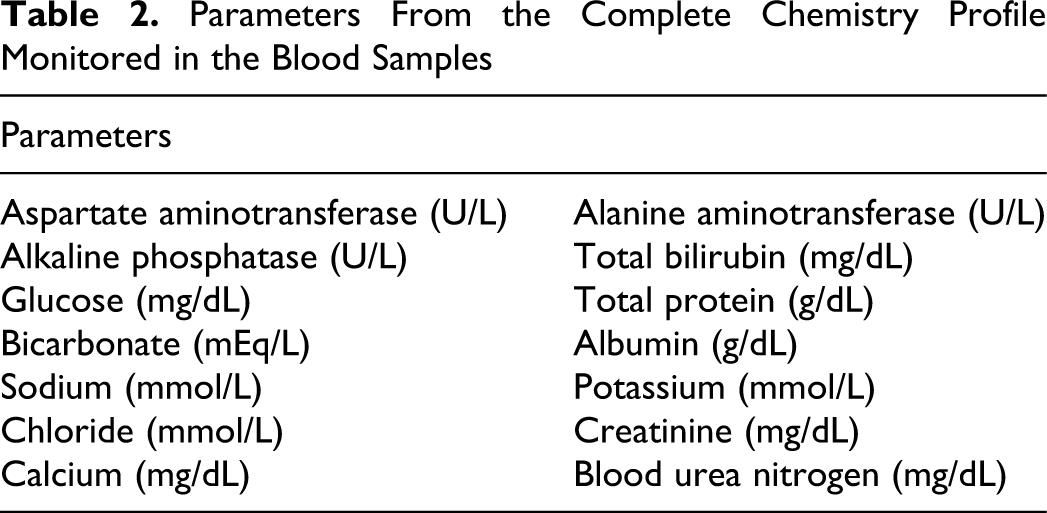
Animals were observed for 14 days after contrast administration with clinical signs evaluated daily and body weight measured on days 2, 7, and 14. Blood samples were acquired 2 and 14 days posttreatment and tested to obtain complete blood counts (CBC), differential, and a complete chemistry profile. A list of the parameters assessed is given in Tables 1 and 2. A urine sample was collected immediately prior to euthanasia on day 14 for analysis (Table 3). Following euthanasia by placing the rats in a CO2 jar, the heart, kidneys, liver, lungs, and spleen were collected and weighed. Macroscopic and microscopic evaluations of the collected tissues were also performed by a veterinary clinical pathologist.
CBC Parameters Monitored in the Blood Samples
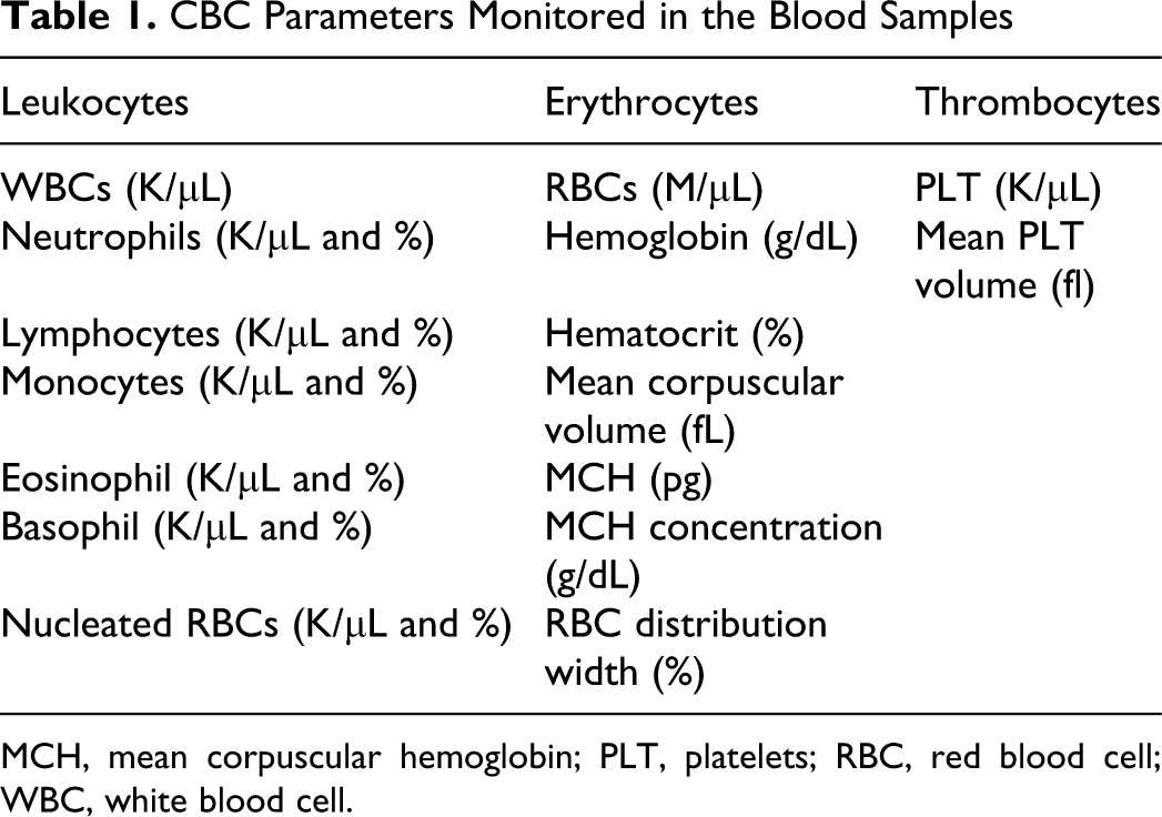
MCH, mean corpuscular hemoglobin; PLT, platelets; RBC, red blood cell; WBC, white blood cell.
Parameters From the Complete Chemistry Profile Monitored in the Blood Samples
Parameters Monitored in the Urine Samples
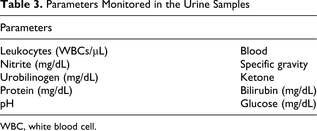
WBC, white blood cell.
The last 4 rats (all males) were used to demonstrate the contrast-enhanced ultrasound imaging possible with the 2 contrast agents. The same dosages as described above (but without saline control injections) were studied. A state-of-the-art commercial ultrasound scanner (Aplio; Toshiba America Medical Systems, Tustin, Calif) equipped with a broad-bandwidth (5-11 MHz) linear array transducer (PLT-704AT) was used to perform power Doppler imaging (PDI), which is a color overlay mode indicating areas of flow in orange hues, and pulse subtraction harmonic imaging (PSHI), which is a nonlinear contrast-specific imaging mode depicting microbubble signals in gray-scale images. 1 Digital images were acquired of the left kidney (in 2 animals in PDI mode and in 2 in PSHI mode) before and after administration of contrast agent. All imaging parameters were kept constant before and after imaging (except that the gain could be reduced post injection to minimize shadowing and blooming artifacts 21 ) and at least 10 minutes passed between injections to ensure a return to baseline conditions.
Data Analysis
Statistical comparison of the survival time (in days) for individual rats following administration of the different contrast agents (including control injections) and dosages was performed using a Cox proportional hazards model 22 and the statistical software package Stata 9.0 (Stata Corporation, College Station, Tex). Because most rats survived exposure to the contrast agents, the Cox regression was conducted with censored data (ie, rats that died were considered “noncensored,” whereas surviving animals were regarded as “right-censored”). The Breslow method for handling tied survival times was used. 22 Moreover, the dose dependency of survival was investigated using a nonparametric test of trend. In both tests P values less than .05 were considered statistically significant.
Because the variables in the blood samples were all continuous, a 3-way factorial analysis of variance (ANOVA) was used to establish differences, with gender, day, and contrast agent considered the dependent variables. Analyses were performed for both contrast agents relative to the PBS control (ie, as a categorical variable) and relative to the dose used (ie, as a continuous variable). A 2-way factorial ANOVA was used to analyze the urine samples (because day was no longer a variable), but otherwise the same tests were carried out. If contrast agent was a significant variable, the underlying regression coefficients were tested for differences between dosages. Tests with P values less than .05 and variables outside their normal range (based on historical control values) were considered significant.
Results
Of the 40 animals that received ST44, 4 animals (10%) died prematurely. All deaths occurred on day 2 of the observation period, and 3 were in the 1.0 mL/kg group (1 male and 2 female rats), whereas the last (female) animal was allocated to the 0.8 mL/kg group. In the group injected with ST68 only, 1 death occurred (on day 2) involving a female rat in the 0.8 mL/kg group. The Cox regression analysis showed that neither contrast agent, the gender of the animal, nor the dose administered had a significant impact on survival (P > .15). However, there was a significant dose-dependency in the ST44 group (ie, higher dosages were more likely to result in death; P = .011), which did not exist for the ST68 group (P = .48).
Not surprisingly, gender, day (when applicable), and their interaction parameters were often significant parameters for the weight, blood, and urine data (P < .05). The weight of rats injected with ST44 showed significant impact due to the test article (contrast agent and control considered together as a categorical variable; P = .0003) but not between individual dosages of ST44 and PBS (P > .08), as demonstrated in Figure 1 . Moreover, when only ST44 was considered (as a continuous variable), there was no effect on weight due to the contrast agent (P = .24). All the male rats increased their weight over the 14 days (on average 39%, range 14%-54%), as did most of the female animals except for 5 rats (including 1 of the controls) that lost weight (average gain 4%; range –5% to 11%). No statistically significant differences in weight were found for animals receiving ST68 (P > 0.41; data not shown).
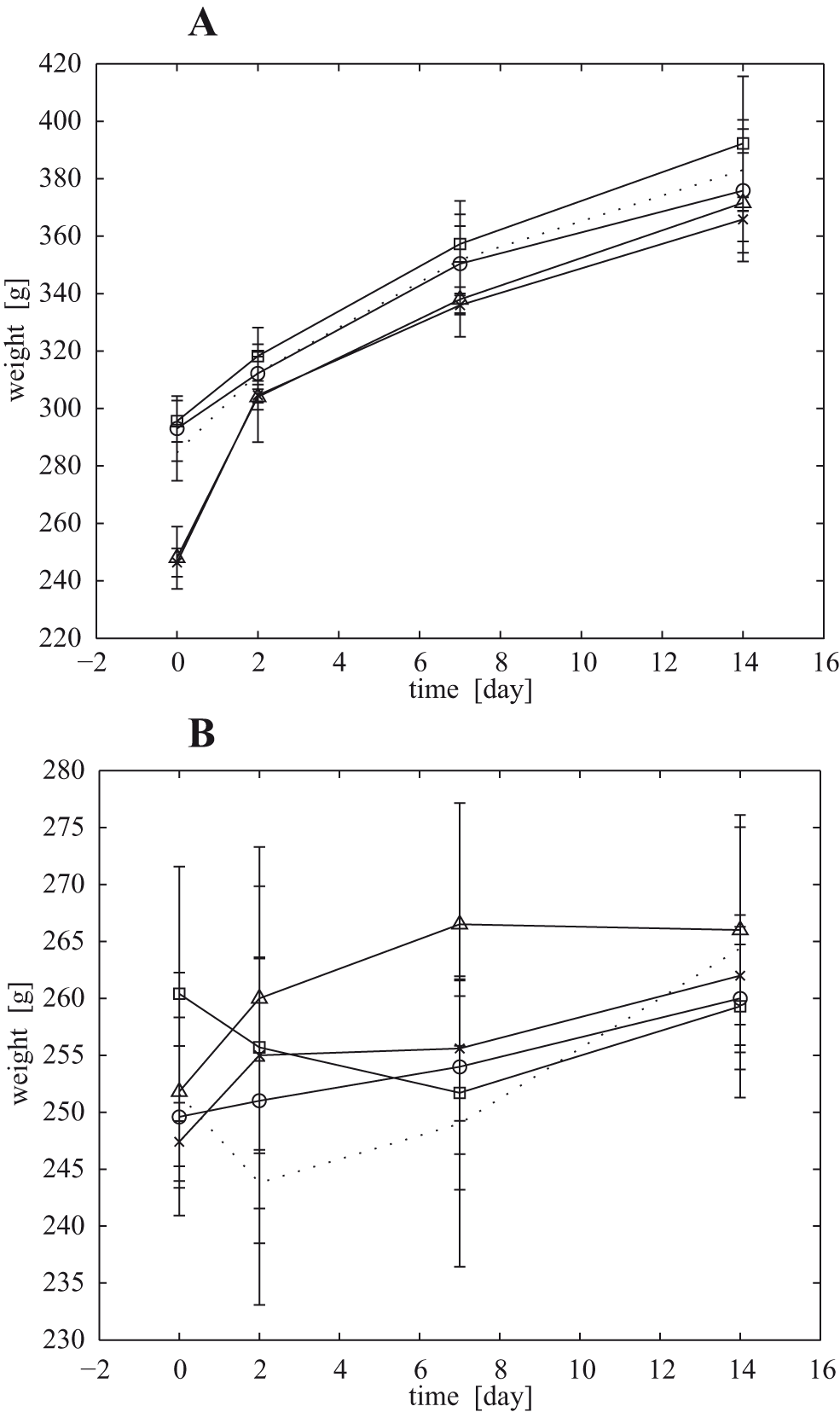
Mean weight (±1 standard deviation) as a function of time for rats injected with ST44 (A, male rats; B, female rats). Dosages are marked as follows: circles, 0.1 mL/kg; ×, 0.2 mL/kg; triangles, 0.8 mL/kg; squares, 1.0 mL/kg. PBS controls are presented as dotted lines. Notice the different y-axes used in parts A and B.
Of the 36 parameters evaluated from the blood samples obtained in the ST44 groups, 4 (lymphocytes, monocytes, hematocrit, and platelets) showed a significant effect on the regression model due to the contrast agent (P < .005; Table 4 ). However, in none of these parameters was a dose dependency detected (P > .15), and only the hematocrit values were outside the normal range, with multiple animals being slightly anemic at the 0.2 and 0.8 mL/kg dose levels. None of the blood samples from the ST68 groups demonstrated significant impact and values outside of their normal range.
Mean Values (±1 Standard Deviation) for the Significant Results Obtained for ST44 in the Blood Samples
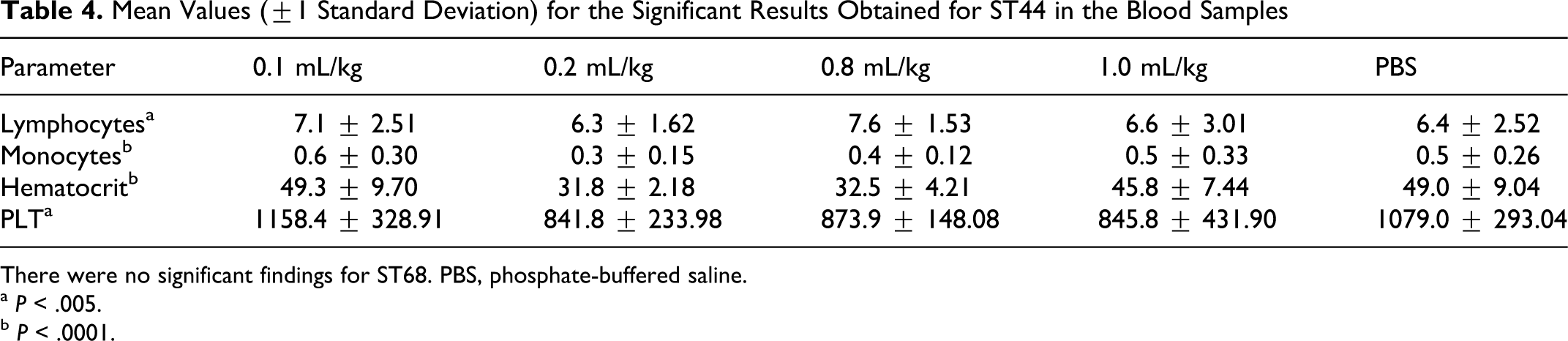
There were no significant findings for ST68. PBS, phosphate-buffered saline.
a P < .005.
b P < .0001.
ST44 also produced more significant regression coefficients in the urine samples than ST68 with 3 and 1 parameters from a total of 10, respectively (P < .05; Table 5 ). However, for both contrast agents the smaller dosages (0.1 and/or 0.2 mL/kg) proved significantly different from PBS. There was a significant dose dependency among leukocytes in the ST68 groups (P = .026) and blood in the ST44 groups (P = .014) but not for the leukocytes and protein expression in the latter (P > .08).
Mean Values (±1 Standard Deviation) for the Significant Results Found in the Urine Samples
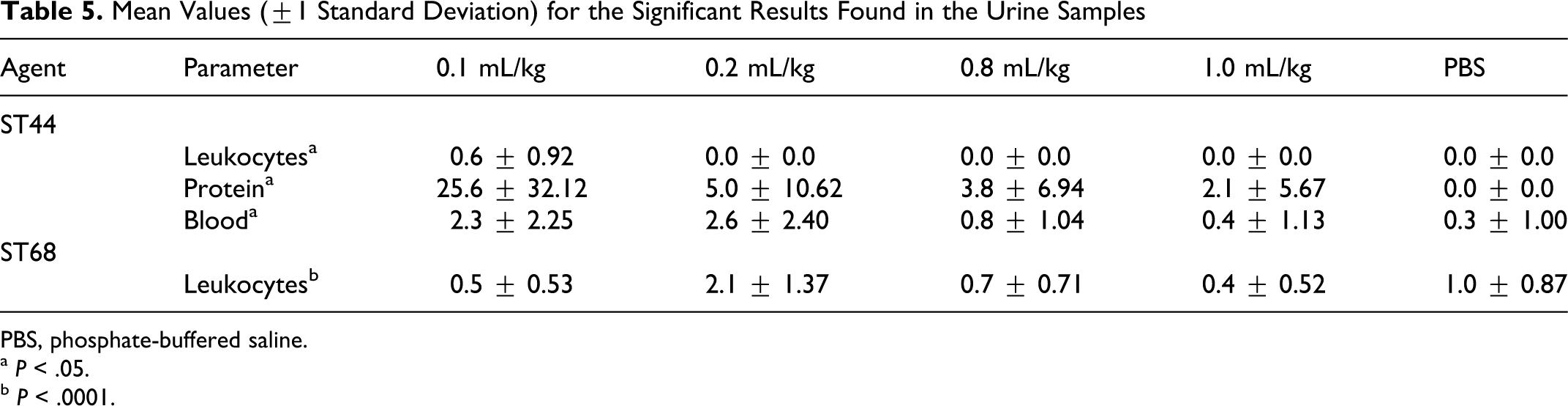
PBS, phosphate-buffered saline.
a P < .05.
b P < .0001.
Multinucleated giant cells (often with granuloma formation), consistent with a foreign body reaction, were found in the lungs of both ST68 and ST44 animals. Following injection of 0.1 mL/kg of ST44, there was a significantly higher degree of giant cell infiltration than in the control animals, with 6 rats demonstrating 1 or a few areas of focal multinucleated giant cells compared with none of the control animals (P = .0023). For ST68, the 0.8 and 1.0 mL/kg dosages produced the significant difference relative to PBS (P < .010), with 14 of 20 animals having multifocal pulmonary giant cell infiltration. Only animals injected with ST68 demonstrated a dose dependency (P = .002), whereas the ST44 groups did not (P = 0.29). No other significant variations relative to histopathology were found (P > .07); although many animals presented with renal medullary mineralization (of varying severity).
Examples of the PDI images obtained with the 2 contrast agents are presented in Figure 2 . Compared with baseline (the pre-injection images; Figures 2a and 2f), marked enhancement was observed for all dosages for both ST44 and ST68. However, the outline of the kidney was better visualized following administration of ST68 than ST44 (eg, Figure 2b vs. 2g or Figure 2c vs. 2h) for the 3 lowest dosages (0.1-0.8 mL/kg). Moreover, more shadowing and blooming artifacts were seen in the images acquired following administration of ST44 and at a lower dose (cf, Figures 2d and 2i). Contrast-specific PSHI examples are shown in Figure 3 , where again marked enhancement could be seen following contrast injection (the anterior margin of the capsule of the kidney is almost invisible prior to contrast; Figures 3a and 3f). Unlike the PDI examples in Figure 2, there are few differences between images produced in PSHI mode by the 2 contrast agents (compare Figures 3b-e with 3g-h). Nonetheless, these results (Figures 2 and 3) support the feasibility of performing clinical ultrasound contrast imaging at the 0.1 mL/kg dose (as predicted in reference 10).
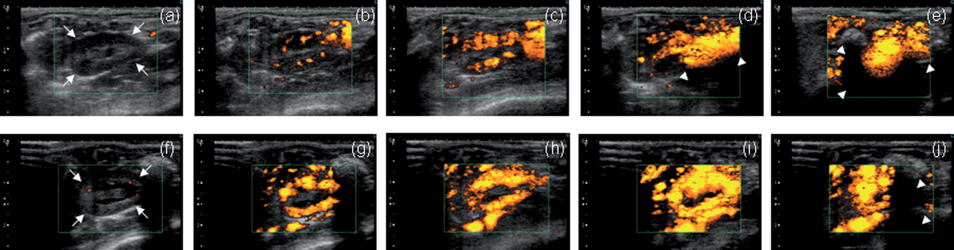
Rat kidneys (arrows) imaged in power Doppler imaging (PDI) mode (the green box delineates the region within which blood flow is measured) before injection of ST44 and ST68 (a and f, respectively) and following injection (b-e and g-j, respectively). Dosages are 0.1, 0.2, 0.8, and 1.0 mL/kg from left to right (ie, b-e and g-j). Notice the excessive contrast enhancement at higher dosages and shallow depths (d-e and j), which prevents sound waves from penetrating deeper (shadowing artifact; arrow heads). All images were acquired 25 seconds after contrast administration. The depth scale on the left of each image is in centimeters.
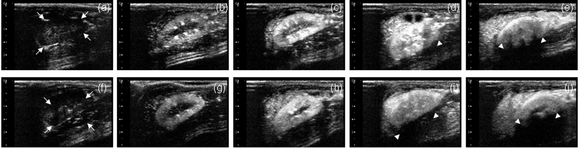
Rat kidneys (arrows) imaged in pulse subtraction harmonic imaging (PSHI) mode before injection of ST44 and ST68 (a and f, respectively) and after injection (b-e and g-j, respectively). Dosages are 0.1, 0.2, 0.8, and 1.0 mL/kg from left to right (ie, b-e and g-j). At lower dosages, the contrast enhancement clearly outlines the entire kidney (b-c and g-h), before becoming excessive and introducing signal loss in the form of shadowing (arrow heads). All images were acquired 6 seconds after contrast administration. The depth scale on the left of each image is in centimeters.
Discussion and Conclusion
We conducted an acute toxicity study of the ultrasound contrast agents ST68 and ST44 in adult rats. Relatively few of the blood, urine, and histopathological parameters evaluated varied significantly with the test article (2 and 8 for ST68 and ST44, respectively) and even fewer demonstrated a dose dependency (2 for ST68 and 1 for ST44). The survival analysis did not demonstrate a statistically significant effect due to the contrast agents. Nonetheless, the fact that 10% of the animals injected with ST44 (4 rats) died compared with none of the controls and 1 of the rats receiving ST68, as well as the dose dependency observed for ST44, indicates that the prudent choice for future nonrodent, toxicology studies and potentially for human clinical trials is ST68 (a conclusion that converges well with the finding that ST68 produced superior images in PDI mode, which is most likely due to differences in the size distribution of the 2 agents, as documented elsewhere. 12
The significant anemia observed in some of the animals injected with ST44 (Table 4) along with the protein and blood in the urine samples (Table 5) warrants further investigation of potential renal complications in a toxicology study in a nonrodent species, especially when the renal medullary mineralization of many animals is considered as well. Moreover, it was not possible to establish a no observable adverse effect level (NOAEL) for ST44 in this project, because even the lowest dose studied (0.1 mL/kg) resulted in significant giant cell infiltration of the lungs. Conversely, ST68 demonstrated minimal to no toxicity below 0.8 mL/kg, indicating that the NOAEL for ST68 appears to be 0.2 mL/kg. Hence, the results of the current toxicology study in a single rodent species and the in vivo imaging studies (eg, Figures 2 and 3 and references 11 and 12) indicate that the surfactant stabilized contrast agent ST68 might achieve similar results in humans with a potentially safe dose of 0.1 mL/kg, where good image enhancement and limited contrast artifacts 21 were observed (assuming that any new areas of concern are not discovered in future nonrodent toxicology studies).
Nonclinical toxicology associated with the constituent chemicals of ST68 and ST44 (ie, Spans and Tweens) is not well studied. Many Spans, including Spans 40 and 60, are dispersible rather than soluble and therefore are less used. The majority of the studies on Span have been for food or cosmetic uses, and Spans are described as generally mild skin irritants but nonsensitizers in animals. 23 Carcinogenic studies of Span 60 were negative. 24 Tween 80 is considered by the FDA as generally recognized as safe (GRAS) and is therefore exempted from the usual food additive tolerance requirement of the Federal Food, Drug, and Cosmetic Act, although Tween 80 has been shown to cause hemolysis and to demonstrate cytotoxicity toward Hepg2 cells in culture. 25 However, to the best of our knowledge, acute toxicity studies such as ours for either surfactant individually or in combination have not been reported in the literature, rendering a direct comparison of the effect of combining them in an ultrasound contrast agent impossible.
There are also relatively few reports in the literature describing preclinical toxicity and safety studies of ultrasound contrast agents. Most studies have been mechanistic in nature, dealing with microbubble uptake in the reticuloendothelial system of the liver 26-28 and lymph nodes 29 or the lack of same. 30 However, the research by Kindberg and colleagues 31 demonstrated in rats that the hepatic clearance of a particular PFC-filled contrast agent (Sonazoid; GE Healthcare, Oslo, Norway) did not impede the liver’s ability to phagocytose other particles. Several groups have described the pharmacokinetics of different commercial ultrasound contrast agents using gas chromatography or radiolabeled microspheres. 32-35 All found that more than 90% of the gas was exhaled within 24 hours, which is in accordance with subsequent results obtained in humans. 36,37
An extensive toxicology study of a commercial ultrasound contrast agent (Optison; GE Healthcare, Princeton, NJ) was performed by Greener and coworkers. 38 They reported on genetic toxicology as well as single-dose and repeated-dose toxicology studies performed in rats, dogs, and monkeys for dosages of 0.25, 5.0, 10, 20, and 25 mL/kg (0.25 mL/kg is the highest possible clinical dose). Compared with controls, there were no adverse effects in the acute, single-dose rat studies (N = 46) at dosages of 0.25, 5.0, and 20 mL/kg. At the highest dose administered (ie, 25 mL/kg) 3 animals died following breathing problems. 38 This is a very similar result to our study, where 1 and 4 animals died (for ST68 and ST44, respectively) and a statistically significant suspected foreign body reaction was observed in the lungs following injection of either contrast agent (P < .01).
It is conceivable that more effects could have been induced in our study by the addition of ultrasound pressure waves (ie, by imaging the animals with ultrasound). Many researchers have reported that the interactions between contrast microbubbles and an ultrasound field can produce bioeffects, in particular for higher acoustic pressures corresponding to a mechanical index greater than 0.4 (see the review by Miller et al 39 ). However, safety data from 5069 consecutive patients who underwent stress echocardiography (2914 with contrast administration) showed that although the contrast group had a higher cardiac risk profile, the rate of major adverse events was very small and no different from controls. 4 The 2 largest safety studies published on the use of ultrasound contrast agents in humans (involving 58 397 and 78 383 subjects, respectively) concluded that these agents have a good safety profile in both cardiac and abdominal ultrasound applications. 2,5
Multiple comparisons, such as those conducted in this study, may create some additional statistical uncertainty, because the more comparisons made, the more likely it is that one will be significant by chance. Some statisticians advocate solving this problem with a Bonferroni adjustment, which assigns the traditional P value of .05 divided by the number of comparisons to be the P value of significance. 40 However, others argue that such adjustments create more problems than they solve, for example, an increased likelihood of finding true differences to be nonsignificant. 41 We decided to adopt the latter strategy in reporting our results to avoid missing significant effects due to these contrast agents.
In conclusion, although this acute-toxicity rat study found relatively few parameters affected by the ultrasound contrast agents ST68 and ST44, there were concerns regarding the lack of a NOAEL and the potential for complications in the lungs and kidneys with ST44. Thus, the prudent choice for future nonrodent toxicology studies and potentially for human clinical trials is ST68, especially given the deaths in the ST44 group and the greater number of parameters that varied significantly following administration of ST44.
Footnotes
Acknowledgment
We gratefully acknowledge the equipment support provided by Toshiba America Medical Systems.