
Abstract
Monoclonal antibodies (mAbs) are an important class of biotherapeutics. Successful development of a mAb depends not only on its biological activity but also on its physicochemical properties, such as homogeneity and stability. mAb stability is affected by its formulation. Among the many techniques used to study the stability of mAbs, differential scanning fluorimetry (DSF) offers both excellent throughput and minimal material consumption. DSF measures the temperature of the protein unfolding transition (Tm) based on the change in fluorescence intensity of the environmentally sensitive dye SYPRO Orange. With DSF adapted to a 96-well plate format, we have shown that low-pH or high-salt concentrations decrease the thermal stability of mAb1, whereas some excipients, such as sucrose, polysorbate 80, and sodium phosphate, increase its stability. The basal fluorescence of SYPRO Orange was enhanced by the presence of detergents, limiting the use of this approach to diluted detergent solutions. Throughput of DSF can be increased further with the use of a 384-well plate. DSF thermograms are in good agreement with the melting profiles obtained by differential scanning calorimetry (DSC). The Tms determined by DSF and DSC were well correlated, with the former being on average lower by 3 °C.
Keywords
Introduction
Antibodies are heterodimers composed of 2 heavy and 2 light polypeptide chains linked by disulfide bonds. The framework of an antibody is divided into the Fc region (fragment crystallizable) and Fab region (fragment antigen binding), which are built of folded domains derived from the heavy- and light-chain proteins. The Fc, for example, contains heavy-chain constant regions 2 and 3, denoted CH2 and CH3, respectively. 1 Antibodies are classified into 5 isotypes based on the identity of the heavy-chain constant region: immunoglobulin A (IgA), IgD, IgE, IgG, and IgM. Of these classes, IgGs constitute the majority of the recombinant antibodies currently under development. There are 4 subclasses of IgGs (1 to 4). 1
The number of therapeutic monoclonal antibodies (mAbs) in development has increased considerably since the approval of the first mAb, muromonab-CD3, in 1986. 1 The fast growth of the therapeutic antibody market is related to the favorable properties of these macromolecules: They are highly specific and effective at low concentrations, cause few side effects, can be modified by attachment of therapeutic or diagnostic molecules, and can be engineered to have a human framework. 1 Despite their popularity, many mAbs display poor biophysical properties, such as low chemical and physical stability, resulting in unwanted modifications, degradation, and aggregation during manufacturing, storage, and dosing. 2 Poor stability can lead to antibody misfolding, resulting in a low product yield and a substantial fraction of inactive material, as well as degradation during storage.1–3 Aggregation can lead to hypersensitive immune response, including anaphylaxis, loss of biological activity, and occurrence of nonresponders, due to the formation of neutralizing antibodies against aggregates or degradants. 3 Therefore, successful development of mAbs depends not only on the attainment of the desired biological activity but also on superior physicochemical properties (e.g., homogeneity and physical and chemical stability). Although antibodies share similar tertiary structure, they display notable differences in their primary sequence, which leads to significant differences in mAb susceptibility to deamidation, isomerization, oxidation, denaturation, and aggregation. 1 These properties can be improved through additional sequence engineering of the mAb and by the development of optimized formulations.3,4 When developing mAb formulations, one needs to consider the therapeutic Ab dosage form (lyophilized or in solution), mAb concentration, ionic strength of the solution, pH, type of buffering substance, and presence of excipients (salts, sugars, polyols, amino acids, surfactants, polymers, antioxidants, preservatives, etc.).1,2,4 Because of the complexity of mAb molecules and the multiple variables to be considered, development of protein formulation is generally an empirical process. Moreover, this process could benefit from fast and miniaturized screening approaches. Techniques typically applied to analyze proteins include size exclusion chromatography, capillary electrophoresis, light scattering, circular dichroism, intrinsic fluorescence spectroscopy, mass spectrometry, and thermal analysis: differential scanning calorimetry (DSC) and differential scanning fluorimetry (DSF).2,3,5,6 Only some of these methods are amenable to high throughput.
DSF is a biophysical technique that is suitable for high-throughput screening with a minimal protein requirement. DSF estimates the temperature of the protein unfolding transition (Tm) by measuring the change in the fluorescence intensity of an environmentally sensitive fluorophore added to the solution. 7 Such molecules display low fluorescence in a hydrophilic environment. In most folded proteins, hydrophobic regions are sequestered from the solution within the protein core. During denaturation, proteins lose their tertiary and secondary structures leading to exposure of the hydrophobic regions. These regions can bind an environmentally sensitive dye leading to an increase in its fluorescence.5,7
Here, we analyzed the impact of formulation on the thermal stability of mAb1 with DSF adapted to a 96-well plate format using SYPRO Orange dye and a real-time PCR system. With this setup, up to 32 conditions could be simultaneously screened in triplicate in less than 75 min. Throughput of DSF could be further increased with a 384-well plate format. Effects of formulation components revealed by DSF are in agreement with general trends for physical stability of Abs reported in the literature.
Material and Methods
Reagents
Reagents were prepared with chemicals of the highest quality commercially available and water treated with a Milli-Q purification system (Millipore, Bedford, MA). SYPRO Orange fluorophore was purchased from Life Technologies (Grand Island, NY) as a 5000× concentrated stock solution in DMSO. Polysorbate 80 (PS80), polysorbate 20 (PS20), sucrose, histidine, sodium chloride, sodium succinate, citrate, and sodium phosphate (di- and monobasic) were obtained from Sigma-Aldrich (St. Louis, MO).
Monoclonal Antibody
Antibodies were manufactured by Gilead Sciences, Inc. (Foster City, CA). The human monoclonal antibody Ab1 (mAb1, subtype IgG4) was purified by protein A capture, low pH elution, viral inactivation, cation and anion exchange chromatography, and viral filtration, and concentrated and filtrated by ultrafiltration and diafiltration to 10 mg/mL in 10 mM sodium phosphate buffer, pH 5.8. Mouse monoclonal antibodies Ab2 (mAb2, IgG2b) and Ab3 (mAb3, IgG1) were purified by MabSelect SuRe (GE Healthcare, Piscataway, NJ) protein A affinity chromatography, followed by concentration by ultrafiltration to 10 mg/mL and dialysis to 20 mM sodium phosphate, pH 6.5, 140 mM sodium chloride, 0.01% PS20. The molecular weights and isoelectric points (pIs) of mAb1, mAb2, and mAb3, calculated based on the amino acid composition, were 145 kDa and 7.9, 146 kDa and 7.7, and 144 kDa and 7.9, respectively.
Monoclonal Antibody Samples Preparation
Samples of mAb1 were prepared in various formulations by dialysis throughout a period of 16 h with 2 buffer changes each with volume exceeding 1000 times the volume of the antibody sample. After dialysis, concentration of mAb1 was measured by ultraviolet-visible spectroscopy using a calculated extinction coefficient of 1.45 (mg/mL)−1. When necessary, samples were concentrated with the use of Amicon Ultra-15 centrifugal filter devices, and their concentrations were adjusted to 10 mg/mL with dialysis buffer.
Differential Scanning Fluorimetry
DSF was carried out in a ViiA7 real-time PCR instrument (Life Technologies) in the respective formulation buffer. Each sample was measured in triplicate. Unless otherwise specified, 96-well MicroAmp Fast reaction plates (Life Technologies) were used with 25 µL sample per well. All concentrations are final after mixing. MAbs were diluted to 1 mg/mL concentration in the respective formulation buffer, unless otherwise stated. SYPRO Orange was diluted 1000-fold from the 5000× concentrated stock to the working dye solution in the appropriate formulation buffer prior to addition to the mAb samples. To prevent bleaching, the working solution of SYPRO Orange was added to the reaction mixture just prior to the experiment. Thermal denaturation was carried out by increasing the temperature from 25 °C to 95 °C at a rate of 0.017 °C per second. Fluorescence intensity (excitation at 490 nm and emission with the use of a ROX filter at 600 to 630 nm) was collected at 0.07 °C intervals and analyzed with Protein Thermal Shift Software (Life Technologies), using the first derivative approach to calculate Tm. In this method, Tm is the temperature corresponding to the maximum value of the first derivative of the DSF melting curve. Delta Tm values (ΔTm) for various formulation buffers were calculated by subtracting the Tm value obtained for mAb1 in a reference formulation from the Tm value obtained for mAb1 in the tested formulation.
Differential Scanning Calorimetry
DSC measurements were performed on a Microcal VP-Capillary DSC platform (GE Healthcare). The mAb1 samples were diluted in respective formulation buffer to a concentration of 1 mg/mL. Matched formulation buffer was used as a reference. The samples were scanned from 20 °C to 95 °C at a rate of 1 °C/min using a filtering period of 8 s and 15 min of pre-equilibration prior to the start of each run. Data were first normalized for protein concentration, then baseline corrected and buffer subtracted using Origin 7.0 AutoSampler DSC software (GE Healthcare). Melting transitions were irreversible and were analyzed with cursor-initiated DSC peak fit function using the non-two-state unfolding model within the Origin 7.0 software.
Results and Discussion
Applicability of DSF for Studying the Thermostability of mAb
One of the important properties of successful mAbs is their conformational stability (i.e., the ability to retain their native secondary, tertiary, and quaternary structures).1,2 Conformational stability can be assessed by measuring thermal stability with the use of techniques sensitive to changes in protein folding, such as circular dichroism, fluorescence spectroscopy, light scattering, DSC, and DSF. A representative thermal unfolding profile (thermogram) of mAb1 obtained by DSF is depicted in Figure 1A . At lower temperatures, at which antibody remains folded, fluorescence intensity was very low, in agreement with the presence of mostly hydrophilic amino acid residues on the surfaces of immunoglobulins under native conditions. Increase in the temperature higher than 55 °C was accompanied by a sharp increase in fluorescence intensity that reached a plateau and ultimately decreased with further increase in the temperature. The initial rise in fluorescence reflects greater exposure of the hydrophobic areas of mAb1 because it undergoes thermal unfolding. Quenching of the fluorescence in the terminal phase of the experiment was shown to be a nonspecific transition associated with a gradual decrease in the inherent fluorescence of SYPRO Orange in a hydrophobic environment at high temperatures. 5 The thermogram for mAb1 displays 2 transitions, which are better visualized in the plot of the first derivative of the fluorescence melting curve (dF/dT; Fig. 1B ). Temperature of the unfolding (melting) transition (Tm) was determined for mAb1 from the value of the local maximum of the dF/dT curve. At 1 mg/mL protein concentration in formulation containing 10 mM sodium phosphate, pH 5.8, 140 mM sodium chloride, 0.02% PS80, the lower and the higher temperature transitions occurred with Tm1 and Tm2 values of 64.9 °C and 71.5 °C, respectively. These multiple temperature-dependent transitions result from the fact that individual domains of the monoclonal antibodies have distinct conformational stabilities and display various levels of the intramolecular interactions with other fragments of the protein, which leads to the differences in cooperativity of domains unfolding. 8
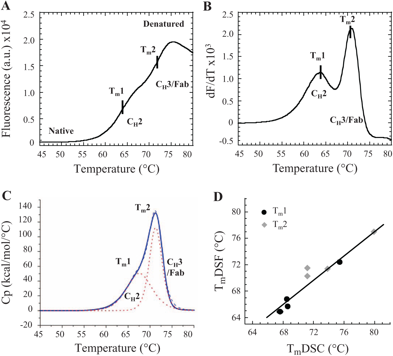
Comparison of thermal unfolding transitions and melting temperatures (Tms) determined for monoclonal antibody Ab1 (mAb1) by (
A thermogram for mAb1 was also obtained by DSC ( Fig. 1C ). Similar to the DSF unfolding profile, the DSC melting curve displayed 2 transitions with Tm1 and Tm2 equal to 67.4 °C and 71.3 °C, respectively (1 mg/mL, in 10 mM sodium phosphate, pH 5.8, 140 mM sodium chloride, 0.02% PS80). Studies have shown that in DSC, mAbs display distinctive unfolding profiles as a result of melting transitions of their individual domains: CH2, CH3, and Fab. The thermal stability of IgG molecules is affected by all of these domains.8–11 DSC curves reported for antibodies of IgG4 subtype display 2 clearly discernible transitions: a lower temperature representing unfolding of the CH2 domain (Tm1), and a higher temperature representing melting of CH3–Fab domains (Tm2).9,10 Fab melting transition is generally well defined with a peak up to 3 times greater than that of CH2 or CH3, which was also true in the case of mAb1.8–10
The temperature for the first transition of mAb1 obtained by DSC was slightly higher than Tm1 determined by DSF. The same trend was observed for mAb1 and other antibodies tested in several additional formulations (
Fig. 1D
,
Adaptation of DSF for mAb Formulation Screening
To evaluate the performance and robustness of Tm determination by DSF, the thermal stability of mAb1 was studied in the presence of increasing concentrations of SYPRO Orange, at various concentrations of antibody, and in the presence of increasing amounts of PS80 ( Fig. 2 ). SYPRO Orange is supplied as a 5000-fold concentrated stock solution in 100% DMSO. Using the dye at concentrations from 1.25- to 10-fold did not change the values of the Tms for mAb1 ( Fig. 2A ). Increasing the concentration of SYPRO Orange increased the fluorescence intensity in the plateau of the melting profile and in the peak of the first derivative curve, resulting in a higher signal-to-noise ratio and better defined transitions. The background fluorescence increased only marginally at the highest SYPRO Orange concentration (data not shown). For the remainder of this study, SYPRO Orange was used at 5-fold concentration.
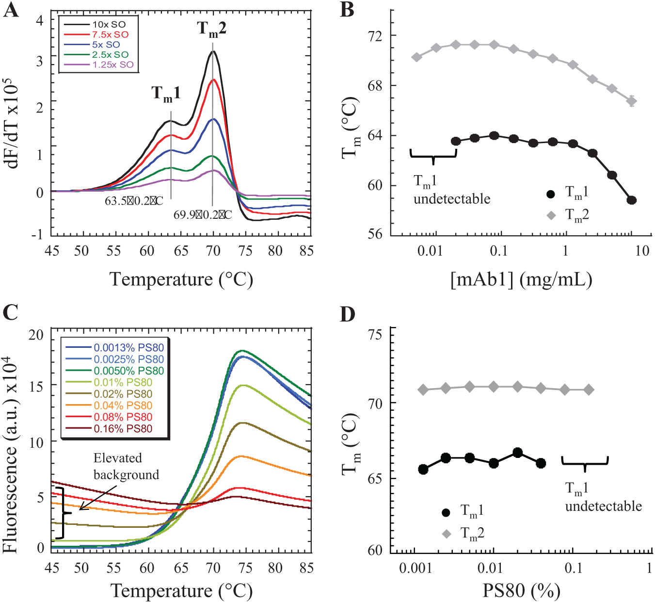
Development of a differential scanning fluorimetry (DSF) thermostability assay to measure the unfolding of monoclonal antibody Ab1 (mAb1). The DSF assay was performed for mAb1 at 1 mg/mL concentration in buffer containing 10 mM sodium phosphate, pH 5.8, 140 mM sodium chloride, 0.005% polysorbate 80 (PS80), and 5× SYPRO Orange (SO) unless otherwise stated. (
The study depicted in
Figure 2B
confirms that DSF can be performed at a wide range of protein concentrations, from 10 mg/mL to as low as 5 µg/mL. At very low protein concentrations (lower than 20 µg/mL), the first melting transition was not well defined, and its midpoint could not be accurately determined; however, the second transition (associated with an overall larger fluorescence change) remained well defined. This shows one of the strengths of DSF: Unlike DSC, DSF can be successfully applied even with limited amounts of material. Dynamic light scattering (DLS) and analytical ultracentrifugation (AUC) showed that mAb1 did not aggregate in the protein concentration range used in these studies and that the main state of the protein is monomeric (
Some antibody formulations contain surfactants, such as Poloxamer 188/407, polysorbate 20/40/80, or sodium lauryl sulfate, to prevent protein aggregation, surface denaturation, and surface adsorption.
4
Surfactants possess hydrophobic groups to which SYPRO Orange could bind, contributing to an increase in fluorescence background. We tested the effect of surfactant on the performance of the DSF assay by varying PS80 from 0.0013% to 0.16% (
Fig. 2C
). With an increase in PS80 concentration, the initial fluorescence (background) increased and the profile of the mAb1 thermal unfolding became distorted (
Fig. 2C
). Nevertheless, the Tm for unfolding transitions could be still determined reliably from the first derivative of the thermograms (
Effects of pH, Buffer Type, and Excipients on Thermal Stability of mAb Revealed by DSF
The effect of sodium chloride concentration on the stability of mAb1 is shown in
Figure 3A
. As the concentration of sodium chloride was increased from 10 mM to 1000 mM, the unfolding transition temperatures shifted to lower values, indicating destabilization of the antibody structure. The first unfolding transition, Tm1, was more sensitive than the second transition, Tm2, to the increase in salt concentration. Increasing sodium chloride to 1000 mM resulted in a decrease in Tm1 and Tm2 by 6.3 ± 0.4 °C and 1.9 ± 0.3 °C, respectively. At low salt concentrations, the thermal unfolding transitions of domains CH2 and CH3–Fab of mAb1 were more separated, suggesting that unfolding of antibody domains is less cooperative (
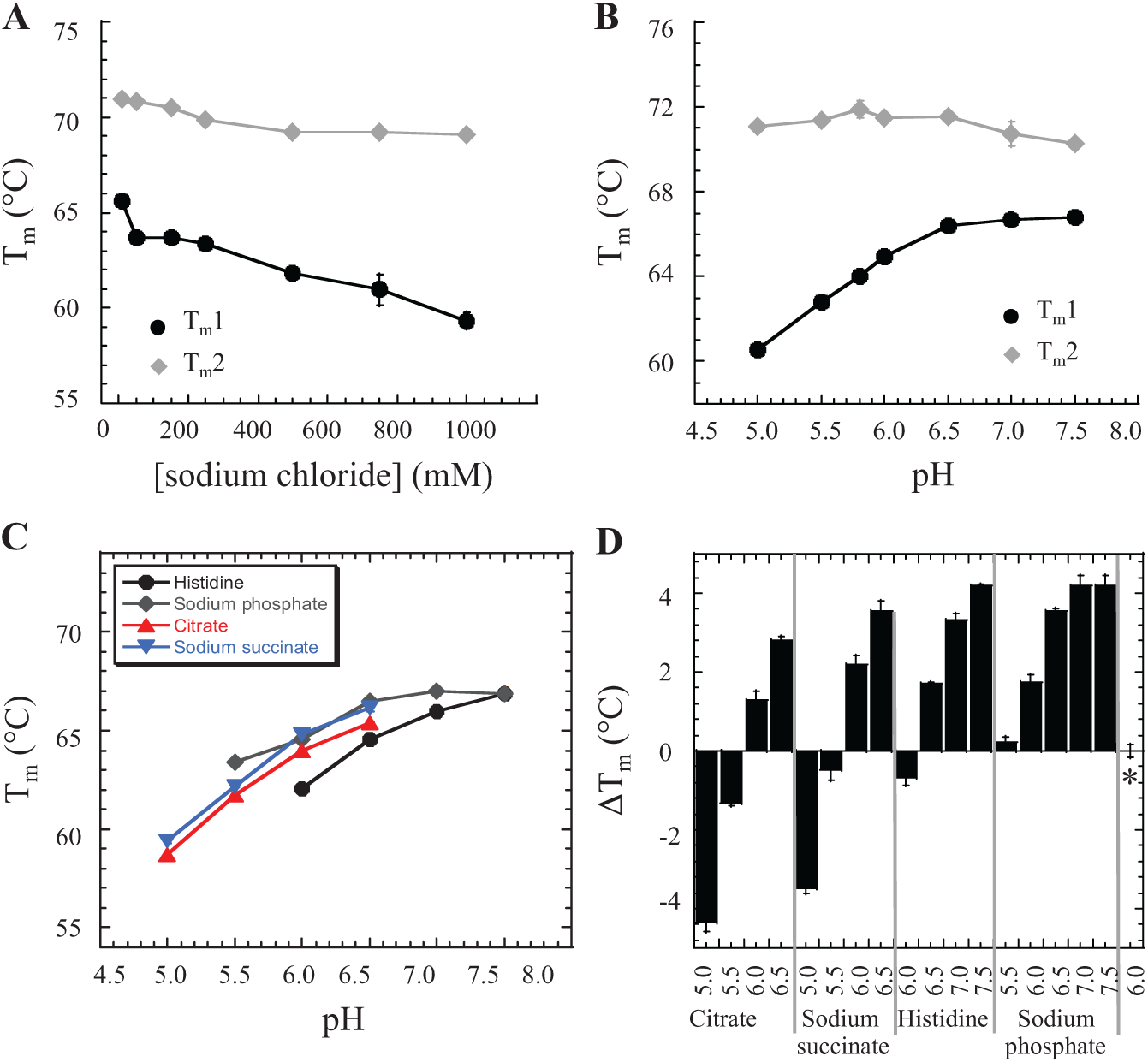
Effects of (
The pH of the formulation also had a substantial influence on the thermal stability of mAb1, especially its CH2 domain. At 1 mg/mL protein concentration in 10 mM sodium phosphate, Tm1 increased by 6.2 ± 0.1 °C and Tm2 decreased by 0.8 ± 0.1 °C as pH increased from 5.0 to 7.5 (
Fig. 3B
and
The effect of excipients such as 0.01% PS80, 10% sucrose, or both on the thermostability of mAb1 is summarized in
Extension of DSF Assay Throughput
The throughput of antibody formulation screening using DSF could be further increased by use of a 384-well plate format. With a 384-well plate setup, up to 128 conditions can be simultaneously screened in triplicate, in less than 75 min, without compromising the sensitivity and reproducibility of the assay.
Figure 4
shows correlations between Tm1 and Tm2 measured for mAb1 in 17 different formulations by DSF in 96-well and 384-well plates. Formulations components and the position of the samples in plates are depicted in
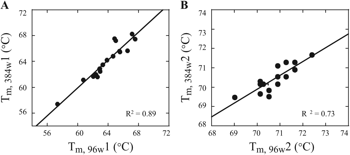
Correlation between melting temperatures (Tms) measured with the use of differential scanning fluorimetry (DSF) in 96-well and 384-well plate formats. Seventeen formulations (
In summary, our study ratifies that DSF is a viable tool for antibody characterization, offering the advantages of rapid method development, a simple format, high-throughput, and minimal sample consumption. The parameters measured by DSF correlate well with those from more established techniques like DSC. DSF can be used productively at multiple stages in the mAb development process, from the screening of early leads in search of the most robust scaffold to the late stage of formulation optimization.
Footnotes
Acknowledgements
The authors would like to thank Dr. Katherine M. Brendza and Dr. Hyock Joo Kwon for critical comments on the manuscript.
Declaration of Conflicting Interests
The authors declared no potential conflicts of interest with respect to the research, authorship, and/or publication of this article.
Funding
The authors received no financial support for the research, authorship, and/or publication of this manuscript beyond normal compensation for employees. The research was supported by internal funding.
References
Supplementary Material
Please find the following supplemental material available below.
For Open Access articles published under a Creative Commons License, all supplemental material carries the same license as the article it is associated with.
For non-Open Access articles published, all supplemental material carries a non-exclusive license, and permission requests for re-use of supplemental material or any part of supplemental material shall be sent directly to the copyright owner as specified in the copyright notice associated with the article.