
Abstract
Tissue factor (TF), the primary initiator of the coagulation cascade, plays a critical role in hemostasis and thrombosis, and inhibition of TF activity appears to be an attractive target for the treatment of cardiovascular diseases. However, few selective small-molecule inhibitors of TF are available, and the present assays for measuring TF activity are relatively expensive and complex. The authors present a simple and high-throughput chromogenic assay for screening TF inhibitors based on using commercial human prothrombin complex instead of purified coagulation factors, reducing the dosage, and performing with a one-stage procedure. In the optimized assay, <45 µL cell lysates was incubated with Tris-CaCl2 buffer (pH 7.3) containing human prothrombin complex at 37°C for 15 min in 96-well or 384-well plates. Tris-EDTA buffer (pH 8.4) containing chromogenic substrate Xa was then added and the absorbance measured at 405 nm. This simplified assay was more sensitive or precise than some reported methods for TF procoagulant activities. Two known active compounds (curcumin and simvastatin) inhibiting TF activity were tested by the simplified assay to validate the screening method. Furthermore, berberine and cryptotanshinone suppressed TF activity induced by lipopolysaccharides in human monocytes by this assay and might be promising new TF inhibitors.
Keywords
Introduction
T
It is known that excessive inhibition of the coagulation cascade at the final stage (factor Xa, thrombin) can lead to bleeding complications. In contrast to classic antithrombotic drugs, inhibition of TF targets the first steps of coagulation while leaving the downstream effectors intact. Therefore, inhibition of TF results in an antithrombotic effect without enhancing bleeding propensity. Recently, small-molecule inhibitors of TF have been the point of much research effort. 8
However, screening TF inhibitors need suitable assays to measure TF activity in vitro. TF procoagulant activity (PCA) is frequently measured by functional methods (i.e., clotting time and chromogenic assays). The clotting methods are simple but time-consuming, are unstable, and necessitate large numbers of cells (1 × 10⁷ cells/mL). 9 Chromogenic assays reported so far seem convenient and sensitive but comparably expensive because purified coagulation factors or specific substrates to factor Xa are needed and/or involve several incubation steps and thus cannot be easily applied to a large number of tests in the same experiment. 10 Some researchers have used prothrombin complex (PPSB, containing coagulation factors II, VII, IX, and X) as a source of coagulation factors to measure TF activity in plasma, 11 cerebrospinal fluid, 12 and monocytes. 13 However, the amounts of reagents were relatively large or the procedures were complicated, which was not ideal for high-throughput screening (HTS) of TF inhibitors in vitro.
Therefore, the purpose of the present study was to optimize a simple, sensitive, and low-cost chromogenic assay on a limited number of cells in microplates, based on the use of commercial human prothrombin complex. The simplified assay could be used conveniently in HTS of TF inhibitors in vitro in 96-well plates and even in 384-well plates.
Materials and Methods
Materials
Ficoll-Hypaque solution (density: 1.077–1.080) was obtained from Haoyang Biological Manufacture Company (Tianjin, China). RPMI 1640 medium was from GIBCO-BRL Life Technologies (Invitrogen, Carlsbad, CA). LPS (from Escherichia coli O55:B5), TNF-α, recombinant human tissue factor, and chromogenic substrate Xa (CH3OCO-D-CHA-Gly-Arg-pNA-AcOH) were obtained from Sigma (St. Louis, MO). Human prothrombin complex (300 IU, 1 IU/mg, containing factors II, VII, IX, and X) was from Hualan Bioengineering Company (Xinxiang, China). Monoclonal antihuman TF antibody was purchased from R&D Systems (Minneapolis, MN). Simvastatin and curcumin were gifts from Professor QiujuanWang of China Pharmaceutical University. Natural products used in this study were obtained from the National Institute for the Control of Pharmaceutical and Biological Products (Beijing, China) or were obtained from our department, and purity was analyzed by high-performance liquid chromatography (HPLC) to be at least 98%. All other reagents were of analytical grade.
Monocyte isolation and culture
The peripheral blood mononuclear cells (PBMCs) were isolated from blood as described earlier. 14 Briefly, 20–50 mL of blood drawn from healthy volunteers into a plastic tube containing heparin (a final concentration of 10 U/mL) was diluted with an equal volume of phosphate-buffered saline (PBS) and then layered over Ficoll-Hypaque solution followed by density gradient centrifugation at 250 g for 15 min at room temperature. The middle buffy layer was collected and then washed twice with PBS by centrifugation at 500 g for 10 min. The cell pellet was suspended in RPMI 1640 medium supplemented with 10% heat-inactivated newborn calf serum and then incubated at 37°C in 5% CO2 for 1 h in 6-well culture dishes (Corning Costar, Corning, NY) allowing monocytes to adhere to the dishes. The cells were then washed twice in Hank’s balanced salt solution (HBSS) and resuspended in 10% RPMI 1640 medium in 6-well plates to yield approximately 80%–90% monocytes as determined by nonspecific esterase staining.
Endothelial cells culture
Human umbilical vein endothelial cells (HUVECs) were isolated from fresh human umbilical cord according to the protocols previously reported, 15 and primary cultures of HUVECs were cultured at 37°C under 5% CO2 in T-75 flasks in M199 medium supplemented with 20% fetal calf serum, 1% penicillin-streptomycin, 1% L-glutamine, 40 mg/mL endothelial cell growth supplement (ECGS), and 15 U/mL heparin. The cells were subcultured by first detaching the cells with trypsin solution and replating them in 24-well culture dishes or in T-75 flasks that were coated with human fibronectin (0.65 mg/cm2). The monolayers were used within 24 h after they reached confluency. Passages between 3 and 6 were used in the present experiments.
Induction of TF and drug treatment
Primary cultures of PBMCs or HUVECs were incubated with control vehicle (PBS) or stimulants such as LPS or TNF-α for 5 h. 16 When testing effects of compounds, they were added at indicated concentrations 1 h before LPS incubation. All incubations were carried out at 37°C and 5% CO2, and at the end of incubation, cells were sedimentated by centrifugation and resuspended in RPMI medium. The cell suspension was frozen at −80°C until measurement of TF procoagulant activity.
Simplified chromogenic assay of TF procoagulant activity and optimization of related factors
The cell lysates were frozen and thawed three times before they were used in the assay. Cell lysates (45 µL) were incubated with Tris-CaCl2 buffer (5 µL, pH 7.3) containing human prothrombin complex in a 96-well plate (Corning Costar) and incubated at 37°C (5–30 min). Then, 50 µL of Tris-EDTA buffer (pH 8.4) containing chromogenic substrate Xa was added to each well, and 3 min later, absorbance at 405 nm was measured with a microplate reader (Sunrise, TECAN Austria GmbH, Grödig, Austria) at 37°C.
Several parameters were defined, including the number of cells (2–4 × 10⁵ cells/well) plated in wells, the concentrations of prothrombin complex (5–40 g/L) and factor Xa chromogenic substrate (0.0625–1 mM), the concentrations of CaCl2 (50, 100, or 200 mM) and EDTA (50, 100, or 200 mM), and the incubation time (5–30 min).
When testing effects of compounds, TF activity obtained in cells pretreated with a control vehicle (0.1% of DMSO) and then stimulated with LPS (100 ng/mL, 5 h) or TNF-α (100 ng/mL, 5 h) was taken as 100%, and the inhibition rate of tested compounds was calculated as (ODmodel – ODsample)/(ODmodel – ODnormal) × 100%. The relative IC50 values of natural compounds inhibiting TF activity were calculated using Origin 7.5 software according to the fitted dose–response curves.
Statistical analysis
Statistical analysis of data was performed using one-way analysis of variance (ANOVA) followed by Student t test. Data are expressed as mean ± SD. A probability value of less than 0.05 was considered statistically significant.
Results
Optimization of TF chromogenic assay based on prothrombin complex
To achieve the most reproducible and sensitive assay, several parameters were defined. The influence of different cell densities on TF activity of monocytes is illustrated in Figure 1a . TF activity was detectable with as few as 1 × 10⁵ human monocytes per well, and the high values were obtained with monocytes of 2–4 × 10⁵ cells/well. The data ( Fig. 1b ) showed that the OD405 increased in proportion to the concentrations of factor Xa chromogenic substrate at the range of 0.0625–1 mM, and 0.5 mM chromogenic substrate provided higher absorbance, which was elevated slightly above this concentration, similar to the previous report. 17
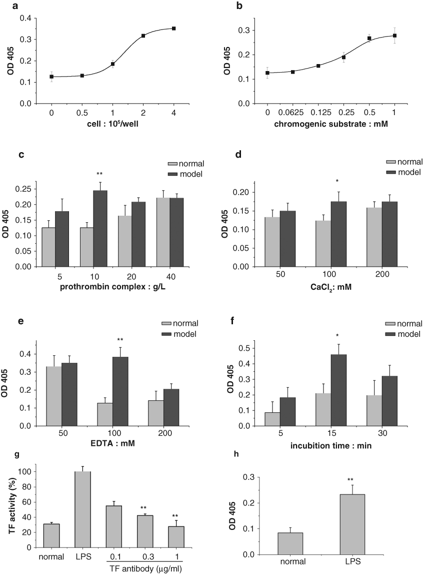
Effects of various factors of the simplified chromogenic assay on monocyte tissue factor (TF) procoagulant activity. (
As for the influence of various concentrations of prothrombin complex, human monocytes were incubated in the absence (normal) or in the presence of LPS (100 ng/mL) (model) for 5 h, and indicated concentrations of prothrombin complex (300 IU) were tested. The results showed that the difference in formation of TF between the normal and model group was significant at the concentration of prothrombin complex at 10 g/L. However, above and below that concentration, the difference was reduced ( Fig. 1c ).
Similarly, the difference of TF activity between the normal and model group was remarkable with the concentration of CaCl2 and EDTA at 100 mM and decreased at other concentrations ( Fig. 1d , e ). The data also showed that the highest value of TF activity was obtained after incubation for 15 min ( Fig. 1f ). In addition, the incubation of monoclonal antihuman tissue factor antibody for 30 min before testing PCA, which is specific and blocks TF, inhibited this PCA in a dose-dependent manner ( Fig. 1g ). Of note, we reduced the volume of reagents in proportion and tested TF activity in 384-well plates. As shown in Figure 1h , the tendency between the normal and model group was similar to that tested in 96-well plates.
Comparison of several assays measuring TF procoagulant activity
This simplified chromogenic assay was compared with a previously reported clotting time assay 18 by establishing a standard curve with purified human TF. The results showed that the linearity range was from 1.0 × 10⁻⁴ to 1.0 × 10⁻² nM TF corresponding to 0.14–0.55 OD405/min (y = 0.090 ln(x) + 0.983, r 2 = 0.996) in the simplified chromogenic assay (as shown in Fig. 2a ). The correlation was not so good in the clotting time assay (as shown in Fig. 2b ), whose linearity range was from 0.0125–0.4 nM TF corresponding to 21.4–40 s of clotting time (y = –4.79 ln(x) + 16.61, r 2 = 0.985) after taking the logarithm of TF concentration. The results showed that TF activity was detectable as low as 1.0 × 10⁻⁴ nM in our simplified chromogenic assay, whereas at least 0.01 nM TF was needed in the clotting time assay. It indicated that the sensitivity of this assay was nearly 100 times higher than that of the clotting time assay.
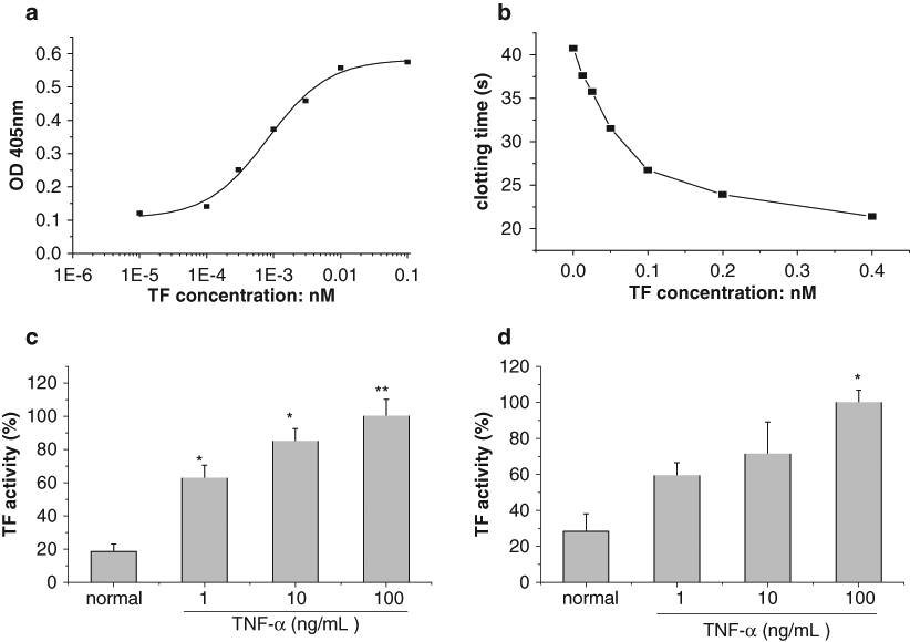
Comparison of several assays measuring tissue factor (TF) procoagulant activity. (
On the other hand, we also compared our modified chromogenic assay with one of the chromogenic assays reported previously 13 by detecting TF procoagulation activity stimulated with TNF-α. The results shown in Figure 2c , d indicate that effects of the two chromogenic assays were similar, but the standard deviation in the modified chromogenic assay was lower than in the latter.
Application of the simplified chromogenic assay: a pilot screen
First, we tested two active compounds (simvastatin and curcumin) that could inhibit TF activity as reported in previous studies 19–21 by our simplified chromogenic assay. The results showed that simvastatin and curcumin markedly suppressed LPS-induced TF activity at the concentrations of 0.01–10 µM (as shown in Fig. 3a , b ). These results were similar to the descriptions of Masato and Pendurthi, 20,21 which provided solid evidence for the validity of the assay.
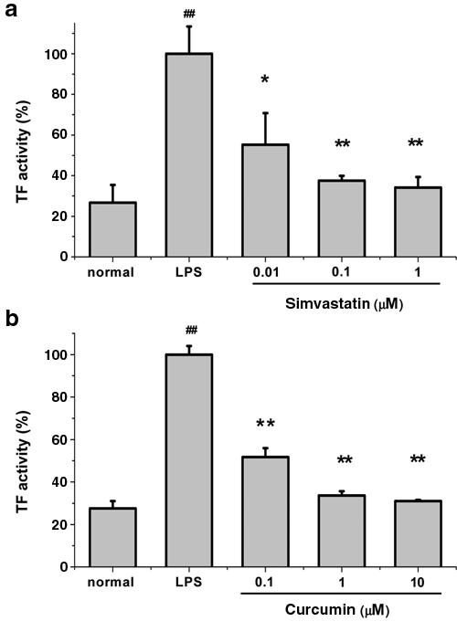
Effects of simvastatin and curcumin on tissue factor (TF) expression induced by lipopolysaccharides (LPS) in human monocytes. Monocytes (3 × 10⁵ cells/well) were preincubated for 1 h with (
Next, we screened from more than 50 kinds of natural compounds and found some new possible TF inhibitors by using this assay. These compounds included alkaloids, flavonoids, saponins, and so on, and they exhibited different potencies due to various structures. Figure 4 shows two representative natural compounds, including berberine and cryptotanshinone, which can significantly inhibit TF procoagulant activity induced by LPS at the concentrations of 0.01–10 µM.
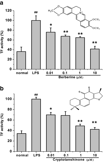
Effects and structures of two representative natural products inhibiting tissue factor (TF) activity in human monocytes. Two representative natural products, (
Furthermore, this modified chromogenic assay could also be easily applied in the measurement of TF activity to other cells. The results showed that LPS or TNF-α could elevate TF activity in a concentration-dependent manner in HUVECs (as shown in Fig. 5 ), which was consistent with previous reports. 22
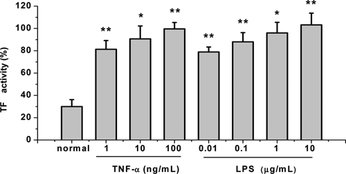
Effects of tumor necrosis factor (TNF)–α and lipopolysaccharides (LPS) on tissue factor (TF) procoagulant activity in human umbilical vein endothelial cells (HUVECs) by the simplified chromogenic assay. HUVECs were incubated with various concentrations of TNF-α and LPS for 5 h. TF activity was measured by the simplified chromogenic assay described in Materials and Methods, and TF activity obtained in cells pretreated with a control vehicle (0.1% of DMSO) and then stimulated with TNF-α (100 ng/mL, 5 h) was taken as 100%. All data presented are the mean ± SD (n = 4) of one representative experiment from two independent experiments. *p < 0.05 and **p < 0.01, compared to normal.
Discussion
The PCA of TF plays an important role in several pathologic conditions, such as disseminated intravascular coagulation, atherosclerosis, arterial and venous thromboembolism, cancer-related hypercoagulability, and immunopathologies, 23 and the assays for analyzing the PCA of TF on either monocytes or endothelial cells are potentially valuable tools, especially if they are simple and inexpensive. In the present study, we described a simple and low-cost chromogenic assay in 96- or 384-well plates to investigate the PCA of TF on monocytes and endothelial cells. The assay will be more suitable for screening of TF inhibitors in microplates.
Most conventional chromogenic assays reported so far use purified coagulation factor VIIa and factor X, which are comparably expensive and not ideal for HTS for TF inhibitors. On the basis of previous methods using commercial human prothrombin complex (PPSB, containing coagulation factors II, VII, IX, and X) as a source of coagulation factor VIIa and factor X, 11–13 we optimized the method and reduced the reagent consumption through investigating the effects of cell counts, the concentrations of prothrombin complex, factor Xa chromogenic substrate, CaCl2, EDTA, and the incubation time ( Fig. 1a – f ). Usually, 50–200 µL of prothrombin complex (the lowest concentration as 3 U/mL) was used in other assays, 11–13 but in our modified assay, we used 5 µL (about 10 U/mL), which was only one-third or one-twelfth of the former, so the total reagent amount and the cost were greatly decreased. Furthermore, we designed a one-stage procedure that involves simultaneous incubation of prothrombin complex containing CaCl2 with cells and then factor Xa–specific substrate, so the procedures were more simple and convenient.
The PCA measured in this assay was related only to a TF-dependent pathway because the TF antibody inhibited this PCA in a dose-dependent manner ( Fig. 1g ), which is specific and blocks TF. Furthermore, the commercial prothrombin complex only includes factors II, VII, IX, and X without thrombin or platelet. Therefore, the assay was specific in measuring TF-dependent PCA. As shown in Figure 1h , this simplified chromogenic assay was also suitable for measuring TF activities in 384-well plates.
Furthermore, we also proved that our modified assay was more sensitive than the clotting time assay, as the lowest limit of detection was 100 times less than that of the latter ( Fig. 2a , b ). This method was similar in detecting TF activity to the previous chromogenic assay, but its precision was higher, partly owing to the simplified operation steps ( Fig. 2c , d ).
To further validate our simplified assay, we also tested two known active compounds, simvastatin and curcumin, which showed similar inhibitory effects on TF procoagulant activity induced by LPS ( Fig. 3a , b ). By our simplified assay, several new possible TF inhibitors have been successfully identified from more than 50 kinds of natural compounds. Here, we showed that berberine, an alkaloid originally isolated from a Chinese herb (Coptis chinensis), and cryptotanshinone from Salvia miltiorrhiza Bge. significantly decreased LPS-induced TF activity with IC50s of 0.025 and 0.11 µM, respectively ( Fig. 4a , b ). These possible TF inhibitors found in our study might be new anticoagulants in clinical therapy in the future, and our previous findings also provide some new explanation for their treatment in cardiovascular diseases. 24 In the meantime, the simplified assay also could be used to measure TF PCA in HUVECs ( Fig. 5 ).
In addition, our recent work demonstrates that this assay can measure imminent TF activity in some tumor cells, such as the human breast cancer cell line MDA-MB-435. 25 It suggests that this assay can test TF activity not only in the cells that can express TF stimulated by cytokines or endotoxin but also in the cells that can highly express TF automatically, such as cancer cells. Recent reports have shown that advanced cancer is associated with a hypercoagulable state, and tissue factor expression by cancer cells has received widespread attention because of its significant contribution to the pathogenesis of cancer progression and metastasis. 26 Therefore, the assay in the present study also shows the possible perspective usage in screening drugs of anticancer metastasis.
In conclusion, we optimized a simple, low-cost, and one-stage chromogenic assay of TF procoagulant activity in microplates, based on the use of commercial human prothrombin complex, which showed more suitable or promising application in the HTS of TF inhibitors in vitro.
Footnotes
Acknowledgements
This work was partially funded by the National Natural Science Foundation of China for Youth (No. 30300451) and the Key Fundamental Research Funds for the Central Universities (No. JKZ200910).
References
Supplementary Material
Please find the following supplemental material available below.
For Open Access articles published under a Creative Commons License, all supplemental material carries the same license as the article it is associated with.
For non-Open Access articles published, all supplemental material carries a non-exclusive license, and permission requests for re-use of supplemental material or any part of supplemental material shall be sent directly to the copyright owner as specified in the copyright notice associated with the article.