
Abstract
Phagocytosis is a critical host defense mechanism that clears invading pathogens, apoptotic cells, and cell debris; it is an essential process for normal development, tissue remodeling, immune response, and inflammation. Here, a functional selection strategy was used to isolate novel phagocytosis-promoting genes. After the retroviral transfer of mouse brain cDNA library into NIH3T3 mouse fibroblast cells, cell sorting was used to select the cells that phagocytosed fluorescent zymosan particles. The cDNAs were retrieved from the selected cells and identified by DNA sequencing as eIF5A, Meg3, Tubb5, Sparcl-1, Uchl-1, Bsg (CD147), Ube2v1, and Pamr1. The phagocytosis-promoting activity for some of these cDNAs was confirmed by transient transfection in the independent phagocytosis assays. Thus, the unbiased selection procedure successfully identified multiple phagocytosis-promoting genes. The selection method can be applied to other cell-based assays where cells with a desired phenotype can be physically separated. Moreover, the new gene targets uncovered in this study could be relevant to biomolecule screening in search of phagocytosis-regulating agents. In a small-scale screen, a series of imidazopyridine compounds was tested to identify the small molecules that modulate eIF5A-mediated phagocytic activity. Several compounds that influenced the phagocytic activity can be further used as chemical-genetic tools to delineate the mechanisms of eIF5A action and be potential drug candidates that are capable of therapeutically modulating phagocytic activity.
Keywords
Introduction
P
Here, we describe the identification of novel phagocytosis-promoting genes based on the functional selection in cultured cells using cell sorting. The unbiased selection procedure successfully identified multiple phagocytosis-promoting genes. Although the isolated cDNAs need to be further characterized, the selection methods may be useful for the initial identification of genes with other physically separable phenotypes. The phagocytosis-promoting genes identified in this study could also be applied to the field of biomolecular screening to search for phagocytosis-regulating small molecules. In a small-scale screen, several imidazopyridine compounds were identified to modulate eIF5A-mediated phagocytic activity.
Materials and Methods
Cell culture
Phoenix Eco cells were cultured in Dulbecco’s modified Eagle’s medium (DMEM) containing 10% fetal bovine serum (FBS; Lonza, Walkersville, MD), hygromycin B (300 µg/mL), and diphtheria toxin (1 µg/mL) at 37°C and 5% CO2. NIH3T3 mouse fibroblast cells and J774 mouse macrophage cells were cultured in DMEM containing 10% FBS, penicillin (10 U/mL), and streptomycin (10 µg/mL) at 37°C and 5% CO2. All cell lines were obtained from American Type Culture Collection (Manassas, VA).
Retrovirus production and infection
A retroviral cDNA library was generated by transient transfection of Phoenix Eco packaging cells 11 with a ViraPort® XR Mouse Brain cDNA Library (Stratagene, La Jolla, CA) using Lipofectamine™ 2000 (Invitrogen, Carlsbad, CA) in accordance with the manufacturer’s instructions. Cell-free supernatants were harvested 2 to 3 days posttransfection and subsequently used to transduce NIH3T3 fibroblast cells in the presence of 8 µg/mL of polybrene. To optimize conditions of viral infection and to monitor virus titer, we used a test construct of the retroviral vector carrying green fluorescent protein (GFP; pFB-hrGFP; Stratagene). For infection of NIH3T3 fibroblast cells, cells (1 × 10⁵ cells/well) were seeded onto 60-mm culture plates 18 h prior to infection and incubated with 1 mL of virus stock for 6 to 8 h in the presence of polybrene (8 µg/mL). Then, 2 mL of fresh DMEM/10% FBS containing polybrene (8 µg/mL) was added to the culture, and the incubation continued. After another 24 h, cells were removed from the plates, and infection efficiency and viral titer was determined by the detection of GFP using fluorescence-activated cell sorting (FACS) or reverse transcription-polymerase chain reaction (RT-PCR; see below) with Ψ signal-specific primers (Ψ is the signal sequence for the initiation of virus packaging). The titer of the viral cDNA library was approximately 1.2 × 10⁶ pfu/mL. NIH3T3 fibroblast cells were infected with the retroviral library of mouse brain cDNAs at a multiplicity of infection (MOI) of 1.
Gene selection procedure
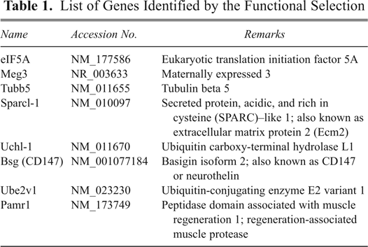
NIH3T3 fibroblast cells (1 × 10⁵ cells/mL) were infected with retroviral cDNA library at MOI of 1 in the presence of polybrene (8 µg/mL). Two days postinfection, the cells were incubated at 37°C for 12 h with 30 µg/mL of zymosan A (Saccharomyces cerevisiae) BioParticles conjugated with Alexa Fluor 594®™ (Molecular Probes, Eugene, OR). After cells were washed 5 times with ice-cold phosphate-buffered saline (PBS) to remove bound particles, phagocytic cells were either visualized under a fluorescent microscope (Axioplan2 imaging, Carl Zeiss, Jena, Germany) or sorted using FACSAria (BD Biosciences, San Jose, CA). Sorted cells were pooled and cultured together for 2 days and subjected to genomic DNA isolation to retrieve the sequences of the chromosomally integrated cDNAs by PCR. In a typical sorting experiment, 90 to 530 cells were isolated (0.002%-0.008%). More than 90% of the isolated cells were successfully grown for further experiments. The integrated cDNA segments were amplified by using a primer set (82F and 84R) that is specific for regions flanking the multiple cloning site of the pFB retroviral vector (backbone of ViraPort® XR Mouse Brain cDNA Library). The primer sequences were as follows: 82F, 5′-CTG CCG ACC CCG GGG GTG-3; 84R, 5′-CAG CTG TTC CAT CTG TTC CTG ACC-3′. PCR was carried out at a 61°C annealing temperature for 20 s and repeated 25 to 30 cycles. The resulting PCR fragments were purified and sequenced, and then identified cDNAs were cloned for further analysis. Eight genes were identified in the screen (
List of Genes Identified by the Functional Selection
Transient transfection
For the evaluation of the phagocytosis-promoting activity of cDNAs identified from functional selection, cDNAs were cloned into pcDNA3.1(+)/myc-HisA (Invitrogen). In brief, cDNAs containing the EcoRI and XhoI site were amplified by using Retro primers. The amplified PCR products were digested with EcoRI and XhoI and cloned into the EcoRI and XhoI site of pcDNA3.1(+)/myc-HisA. The transient transfection of NIH3T3 fibroblast cells or J774 macrophage cells with the constructed plasmids was performed using Lipofectamine™ 2000 (Invitrogen) according to the manufacturer’s instructions. Two days after the transfection, transiently transfected cells were used for the experiments.
Phagocytosis assay
Phagocytosis-promoting activity of the individual cDNAs was evaluated by a phagocytosis assay followed by either microscopic observation or flow cytometric analysis. Trans-fected cells were incubated with 30 µg/mL of zymosan A (S. cerevisiae) BioParticles conjugated with Alexa Fluor 594™ for 12 h (NIH3T3 cells) or 3 h (J774 cells). After thorough washing, cells were either visualized under a fluorescent microscope (Axioplan 2 imaging, Carl Zeiss) or subjected to flow cytometry using FACSCalibur™ (BD Biosciences). A minimum of 300 cells were counted for the detection of phagocytic cells by fluorescence microscopy, and 10,000 cells were analyzed for flow cytometry, respectively.
Western blot analysis
Cells were lysed in triple-detergent lysis buffer, and the protein concentration of cell lysates was determined using Quant-iT™ Protein Assay kit (Invitrogen) after centrifugation for 20 min at 4°C. The samples were boiled for 5 min, subjected to sodium dodecyl sulfate–polyacrylamide gel electrophoresis (SDS-PAGE; 10% gel), and transferred to Hybond™ ECL nitrocellulose membranes (Amersham Biosciences, Piscataway, NJ). The membranes were sequentially incubated with appropriate primary and secondary antibody after blocking. Secondary antibody coupled with horseradish peroxidase was detected by ECL (Amersham Biosciences). Antibody against myc-tag was purchased from Cell Signaling (Beverly, MA).
Reverse transcription-polymerase chain reaction (RT-PCR)
Total RNA was extracted from NIH3T3 fibroblast cells using TRIzol®™ reagents (Molecular Research Center, Cincinnati, OH). Reverse transcription was carried out using Moloney murine leukemia virus (M-MLV) reverse transcriptase (Promega, Madison, WI) and oligo-dT primer. PCR amplification using primer sets specific for the Ψ signal was carried out at a 55°C annealing temperature for 25 cycles. Nucleotide sequences of the primers: Ψ signal forward, GTC TGT CCG ATT GTC TAG TGT; Ψ signal reverse, AGG TTC TCG TCT CCT ACC AGA.
Chemical-genetic screen
Imidazo [4, 5-b] pyridine compounds were obtained from the Korea Research Institute of Chemical Technology (Daejeon, Korea). J774 cells were transfected with either empty vector or eIF5A and treated with the compounds (1-2 µM). Phagocytic activity of the transfectants was analyzed as described above. The compounds were solubilized in dimethyl sulfoxide (DMSO) and added to cell cultures to the desired concentration. DMSO was used at a concentration less than 0.1%, which was without effect on either cell viability or phagocytosis.
Cell viability assessment
Cell viability was assessed by a modified 3-(4,5-dimethylthiazol-2-yl)-2,5-diphenyltetrazolium bromide (MTT) assay. After treatment with various compounds for 24 h, the culture media were aspirated, and MTT (0.5 mg/mL in PBS) was added to cells and then incubated at 37°C for 3 h. The resulting formazan crystals were dissolved in DMSO. Absorbance was determined at 570 nm using a microplate reader.
Statistical analysis
All data were presented as mean ± SD from 3 or more independent experiments. Statistical comparison between different treatments was done by either Student’s t-test or 1-way analysis of variance (ANOVA) following the Student Newman Keul’s post hoc analysis using the SPSS program (version 12.0; SPSS, Inc., an IBM Company, Chicago, IL). A value of p < 0.05 was considered statistically significant.
Results
To identify novel phagocytosis-promoting genes, NIH3T3 fibroblast cells were infected with a retroviral mouse brain cDNA library at MOI of 1. Two days postinfection, cells were subjected to phagocytic uptake of zymosan particles and cell sorting (
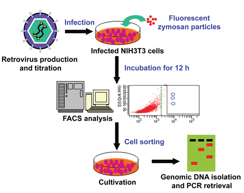
A gain-of-function cell sorting–based selection for phagocytosis-promoting genes. NIH3T3 cells were infected with retroviral cDNA library. Two days postinfection, cells were incubated with zymosan particles conjugated with Alexa Fluor 594™ (30 µg/mL) at 37°C for 12 h. Cells that engulfed zymosan particles were selected by cell sorting. Genomic DNA was isolated from the selected cells. The chromosomally integrated cDNAs were identified by PCR and sequencing. Finally, identified cDNA fragments were cloned and further analyzed.
Among the 8 cDNAs identified by the selection procedure (
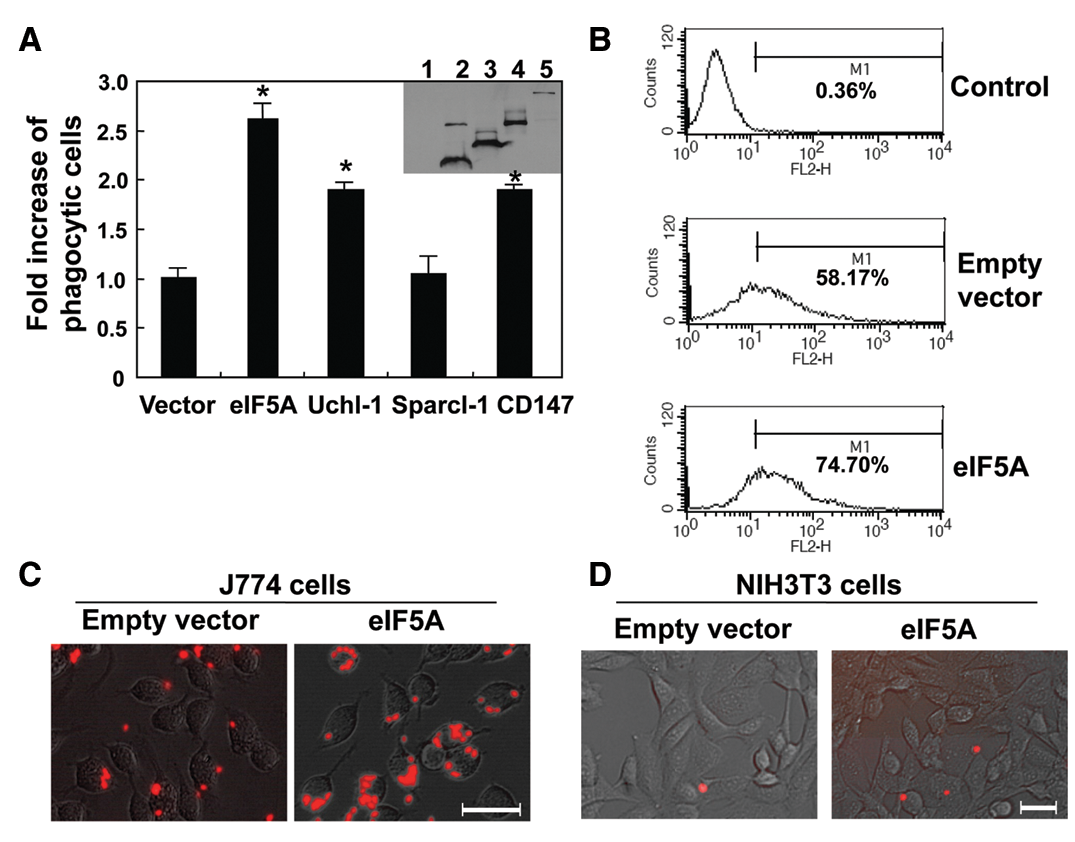
Functional validation of the genes selected by the method. (
Comparison of Phagocytosis-Promoting Activity of the Identified Genes in NIH3T3 Cells versus J774 Cells
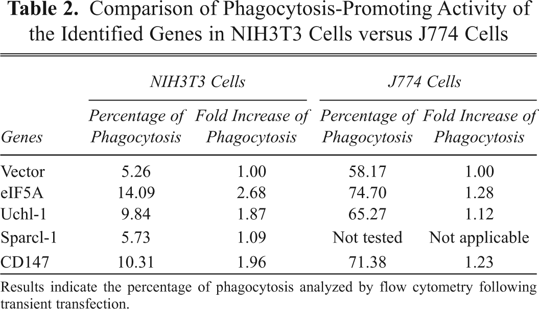
Results indicate the percentage of phagocytosis analyzed by flow cytometry following transient transfection.
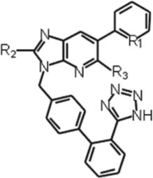
In the next set of experiments, we conducted a chemical-genetic screen using one of the novel targets identified in this study to link the targets mechanistically to phagocytosis and/or demonstrate the utility of the method for drug discovery. The chemical genetics aims were identifying which proteins regulate different biological processes, understanding in molecular detail how proteins perform their biological functions, and identifying small molecules that may be of medical value. We used eIF5A-transfected cells for the chemical-genetic screen of a series of imidazopyridine compounds (
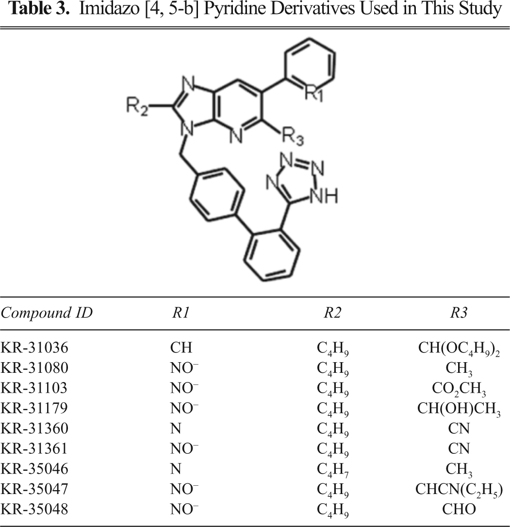
Imidazo [4, 5-b] Pyridine Derivatives Used in This Study
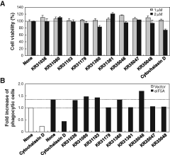
Chemical-genetic screens using eIF5A-transfected cells. The eIF5A-transfected J774 macrophage cells were pretreated with the imidazopyridine compounds (1-2 µM) for 1 h. Cytochalasin D (1-2 µM), which is known to inhibit phagocytosis, was used for comparison purposes. Cytotoxicity of the compounds was measured by the 3-(4,5-dimethylthiazol-2-yl)-2,5-diphenyltetrazolium bromide (MTT) assay (
Discussion
Here, we used cell sorting–based functional selection to identify the novel genes that contribute to phagocytosis. Previously, phagocytosis-related genes that enhance the engulfment of apoptotic cells were isolated by the retrovirus-mediated expression cloning system. 12 The screening method, however, required repeated phagocytosis assays to identify positive clones through the sibling procedure; the cDNA library was divided into small pools followed by the repeated flow cytometric analysis. A retroviral cDNA library screening was also performed to identify dectin-1 as a β-glucan receptor. 13 In comparison with these screening methods, the selection procedure described here efficiently identified multiple phagocytosis-promoting genes by a single cell-sorting step. A similar cell sorting–based selection has been used to identify murine-paired Ig-like receptor (PIR)–B as a novel receptor for Staphylococcus aureus. 14 In that report, the selection procedure was used to isolate cDNAs that bind to specific microbial species. A functional RNAi screen was done in Drosophila melanogaster S2 cells to study the phagocytosis of Candida albicans, 15 Escherichia coli, 16 Mycobacterium fortuitum, 17 or apoptotic cells. 18 A combination of gain-of-function selection and RNAi-based loss-of-function screen may provide even more powerful tool for the phagocytosis research.
Some of the genes selected by the current method were previously involved in the phagocytosis-related biological processes, therefore giving credence to the selection method. The identified genes, such as CD147, eIF5A, and Uchl-1, were also functionally validated by the separate phagocytosis assay. CD147, a member of the immunoglobulin superfamily, is also known as EMMPRIN, M6, neurothelin, OX47, and basigin. 19 CD147 interacted with monocarboxylate transporter proteins, integrins, cyclophilins, and caveolin-1. CD147 induced the expression of matrix metalloproteinases. 20 CD147/integrin interactions were essential for lamellipodia formation. 21 CD147 expressed on immune cells was involved in cell migration and activation—phenotypes that are not unrelated to phagocytosis. 22,23 Although eIF5A was originally identified as a translation initiation factor, the protein has also been implicated in nuclear export of HIV-1 Rev and mRNA decay as well as cell wall integrity and actin cytoskeleton polarization in yeast. 24 A recent study indicated that eIF5A, as the only known protein to contain the atypical amino acid hypusine, is required for cell growth and affects autophagy and protein synthesis. 25 Ubiquitin carboxy-terminal hydrolase L1 (Uchl-1) is a deubiquitinating enzyme that is involved in the mono-ubiquitin recycling. Mutations of the Uchl-1 gene and alterations of its activity have been associated with the pathogenesis of neurodegenerative diseases such as Parkinson’s disease, Alzheimer’s disease, and Huntington’s disease. 26,27 Identification of Uchl-1 as a phagocytosis-promoting gene in the current work suggests that the ubiquitin-proteasome system (UPS) may be involved in the regulation of phagocytic processes. It is also speculated that the impaired phagocytic activity may be linked to the pathogenesis of neurodegenerative diseases, and Uchl-1 may form the molecular basis that underlies this link.
The functional genetic selection or screen is a powerful tool used for the identification of genes that are involved in a variety of cellular processes/phenotypes/behaviors such as cell proliferation, 1 protein transport, 3 protein phosphorylation, 4 gene transcription, 5,28 tumor suppression, 29 apoptosis, 2 metastasis, 9 and bacterial virulence. 30 Recently, we reported a functional genetic selection of novel cell migration–promoting determinants. 8 Cell migration assay combined with retroviral transfer of the brain cDNA library successfully identified novel cell migration–promoting genes. Thus, the current functional genetic selection strategy can be easily adapted to a wide variety of cell-based assays. If cells of a given phenotype can be physically separated, for instance, by cell sorting or migration, causative genes can be identified from the selected cells of that phenotype.
In conclusion, our in vitro selection strategy successfully identified phagocytosis-promoting genes such as eIF5A, Uchl-1, and CD147. Further studies are necessary to gain a better understanding of the role of these genes in phagocytosis, which involves multistep processes requiring the coordinated action of many genes. Our in vitro genetic selection method may assist in the systematic identification and characterization of novel genes that are related with phagocytosis. This will advance our understanding of the phagocytosis process and ultimately provide a new means of modulating the cellular process. Furthermore, the novel target genes identified in this study could be used in the field of biomolecular screening or development of secondary assays/tools for screening.
Footnotes
Acknowledgements
The authors thank Dr. Kyu-Yang Yi (Korea Research Institute of Chemical Technology, Daejeon, Korea) for generously providing imidazo [4, 5-b] pyridine compounds.
This work was supported by Mid-Career Researcher Program through NRF grant funded by the MEST (No. 2010-0000337) and by the Bio R&D program through the Korea Science and Engineering Foundation funded by the Ministry of Education, Science and Technology (2008-04090).