
Abstract
The tyrosine kinase Wee1 is part of a key cellular sensing mechanism that signals completion of DNA replication, ensuring proper timing of entry into mitosis. Wee1 acts as an inhibitor of mitotic entry by phosphorylating cyclin-dependent kinase CDK1. Wee1 activity is mainly regulated at the protein level through its phosphorylation and subsequent degradation by the ubiquitin proteasome pathway. To facilitate identification of small molecules preventing Wee1 degradation, a homogeneous cell-based assay was developed using HeLa cells transiently transfected with a Wee1-luciferase fusion protein. To ensure ultra-high-throughput screening (uHTS) compatibility, the assay was scaled to a 1536-well plate format and cells were transfected in bulk and cryopreserved. This miniaturized homogeneous assay demonstrated robust performance, with a calculated Z′ factor of 0.65 ± 0.05. The assay was screened against a publicly available library of ~218,000 compounds to identify Wee1 stabilizers. Nonselective, cytotoxic, and promiscuous compounds were rapidly triaged through the use of a similarly formatted counterscreen that measured stabilization of an N-cyclin B-luciferase fusion protein, as well as execution of viability assessment in the parental HeLa cell line. This screening campaign led to the discovery of 4 unrelated cell-permeable small molecules that showed selective Wee1-luciferase stabilization with micromolar potency. One of these compounds, SID4243143 (ML 118), was shown to inhibit cell cycle progression, underscoring the importance of Wee1 degradation to the cell cycle. Results suggest that this uHTS approach is suitable for identifying selective chemical probes that prevent Wee1 degradation and generally applicable to discovering inhibitors of the ubiquitin proteasome pathway.
Keywords
Introduction
W
In this report, we describe a novel homogeneous 1536-well plate assay to monitor Wee1 degradation using cryopreserved transiently transected cells. We also demonstrate the excellent performance of this assay in the context of an ultra-high-throughput screening (uHTS) campaign that led to the identification of potential selective cell-permeable Wee1-Luc stabilizers.
Materials and Methods
Vector construction
The construct allowing expression of the K328M (kinase inactive) mutant of the Wee1-luciferase fusion protein (K328M-Wee1-Luc) was created by standard cloning methods as previously described. 7 The N-cyclin B-Luc-expressing construct has also been previously reported. 13,14
Cell culture
HeLa cells (American Type Culture Collection, Manassas, VA) were routinely cultured in T-175 flasks (Corning Life Sciences, Acton, MA) in Dulbecco’s modified Eagle’s media (DMEM; GIBCO, Carlsbad, CA) supplemented with 10% (v/v) fetal bovine serum (FBS; Hyclone, Logan, UT) and 1% penicillin/streptomycin/neomycin mix (PSN; GIBCO) at 37°C, 5% CO2, 95% relative humidity (RH). For small-scale experiments, HeLa cells were transiently transfected with the K328M-Wee1-Luc expression vector by batches of 6 × 10⁶ cells prepared in T75 flasks (Corning) containing 24 mL of a 1:1 ratio of OptiMEM and DMEM supplemented with 10% FBS, 1% PSN, 29 µg of K328M-Wee1-Luc plasmid, and 87 µL of TransIT-LT1 reagent, according to the manufacturer’s protocol (Mirus Bioproducts, Madison, WI). Cells were then incubated for 2 days at 37°C, 5% CO2, 95% RH. For large-scale experiments, cells were prepared in larger quantities (≈0.5-2 × 10⁹) and transfected using the same transfection reagents and media quantities relative to the cell number. Two days posttransfection, cells were cryopreserved. 15 Briefly, cells were trypsinized, counted, and resuspended in freezing medium (DMEM supplemented with 10% DMSO, 10% FBS, and 1% PSN) at a concentration ranging from 1.5 to 2 × 10⁷ cells per mL. Cells were then dispensed into 1.8-mL cryovials (Nalgene, Rochester, NY) and transferred into a −80°C freezing unit using a device allowing a cooling rate of roughly 1°C per minute (Mr. Frosty, Nalgene).
Wee1 degradation uHTS assay
When using cryopreserved cells, frozen stocks of transiently transfected cells were rapidly thawed prior to the assay by transferring them in a centrifuge tube containing preheated media composed of phenol red–free DMEM supplemented with 10% FBS and 1% PSN (1:50 v/v final dilution of frozen stock). Transiently transfected cells, either from frozen or regular stocks, were then centrifuged, counted, and resuspended at a concentration of 800,000 cells per mL. A stepwise assay protocol is presented in
Wee1 Degradation Assay Protocol in 1536-Well Plate Format
uHTS screen using the Wee1 assay
Details relative to each step of the uHTS campaign are shown in
Ultra-High-Throughput Screening Campaign Summary and Results
NA, not applicable; S/B, signal to background.
PubChem AIDs are accessible online at http://www.ncbi.nlm.nih.gov/sites/entrez?db=pcassay&term=xxxx, where xxxx represents the PubChem AID number listed in the table.
The primary screen hit cutoff was calculated as the average percent activation of all test compounds plus 3 times the standard deviation.
The hit cutoff calculated for the primary run was also applied to the confirmation run.
N-cyclin B counterscreen assay
The N-cyclin B assay has previously been reported elsewhere. 13,14 To be used as a counterscreen for potential Wee1 degradation inhibitors, we modified the original protocol to match the one followed for the Wee1 assay as described above.
Cell viability assay
In total, 5 µL of nontransfected HeLa cell suspension (100,000 cells per mL) in DMEM supplemented with 10% FBS and 1% PSN was dispensed into each well of a 1536-well plate. Four hours after dispensing, 25 nL of test compounds prepared as 10-point, 1:3 serial dilutions was added to the cells using the Pin Tool. Plates were incubated for 20 h at 37°C, 5% CO2, 95% RH, and 5 µL of CellTiter-Glo (Promega, Madison, WI) was then added to each well. After a 15-min incubation at room temperature, luminescence was measured for 30 s using the ViewLux reader (PerkinElmer Life and Analytical Sciences). Cell survival was expressed as a percentage relative to wells containing media only (no cells, 0% survival) and wells containing cells treated with DMSO at 0.5% final only (100% survival).
Screening data acquisition, normalization, representation, and analysis
Raw fluorescent data from the ViewLux plate reader were uploaded into our institutional HTS database (MDL Information Systems, San Ramon, CA). Activity of each well was normalized on a per-plate basis using the following equation:

where high control represents wells from the same plate containing 30 µM MG132 (100% activation control, n = 24), and low control represents wells from the same plate treated with DMSO only (0% activation, n = 24).
A Z′ value greater than 0.5 was required for plate validation during the quality control process. 18 For dose-response experiments, triplicate percentage activations were plotted against compound concentration. A 4-parameter equation describing a sigmoidal dose-response curve was then fitted with an adjustable baseline using Assay Explorer software (MDL Information Systems, Santa Clara, CA). All data showed here were represented using Prism version 4.03 (GraphPad Software, San Diego, CA). Fitting of dose-response curves and EC50 determination were performed using the variable slope sigmoidal curve analysis tool of Prism. Results of screening assays presented in this text can be found at the National Institutes of Health’s (NIH’s) PubChem Web site (http://pubchem.ncbi.nlm.nih.gov/).
Chemicals
The MLSMR library was provided by BioFocus DPI (South San Francisco, CA) through the NIH’s Roadmap Molecular Libraries Initiative. Details regarding compound selection for this library can be found online at http://mli.nih.gov/mli/ compound-repository/mlsmr-compounds/. Briefly, the MLSMR library is a highly diversified collection of small molecules (more than 50% of compounds exhibit molecular weights between 350 and 410 g/mol) comprising both synthetic and natural products, from either commercial or academic sources, that can be grouped into the 3 following categories: (1) specialty sets of known bioactive compounds such as drugs and toxins, (2) focused libraries aimed at specific target classes, and (3) diversity sets covering a large area of the chemical space. Reference control MG132 (Z-Leu-Leu-Leu-CHO) was purchased from the American Peptide Company (Sunnyvale, CA). Synthesis of SID4243143, SID3713089, and SID4256064 is detailed in the supplemental section of this article.
FACS analysis
Compound-treated HeLa cells were harvested by collecting culture media, washing once in phosphate-buffered saline (PBS), and trypsinizing. Cells were centrifuged at 400 g for 5 min at 4°C. Cells were then washed with cold PBS, centrifuged at 400 g for 5 min at 4°C, and rapidly resuspended with 7.5 mL 70% ethanol and incubated at −20°C overnight. Cells were then centrifuged at 400 g for 10 min at 4°C and washed with 10 mL cold PBS. The supernatant was removed and the cell pellet resuspended in 38 mM sodium citrate containing 69 mM of propidium iodide (Invitrogen, Carlsbad, CA) and 19 mg/mL of Rnase A. Fluorescence-activated cell sorting (FACS) analysis was performed on a BD Biosciences (San Jose, CA) LSR II system and analyzed using Flowjo 8.7.3 software.
Kinase profiling
The kinase profiling assay was performed by Reaction Biology Corporation (Malvern, PA) using a radiometric-based filtration binding assay as previously described. 19 Briefly, compound SID4243143 was tested in duplicate at 10 µM on a panel of purified kinases in the presence of 32P-γ-ATP. Incorporation of 32P-γ-ATP into peptide substrates was measured and compared to DMSO controls. The kinase pan-inhibitor staurosporine was used as a positive control for these experiments.
Results
Development, validation, and miniaturization of a cell-based HTS assay to monitor Wee1 stabilization
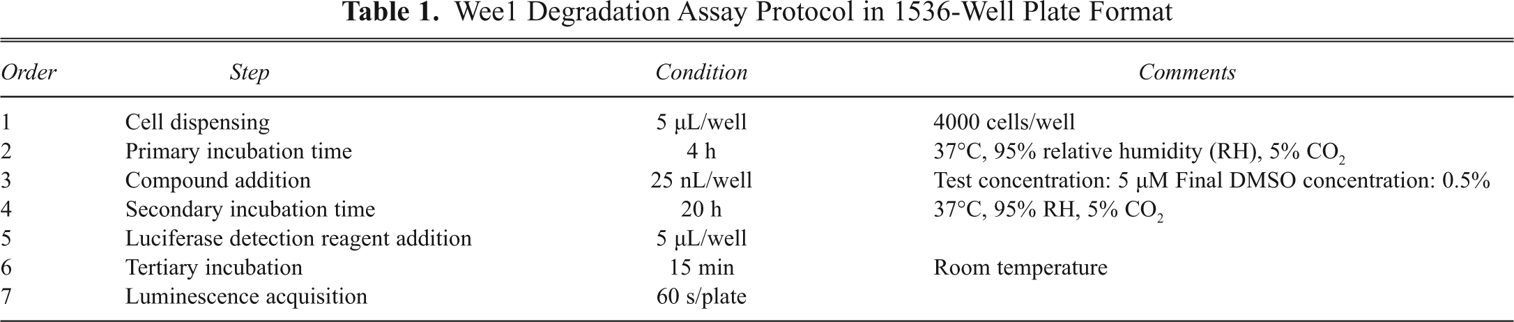
The primary screening assay was designed to measure the effect of test compounds on the stabilization of intracellular Wee1. To facilitate Wee1 protein level monitoring in cells, the firefly luciferase protein was fused in frame with the C-terminal end of Wee1. We used a kinase inactive mutant of Wee1, Wee1K328M, to prevent known toxicity issues associated with Wee1 overexpression.
6
Prior studies demonstrated that this fusion protein, K328M-Wee1-Luc, had similar degradation characteristics as wild-type endogenous Wee1.
7
HeLa cells transiently transfected with the K328M-Wee1-Luc encoding plasmid were incubated with test compounds for 20 h and lysed to measure their luciferase content via the use of a light-emitting D-luciferin substrate (
(
To reduce cost per well and allow higher throughput, we miniaturized the assay to a 1536-well plate format in a final volume of 10 µL/well. Critical variables of the assay, such as transfection conditions, cell seeding density, DMSO tolerance, and primary and secondary incubation times, were systematically assessed and optimized during the miniaturization phase to provide the best compromise between assay performance, reagent consumption, and protocol compliance with the robotic platform.
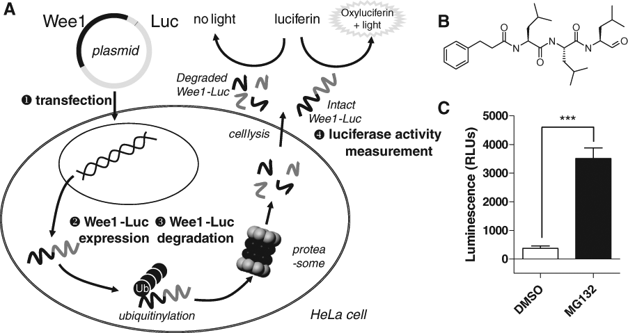
Wee1 degradation assay optimization and validation. (
To uncouple the transfection step from the rest of the assay, large amounts (>0.5 × 10⁹ cells) of K328M-Wee1-Luc-expressing cells were produced and cryopreserved. Cells were transfected and cultured as described above, the only difference being that they were frozen down 48 h posttransfection. Upon thawing, plating, and treatment with a serial dilution of MG132, the thawed cells displayed pharmacological properties comparable to those of noncryopreserved suspensions (4540 ± 440 nM vs. 5330 ± 360 nM;
K328M-Wee1-Luc uHTS assay performance
Three types of controls (n = 24 each) were placed on every assay plate: a positive control (30 µM MG132, serving as a 100% activation reference), a negative control (DMSO only, 0% activation reference), and a 50% activation control (using a MG132 concentration equivalent to its EC50 in the assay; i.e., 5 µM). These controls were used to (1) ensure cell responsiveness upon MG132 treatment, (2) monitor quality control through Z′ and S/B calculation, (3) verify proper compound dispensing, and (4) normalize data on a per-plate basis. A scattergram of the raw luminescent counts for the different controls from a typical assay plate is shown in
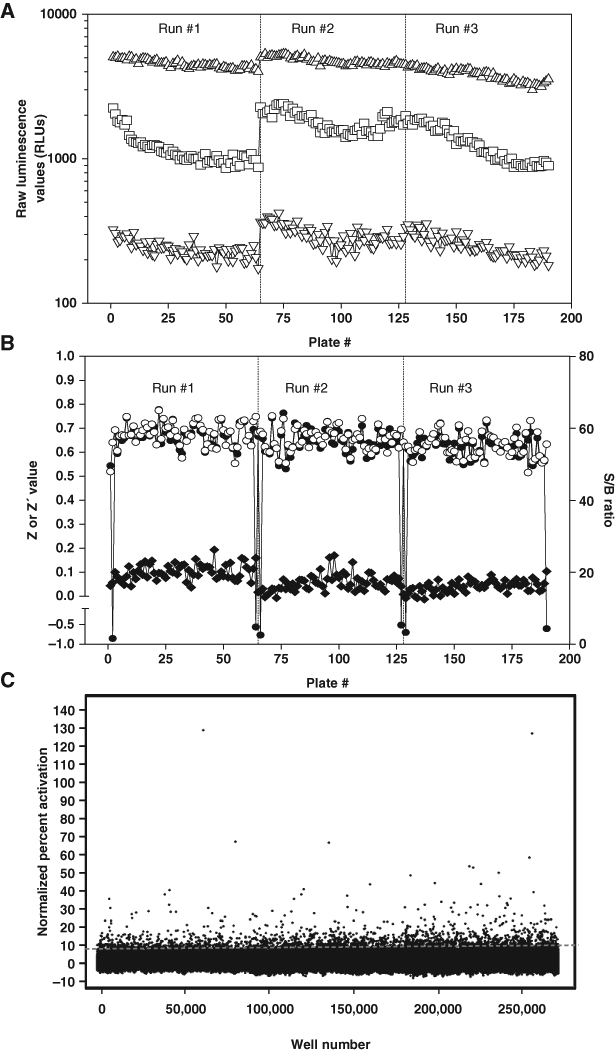
Wee1-Luc primary ultra-high-throughput screening (uHTS) campaign performance. (
K328M-Wee1-Luc screening campaign results
A stepwise description of the uHTS funnel employed to identify Wee1 degradation inhibitors is presented in
The N-cyclin B-Luc counterscreen. (
To eliminate compounds affecting Wee1 turnover in a nonselective manner, all 1090 available compounds were retested in triplicate at 5 µM in both the original K328M-Wee1-Luc assay and in a similarly formatted counterscreen assay employing a N-cyclin B-luciferase fusion protein.
14
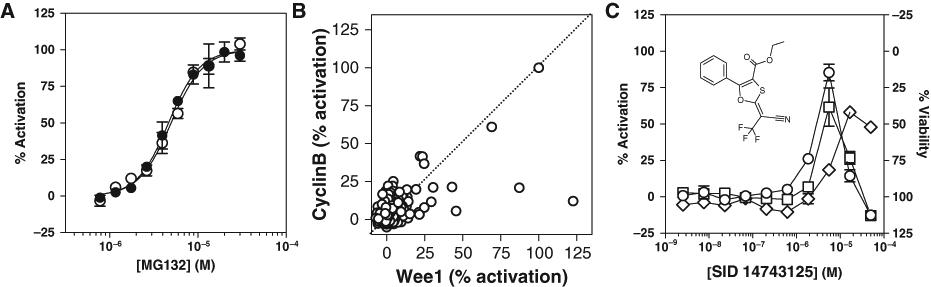
Consistent with its ability to block the ubiquitin-proteasome pathway, the reference control MG132 exhibited comparable dose-response curves in both the K328M-Wee1-Luc and the N-cyclin B-Luc assays (4.7 µM and 5.0 µM EC50, respectively), hence facilitating the comparison of normalized results between the 2 assays (
From triplicate screening results, 39 compounds confirmed activity (i.e., yielded in average percentage activation greater than the primary hit cutoff of 8.82%) and exhibited selective activity in the K328M-Wee1-Luc assay (
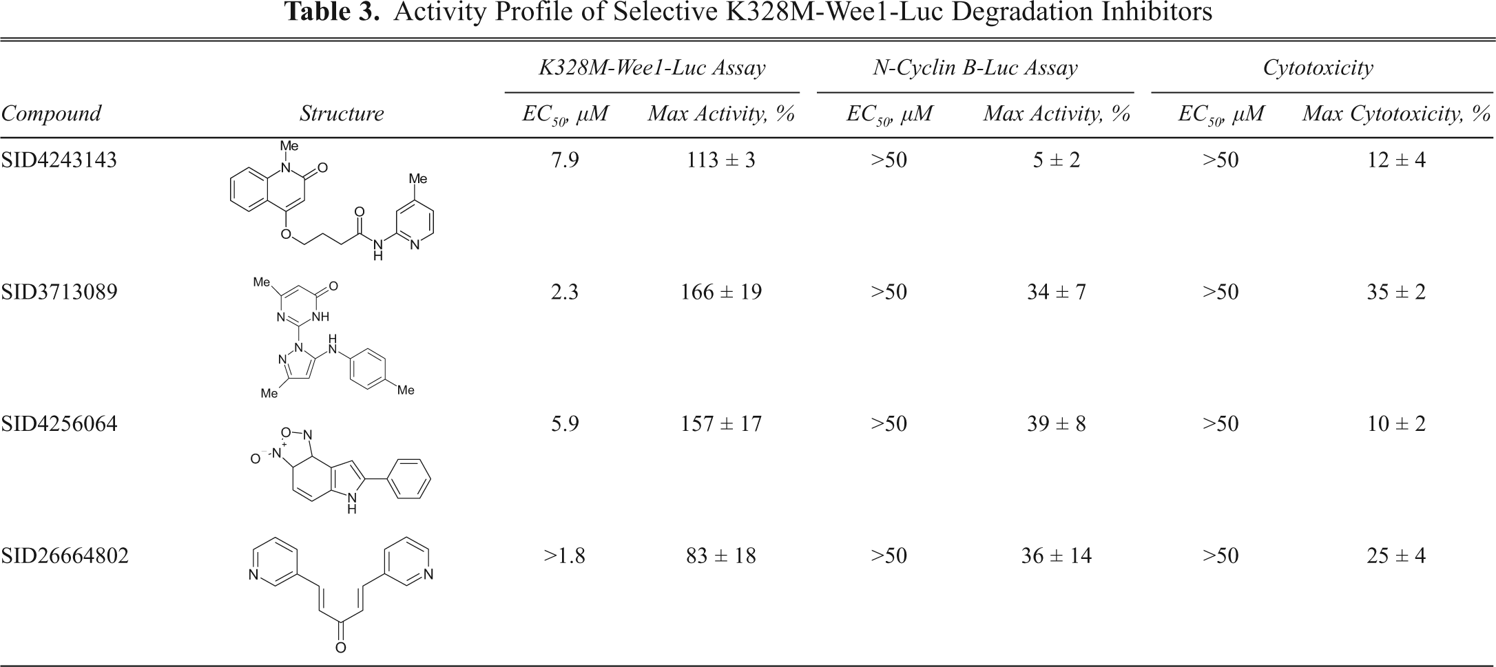
Wee1 selective compounds were chosen by applying the following set of criteria: (1) a maximal percent activity greater than 50% and an EC50 below 10 µM in the K328M-Wee1-Luc assay and (2) a maximal percent activity lower than 50% in the N-cyclin B-Luc assay. Four structurally unrelated compounds passed these criteria with EC50 values ranging from ~2 to 8µM—namely, SID26664802, SID4256064, SID3713089, and SID4243143 (
Activity Profile of Selective K328M-Wee1-Luc Degradation Inhibitors
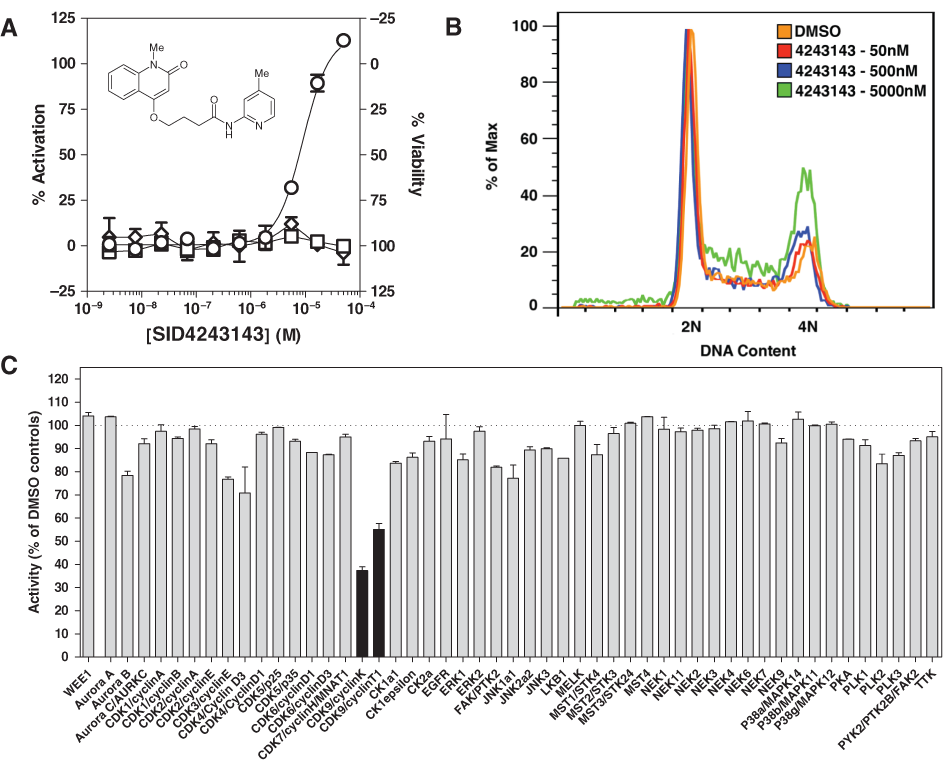
Characterization of SID4243143. (
Profiling SID4243143 activity
Our prediction was that a small-molecule inhibitor of Wee1 degradation would inhibit cell cycle progression because overexpression of nondegradable Wee1 induces a G2-M arrest.
6
FACS analysis was performed to test the effect of SID4243143 on the cell cycle (see Materials and Methods for details). Identical to the protocol that was used during the course of the screen, HeLa cells were treated for 20 h with different concentrations of SID4243143. As shown in
Wee1 degradation has been shown to be regulated by complex pathways involving kinases and phosphatases.
22
In an attempt to identify potential new proteins involved in Wee1 phosphorylation, we profiled SID4243143 against a panel of kinases involved in cell cycle progression, including Wee1 itself. As shown in
In addition to characterization of the biological properties of SID4243143, a chemical optimization effort was initiated. Preliminary results of a structure-activity relationship (SAR) study revealed an unusually stringent profile of compound SID4243143 (see supplemental section). Indeed, initial SAR efforts focused on moving the methyl to the 2-position of the pyridine ring resulted in complete loss of activity. Furthermore, replacement of the pyridine ring with various phenyl derivatives resulted in loss of activity in the Wee1 assay. Finally, amides based on 2-amino-6-methylpyridine and 2-amino-5-methylpyridine with only 1 methylene between the phenolic oxygen and carbonyl group were also found to be inactive.
Discussion
We report here the development of a simple homogeneous screening protocol for identifying small-molecule inhibitors of Wee1 turnover using a luciferase reporter system. With a Z′ value greater than >0.6 and a throughput of ~15,000 compounds per hour, the K328M-Wee1-Luc 1536-well plate assay proved to adapt very well in the context of automated uHTS and demonstrated excellent assay performance over the course of screening a small-molecule collection encompassing ~218,000 distinct chemical entities.
In optimizing the assay, we found that the use of cryopreserved transiently transfected cells was beneficial to the screening process, as previously reported by others. 15,23,24 In the case of the research presented here, it uncoupled the labor and time-consuming transfection step from the rest of the assay, greatly facilitating the organization of the screen and allowing measurement of results as fast as 20 h after initiating the assay instead of 72 h (i.e., if the transfection step was kept part of the process). In addition, the use of cryopreserved cells, as long as they are prepared in sufficient quantities, samples a single batch of transiently transfected cells for all the different steps involved in the lead discovery and optimization effort, hence increasing assay data consistency along the entire process.
As judged by the amount of compounds that reproduced activity in the secondary assay (
Another explanation for the low confirmation rate lies in the fact that activation profiles observed for all active compounds, including the reference control MG132, displayed very steep dose-response curves with Hill slopes >2.5. In some instances, compounds displayed a bell-shaped dose-response curve, as exemplified by compound SID14743125 (
The use of the N-cyclin B-Luc assay as a counterscreen proved to be an effective way of rapidly eliminating nonselective and promiscuous compounds early in the probe discovery process, allowing us to focus on a limited set of compounds that reduced Wee1 degradation. Among them, SID4243143 appeared to be the most attractive. Although this compound has also been reported as active in a nontrivial number of other cell-based assays available on PubChem (13 assays of 359, i.e. 3.6%), our results clearly show a selective effect on the K328M-Wee1-Luc fusion protein over the N-cyclin B-Luc construct, ruling out the possibility that SID4243143 acts broadly as a proteasome inhibitor or any other mechanism that is not specific to the Wee1 degradation pathway. Moreover, SID4243143 did not affect HeLa cell viability at concentrations up to 50 µM. Results of the FACS analysis indicated that SID4243143 inhibits cell cycle progression in HeLa cells (
The confirmed robustness of the screening platform and its ability to rapidly identify and triage promising selective, nontoxic and cell-permeable compounds encourages us to further screen other chemical libraries. In addition to the possibility of identifying other potent, selective scaffolds that will be useful in cell cycle research or as possible starting points for chemotherapeutic drug development, our long-term goal is to uncover potential phospho-transferases involved in Wee1 degradation through the screen of a focused library designed at targeting kinases. Taken together, the assays and automated protocol described herein proved to be an excellent platform for interrogating large-compound libraries for inhibitors of Wee1 degradation.
Footnotes
Acknowledgements
We thank Pierre Baillargeon, Lina DeLuca, and Louis Scampavia (Lead Identification Division, Scripps Florida) for compound management and quality control of compounds by LC-MS.
The work of NGA, PSH, and SS was supported by the National Institutes of Health grant #NS05699. The National Institutes of Health Molecular Library Screening Center Network (5U54MH074404) supported the research efforts of PC, PSH, FM, JKM, and WRR.
References
Supplementary Material
Please find the following supplemental material available below.
For Open Access articles published under a Creative Commons License, all supplemental material carries the same license as the article it is associated with.
For non-Open Access articles published, all supplemental material carries a non-exclusive license, and permission requests for re-use of supplemental material or any part of supplemental material shall be sent directly to the copyright owner as specified in the copyright notice associated with the article.