
Abstract
Objectives
Preeclampsia (PE) is a serious complication of pregnancy. The fibrinolytic system play crucial roles regarding placentation and evolution of PE.
Aim
To study comprehensively components of the fibrinolytic system and fibrin lysability in women with PE.
Design and Methods
117 women with PE and matched controls were included. Tissue type plasminogen activator (t-PA), plasminogen, PAI-1, plasmin inhibitor (PI), D-dimer, the fibrinolytic potential of dextran sulphate euglobulin fraction (DEF), PAI-2, polymere PAI-2, fibrin clot lysability, thrombin activatable fibrinolysis inhibitor (TAFI) and fibrinogen were assessed.
Results
Women with PE had significantly increased concentrations of t-PA and PAI-1, whereas the plasma concentration of PAI-2 was significantly lower compared to controls, p < 0.0001. Polymere PAI-2 was detected in both groups. DEF, TAFI and fibrinogen were not different between the groups. D-dimer was significantly increased and plasminogen/PI together with fibrin clot lysability time decreased in the PE-group, p = 0.0004 p = 0.04, p = 0.03, p < 0.0001 respectively.
Conclusion
This study demonstrates that PE is associated with an affected t-PA/PAI-1 system, decreased PAI-2 and increased fibrin lysability. Furthermore, PAI-2 has the potential to polymerize during pregnancy.
Introduction
Preeclampsia (PE) is a serious complication of pregnancy, contributing worldwide considerably to maternal morbidity and mortality, and can cause severe perinatal complications. PE affects approximately 5% of all pregnancies. 1 The incidence of preterm PE (before 37 weeks of gestation) has remained unchanged during the last decade, 2 and the pathophysiology of PE is only partly understood. However, placental dysfunction, reduced maternal cardiovascular adaption, or a combination of these conditions may contribute to the pathophysiology of PE.
Pregnancy presents a hyper-coagulable state, and even small changes in coagulation and fibrinolysis can contribute to adverse pregnancy outcomes such as PE. 3 Changes in the fibrinolytic system play crucial roles regarding placentation. 4 Incomplete remodelling of the spiral arteries in the uterus is related to PE, causing increased vascular resistance and hypo-perfusion of the placental tissue. Fibrinolytic activators and inhibitors affect the development of the spiral arteries, and even small variations in these factors can lead to adverse pregnancy outcomes. 3
The fibrinolytic process involves the activation of plasminogen into plasmin, which is the ultimate proteolytic enzyme of the fibrinolytic system. The subsequent plasmin induced resolution of fibrin leads to formation of fibrin degradation products, e.g. D-dimer. Tissue type plasminogen activator (t-PA) is considered the most important plasminogen activator and the plasminogen activating capacity of t-PA depends on the presence of fibrin. Studies have demonstrated that PE is associated with disturbance of t-PA induced activation of plasminogen. 5 Plasminogen can also be converted to plasmin by the contact activation pathway and by urokinase-type plasminogen activator (u-PA).6–8 These pathways are incompletely studied in PE.
The activation of the fibrinolytic system is controlled by a number of inhibitors, of which plasminogen activator type 2 (PAI-2) is placenta derived and thus unique for pregnancy. This inhibitor has been investigated widely in pregnancies complicated by PE, and a decrease in the plasma concentration of PAI-2 is observed in women with PE compared to normal pregnant women. This reduction may be related to impaired placental function in women with PE, 9 but misfolding of PAI-2 could also contribute to the reduced plasma concentration. Previously, studies have shown that misfolded proteins are present in the placenta and urine from women with PE. 10 In this context, it is of interest that PAI-2 has the ability to polymerize or misfold spontaneously.
We have performed a comprehensive, cross-sectional study on plasminogen activation and inhibition in pregnant women suffering from PE and healthy matched controls. Particular focus was put on the different fibrinolytic pathways. In addition, we studied the presence of PAI-2 polymer forms in pregnancies complicated by PE and in pregnant controls. Finally, we compared the lysability of fibrin generated in plasma from women with PE and matched pregnant controls without PE.
Materials and Methods
Cohort
The patient cohort is described previously. 11 In brief, the Preeclampsia and ContAct System study (PreCAS) is a matched cross-sectional trial conducted from January 2020 to October 2021. Patients were included at Departments of Obstetrics and Gynecology at University Hospital of Southern Denmark, Esbjerg and Odense.
PE was defined by a national guideline from 2018 12 as a combination of hypertension and proteinuria after 20 weeks of gestation or hypertension accompanied by one of the following: hematological or neurological complications, liver dysfunction, renal failure, pulmonary edema or utero placental insufficiency. The National Guideline is generally in accordance with the ISSHP guideline from 2018. 13 Pregnant women above the age of 18 fulfilling the diagnostic criteria of PE were enrolled in the study either in the outpatient clinic or when admitted to the hospital. One-hundred and seventeen women were asked to participate and all accepted.
For each women with PE, a healthy pregnant woman was included. The control subjects were matched with cases with respect to gestational week (GA) (± 1 week), age (± 1 year) and pregestational body mass index (BMI) (± 1 kg/m2). One thousand and forty four pregnant women consented to be possible controls. Controls with the best match on all three criteria were contacted by telephone and invited to participate in the study.
In the pilot study addressing the presence of polymerized PAI-2, ten random patients and their controls were included.
Blood Sampling
In brief, blood samples were collected from the patients at inclusion when the patients were diagnosed with PE and from the controls in a comparable week of gestation. The collection and handling of blood specimens followed the G41 guideline from Clinical and Laboratory Standards Institute (CLSI) 14 and the H21-A5 guideline from CLSI. 15
16.8 mL blood was collected from an antecubital vein in four evacuated 2.7 mL tubes containing 0.105 mol/L sodium citrate (Vacutainer 9NC, Becton Dickinson, Plymouth, UK) and two evacuated 3 mL tubes containing 5.4 mg dipotassium-ethylene-diamine-tetraacetate (EDTA) (Vacutainer K2E, Becton Dickinson). Platelet poor plasma was collected after centrifugation for 20 min at 2000 × g. The citrate and EDTA-stabilized plasma samples, respectively, were subsequently stored at − 80 °C in tightly capped cryotubes (Sarstedt, VWR-Bie & Berntsen, Søborg, Denmark). Before analysis, the samples were thawed for 5 min at 37 °C, kept at room temperature, and analyzed within one hour.
Biochemical Methods
In-house prepared enzyme linked immunoassays (ELISA) employing specific monoclonal antibodies were used to determine the plasma concentration of t-PA and PAI-1. 16 Plasma levels of fibrinogen was determined with the Dade® Thrombin reagent Kit using the Sysmex CS-5100 kit, based on the Clauss Method. 17 Kit was from Siemens Healthcare Diagnostics Products GmbH, Marburg, Germany. Plasminogen and plasmin inhibitor were determined with the Plasminogen kit, and the HemosIL Plasmin Inhibitor kit, respectively, using the ACL TOP 350 analyzer. Kits and analyzer were from Instrumentation Laboratories, Milan, Italy. D-dimer was determined using the INNOVANCE® D-dimer particle-enhanced immunoturbidimetric assay from Siemens Healthcare Diagnostics Products GmbH. Thrombin activatable fibrinolysis inhibitor (TAFI) was determined with the VisuLize TAFI antigen kit from Affinity Biologicals, Ontario, Canada. 18
The protein concentration of PAI-2 was assessed by an in-house prepared ELISA employing specific monoclonal antibody (mAb) as capture and polyclonal antibody (pAb) as detection. Briefly, MaxiSorp plates (NuncTM MaxisorpTM, Roskilde Denmark) were coated with 2.5 µg/mL mAb clone 19-26-3 against PAI-2 in coating buffer overnight at 4 °C. All the incubation steps were performed with constant agitation at room temperature. The plates were washed thrice in PBS-Tween and incubated for one hour in wash buffer. Subsequently, plasma was added 1:50, diluted in wash buffer with 50 mM EDTA and recombinant PAI-2 (R&D Biosystems®, 9206-PI-025) was used as calibrator. Plates with plasma samples were incubated for one hour, after which the plates were washed thrice. The pAb (antibodies-online.com, ABIN349627) was diluted to a concentration of 2 mg/mL in wash buffer, added to the plates and incubated for one hour. The plates were washed thrice, and HRP-conjugated swine-anti-rabbit (Dako A/S, P0217) was added to the plates, diluted 1:1000 in wash buffer. After incubation for 30 min, the plates were washed thrice and developed for 10 min with TMB Ultra (Thermo ScientificTM 1-StepTM Ultra TMB-ELISA Substrate Solution, 34029). The reaction was stopped with 100 mL 200 mM H2SO4 and the plates were read at 450 nm.
Fibrin clot lysis was determined as described previously (19, 20). In brief, fibrin clot lysis induced by tPA was investigated by mixing 60 µL of citrated plasma with 120 µL of a reaction mixture containing thrombin (1.0 IU/mL), calcium (15 mmol/L), Tris-HCl (50 mmol/L), tPA (300 ng/mL), and NaCl (150 mmol/L). Fibrin clot lysis was subsequently calculated as the time until 50% of the fibrin clot was lysed.
The fibrinolytic activity of the dextransulphate euglobulin fractions (DEF) of plasma was measured using a microtiter plate assay as previously described. 19
All experiments were performed in duplicate.
Immunoprecipitation was used to visualize the degree of polymerization of PAI-2 in plasma samples. DynabeadsTM M-280 Sheep Anti-Mouse IgG (InvitrogenTM, 11202D) was re-suspended and transferred to Eppendorf tubes, and subsequently washed trice in PBS-HSA, the supernatant was removed by placing the tubes in a magnetic separation rack. A PAI-2 specific antibody, mAb 19-26-3, was added to the tubes in a concentration of 2 µg/mL. The tubes were then incubated for one hour at 4°C in an end-over-end rotator, after which the beads where washed trice in PBS-HSA. Ten plasma samples from PE patients and matched controls, were diluted to the concentration of 40 ng/mL PAI-2 and added to the beads. The Eppendorf tubes were incubated for one hour at 4°C in an end-over-end rotator. After the incubation, the beads were washed trice and 50 µL 0.5% citric acid was added and incubated for 5 min and then placed in the magnetic rack until the supernatant was clear. Subsequently, the supernatant was collected and subjected to Sodium dodecyl sulfate–polyacrylamide gel electrophoresis (SDS-PAGE), followed by western blotting as as previously described. 20
SDS-PAGE was performed on Novex® BoltTM 4-12% Bis-Tris Plus Gels (Invitrogen, #NW04127BOX) inserted in an Invitrogen Novex® Mini-Cell electrophoresis. NovexTM Sharp Pre-stained Protein Standard (InvitrogenTM, LC5800) was used for molecular weight estimation.
Proteins were electro-blotted onto a PVDF membrane using the Trans-Blot® TurboTM Transfer System (Bio-Rad). The membrane was incubated washed and blocked in PBS-Tween and incubated with biotinylated polyclonal antibody against PAI-2 (Abcam, 137588) in PBS-tween. After washing the membrane was incubated with HRP-conjugated streptavidin (GenScript, M00091) PBS-tween. Lastly, a SuperSignalTM West Dura Extended Duration Substrate kit (Thermo Scientific, WJ333949) was used to develop the membrane in a Bio-Rad Molecular Imager® ChemiDocTM XRS Imaging System.
Statistics
Statistical calculations were performed by GraphPad Prism version 9.1.2 (GraphPad Software, San Diego, CA, USA). The distribution of the results were verified by a QQ plot and the Kolmogorov-Smirnov test. Paired t-test, Wilcoxon matched-pairs signed rank test, unpaired t-test, Mann-Whitney or Chi2-test were applied as appropriate. Results are presented as mean ± standard deviation or median and 25-75 percentile range, as appropriate. A p-value < 0.05 was considered statistical significant.
Results
Women with PE were comparable with controls with respect to the matching criteria, maternal age, pregestational BMI and gestational age.
The characteristics of the study participants are shown in Table 1.
Characteristics of the Study Participants.

The results are presented as mean ± SD or median (min-max), as appropriate
In early pregnancy the blood pressure was higher in the women developing PE, than in the control women, p < 0.0001. The weight of the newborn was lower, and the duration of pregnancy was shorter in women with PE compared to their controls, p < 0.0001 and p < 0.0001, respectivly. No difference was seen in the number of pregnant women who had received assisted reproductive treatment, p = 0.54.
As shown in Table 2 the concentration of t-PA and PAI-1 were significantly higher, whereas PI and plasminogen concentrations were significantly lower in the women with PE compared to the controls, p < 0.0001, p < 0.0001, p = 0.03, p = 0.04, respectively.
t-PA/PAI System

t-PA; tissue plasminogen activator, PAI-1; plasminogen activator inhibitor 1, PI; plasmin inhibitor, DEF; dextran sulphate euglobulin fraction. The results are presented as mean ± SD or median (25-75 percentiles), as appropriate.
The women in the PE group had significantly higher D-dimer concentration than the controls, p = 0.0004.
No difference was observed in DEF induced lysis between the PE women and the controls, p = 0.43.
The plasma concentration of PAI-2 was significantly lower in the women with PE compared to the controls (figure 1), p < 0.0001.

PAI-2 (ng/mL). The plasma concentration of plasminogen activator inhibitor 2 (PAI-2) in women with preeclampsia and controls.
In the pilot study of 10 women with PE and their controls, the band representing polymerized PAI-2 was detected in patients PE-27, PE 9, PE-10ALL and control con-86 and con-71 (figure 2).

Polymerized PAI-2. PE; patient with preeclampsia, con; control, p-PAI-2; polymer forms of PAI-2. Pilot study of 10 women with PE and their controls, the band representing polymerized PAI-2 was detected in patients PE-27, PE 9, PE-10ALL and control con-86 and con-71.
Table 3 shows a significant reduction in the time to lyse 50% of the fibrin clot in the women with PE compared to controls, p < 0.0001. The plasma concentration of TAFI and fibrinogen were not significantly different between women with PE and control subjects, p = 0.15 and p = 0.12, respectively.
Fibrin-Lysability
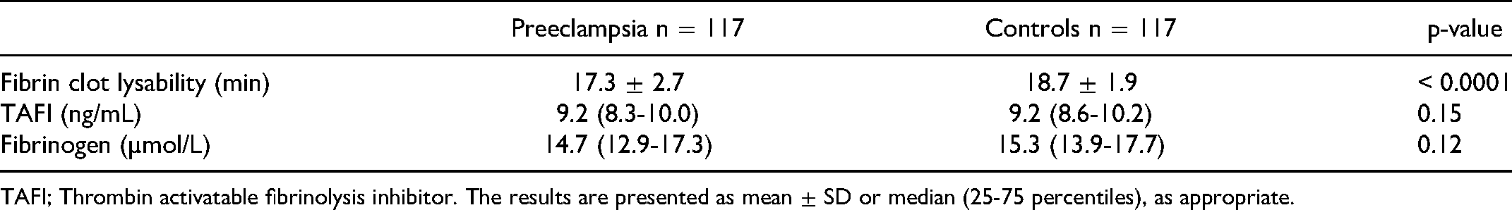
TAFI; Thrombin activatable fibrinolysis inhibitor. The results are presented as mean ± SD or median (25-75 percentiles), as appropriate.
Discussion
Main Findings
The present trial is a thoroughly matched cross-sectional study addressing the fibrinolytic system and fibrin lysability in pregnant women with PE. A number of studies have reported an association between PE and alterations in the fibrinolytic proteins in plasma, though only few markers have been investigated simultaneously. We investigated the t-PA/PAI-1 system and observed a significant increase in the protein concentration of t-PA and PAI-1 in women with PE compared to controls. This is in line with most former studies.4, 21–58 Both t-PA and PAI-1 are synthesized in the vascular endothelial cells, and the increase in women with PE indicates a systemic endothelial activation. In agreement with our results most studies have reported that PE is characterized by an increased concentration of PAI-1. 39 Few small studies describe a decrease or no significant differences in PAI-1 among women with PE and women without PE.22, 23, 39, 59, 60
PAI-2 is unique for pregnancy. This inhibitor regulates the plasminogen activating activity of both t-PA and urokinase. PAI-2 controls the fibrinolytic activity in trophoblasts, regulating the adhesive, tissue modelling and invasive processes characterizing the activity of trophoblasts, which is prerequisite for the development of normal spiral arteries in the uterus. 61 Disturbances in the level of PAI-2 may compromise the development of the spiral arteries and by that contribute to the development of PE. The majority of studies, including the present study, reveals a significant decrease in PAI-2 in women with PE compared to pregnant women without PE.
Notably, PAI-2 is the only serpin that polymerizes spontaneously under physiological conditions. The polymerization is dependent on the oxidative processes characterizing the extracellular milieu. 62 At the cellular level, PE is characterized by release of free radicals from placenta. 63 The induced increased oxidative stress may potentially cause uncontrolled polymer forms leading to misfolding of PAI-2, which may lead to impaired inhibitory activity and challenge the development of the spiral arteries. In the present study we demonstrated that PAI-2 polymers were present both in plasma from women with uncomplicated pregnancies as well in plasma from women with PE (Fig 2). This pilot observation needs further consideration.
We also report on different pattern response in PAI-1 and PAI-2 in PE. The increased concentration of PAI-1 in PE is probably related to an endothelial dysfunction, whereas the decreased plasma concentration of PAI-2 might be related to the reduction in placental size due to impaired placental development. It could also be speculated, that the decreased level of native PAI-2 in women with PE are caused by uncontrolled polymerization of the protein.
A global test of the total fibrinolytic capacity of plasma, including the contact activation and the u-PA dependent pathways (DEF) was applied. Dextran sulphate, a large negatively charged molecule, can induce plasminogen activation 64 through these two pathways. The method reduces the influence of inhibitors. From former studies, it is recognized that the concentration of u-PA is reduced in PE women.22, 65 In the present study, we did not observe a difference in dextran sulphate induced fibrinolysis between the two groups.
D-dimer is repeatedly reported to be increased in PE women 66 as also reported in this study. In addition, the present study demonstrates a significant decrease in plasminogen in PE women. The plasma concentration of plasminogen have been studied in relation to PE and diverse results are seen,46, 52, 67, 68 but methods and the number of participants makes the comparison of studies difficult. The decrease observed in this study, could represent a consumption of plasminogen caused by increased plasmin formation. The activity of PI is similarly decreased in women with PE compared to controls, supporting increased plasmin formation and thereby consumption of PI.
Finally, we studied the lysability of fibrin, which have not been elucidated before in PE. This assay was performed with addition of high amounts of t-PA to a preformed clot from each study participant. As such, genuine t-PA do not play a significant role on the lysability results. We observed a decrease in time when 50% of the fibrin clot has lysed. TAFI and fibrinogen are both determinants of fibrin lysability, and results from these did not deviate between the two groups. Fibrinogen concentration in plasma is of importance for fibrin fiber density while TAFI removes the c-terminal lysin from fibrin, thereby reducing binding of t-PA and plasminogen causing reduced lysis. 69 Taken together our results on fibrin lysability indicate that women with PE produces fibrin more susceptible to lysis compared to pregnant women without PE, but additional studies are needed to elucidate the mechanisms involved.
Strengths and Limitations
The strength of this study is the matched design. The fibrinolytic system might be dependent on the maternal and gestational age and the maternal BMI. Conversely, our design prevents the potential bias caused by these factors, and in addition, the number of participants in our study are considerably higher compared to many former studies. On the other hand, the cross sectional design might be a limitation. It would have been profitable to use a prospective study design to reveal the changes in fibrinolysis and presence of polymerized PAI-2 even before onset of PE, but this would require a much larger number of participants. This could also make it possible to sub-divide the cases in eg preterm/term PE and PE complicated by fetal growth restriction.
Conclusion
The present study demonstrates that PE is characterized by changes in the t-PA/PAI-1 system indicating an endothelial dysfunction. The PAI-2 concentration is decreased in women with PE and PAI-2 has a potential to polymerize/misfold in both normal and PE pregnancies. Dextran sulphate induced fibrinolysis is not affected in women with PE indicating an unchanged contact activation pathway and u-PA dependent fibrinolysis. Increased concentrations of fibrin D-dimer and decreased concentrations of plasminogen/PI are demonstrated. In addition Increased lysability of fibrin characterizes women with PE compared to controls. These changes of the fibrinolytic system may contribute to the pathophysiology or progress of PE.
Footnotes
Acknowledgments
All the pregnant women included in the PreCAS study.
Clinical staff enrolling patients at the Departments of Gynecology and Obstetrics, University Hospital of Southern Denmark, Esbjerg and Odense, Denmark.
Kathrine Overgaard, Anette Larsen, Gunhild Nielsen and Lars Christian Nielsen, Unit for Thrombosis Research, Department of Regional Health Research, University of Southern Denmark, Esbjerg, Denmark for managing blood samples and biochemical analysis.
Funding
Lida and Oskar Nielsens Foundation, Esbjerg Foundation, Gangsted Foundation, The Research Foundation of Southern Denmark and “Et Sundere Syddanmark”.
Ethics Approval
Ethical approval to report this case was obtained from the regional ethics committee in the Region of Southern Denmark (S-20190142) approved the project. The project was registered as NCT04825145 at clinicaltrials.gov.
Informed Consent
Written informed consent was obtained from the patient(s) for their anonymized information to be published in this article. The study was conducted according to the Helsinki declaration including study approval by the Danish Data Protection Agency.
Declaration of Conflicting Interests
The author(s) declared no potential conflicts of interest with respect to the research, authorship, and/or publication of this article.
Funding
The author(s) disclosed receipt of the following financial support for the research, authorship, and/or publication of this article: This work was supported by the Lida and Oskar Nielsens Foundation, Gangstedfonden, The Research Foundation of Southern Denmark, Esbjerg Foundation, Et Sundere Syddanmark,