
Abstract
Myocardial ischemia–reperfusion (IR) injury is associated with high disability and mortality worldwide. This study was to explore the roles of dioscin in the myocardial IR rats and discover the related molecular mechanisms. Rats were divided into 5 groups: sham, IR, IR + 15 mg/kg dioscin, IR + 30 mg/kg dioscin, and IR + 60 mg/kg dioscin. Heart rate (HR), mean arterial blood pressure (MAP), and rate pressure product (RPP) were evaluated at 10 minutes before ischemia, immediately after ischemia, and at the beginning, middle, and end of reperfusion. Arrhythmia score and myocardial infarct size were examined in rats of all groups. The serum creatine kinase-muscle/brain (CKMB) and cardiac troponin I (cTnI) levels were analyzed via enzyme-linked immunosorbent assay. Protein amount of total connexin 43 (T-Cx43) and phosphorylated connexin 43 (P-Cx43) was evaluated by Western blot. Ischemia reperfusion significantly decreased HR, MAP, and RPP of rats compared to the sham group. However, dioscin significantly attenuated the above phenomena in a dose-dependent manner. Dioscin markedly inhibited IR-induced increase in arrhythmias score, infarct size, and serum CKMB and cTnI levels. In addition, dioscin strikingly induced IR-repressed expression of T-Cx43 and P-Cx43. Our results suggested that dioscin pretreatment exhibited protective effects against myocardial IR injury. Moreover, we found that dioscin attenuated myocardial IR-induced ventricular arrhythmias via upregulating Cx43 expression and activation.
Introduction
Coronary heart disease, the incidence of which is growing at an alarming rate in most countries, affects large number of population. 1,2 At present, timely coronary artery reperfusion has become the most effective method to improve myocardial ischemia and reduce mortality after an acute myocardial infarction. 2,3 However, more and more evidences prove that myocardial ischemia–reperfusion (IR) can itself cause increased infarct size, ventricular arrhythmias, myocardial stunning, left ventricular remodeling, and other events, which reduce the benefit of reperfusion. 1,3,4 Thus, developing a powerful strategy to limit the IR-induced myocardial injury is an important mean to salvage the healthy myocardium.
It has been validated that many natural occurring products have cardioprotective activities. For example, Li et al showed that eriodictyol from citrus had protective effect against myocardial IR injury via activating Janus kinase 2. 5 Song et al proved that polysaccharide from Salvia miltiorrhiza had beneficial effects on myocardial IR injury via reducing oxidative stress and repressing apoptosis. 6
Dioscin is a natural steroidal saponin isolated from many different herbs such as Polygonatum zanlanscianense and Dioscorea nipponica Makino. 7,8 Beyond the well-documented antitumor, 9 anti-inflammatory, 10 antifungal, 11 and antioxidant 12 properties, it has also been demonstrated that dioscin possessed protective activity against IR injury of various organs, such as kidney, 13 brain, 14 liver, 15 and stomach. 16 However, the action of it on myocardial IR injury has not yet been reported.
We aimed to evaluate the preventive effects and molecular mechanisms of dioscin against myocardial IR injury. In this study, the rat model of myocardial IR was established via the ligation of the left anterior descending coronary artery (LAD) for 30 minutes and then reperfusion for 120 minutes. Dioscin was administered daily through oral gavage at dosages of 15, 30, and 60 mg/kg for 1 week before surgery. Subsequently, hemodynamic changes, arrhythmia score, myocardial infarct size, serum creatine kinase-muscle/brain (CKMB) and cardiac troponin I (cTnI) levels, and the protein levels of total connexin 43 (T-Cx43) and phosphorylated connexin 43 (P-Cx43) were analyzed in rats of all the 5 groups.
Materials and Methods
Myocardial IR Injury Rat Model
All the experiment protocols were reviewed and approved by the institutional ethical review board of Tangdu Hospital, Air force Military Medical University. Eight to 12-week-old male Sprague Dawley rats (250-320 g) were housed on a 12-hour light–dark circadian cycle at 21°C to 23°C and a humidity of 55%. All the rats were provided food and water ad libitum.
The rats of acute myocardial IR were established as described previously. 17 In brief, anesthesia was conducted through intraperitoneal injection of 3% sodium pentobarbital, and the rats were supine on the operating platform. After endotracheal intubation, rats were connected to a small animal ventilator. The body temperature was maintained at 36.5°C to 37.5°C with a heating pad. A heparin-filled intravenous catheter was placed into the right carotid artery to record arterial pressure. On the other hand, the left jugular vein was also inserted by intravenous catheter for fluid or drug administration. The arterial pressure and lead II electrocardiogram (ECG) were recorded via the biological signal acquisition system (BL-420F; Chengdu Taimeng Software Co Ltd, China). Electrocardiogram and heart rate (HR) were monitored via needle electrodes subcutaneously placed on rat limbs. Rat hearts were exposed via left thoracotomy and pericardotomy.
The LAD branch of the left main coronary artery was ligated with a 4/0 silk ligature 1∼2 mm away from the left auricle. Successful LAD occlusion was confirmed by ECG showing typical ST-segment elevation, cyanosis below the silk ligature, and the weakened movement of ischemic region. After 30-minute regional ischemia, animals were subjected to 120-minute reperfusion. Rats in the sham group received exactly the same surgery without LAD ligation.
Dioscin (>98% purity, Xi’an Tonking Biotech) was dissolved with 0.5% carboxymethylcellulose sodium (Sigma). To explore the role of dioscin on myocardial IR, myocardial IR rats were intragastrically fed with dioscin at various concentrates (15, 30, or 60 mg/kg) once daily for continuous 7 days before the operation. To test the safety of dioscin, rats in sham group were also pretreated with various concentrates of dioscin (15, 30, or 60 mg/kg) as described earlier.
Arrhythmia Score Evaluation
Arrhythmia score was evaluated according to the method described previously. 2 Score 0, no ventricular arrhythmia; 1, accidental premature ventricular contraction (PVC; <3 times per minute); 2, frequent PVC (≥ 3 times per minute); 3, accidental ventricular tachycardia (VT; <3 times per minute); 4, frequent VT (≥ 3 times per minute) or accidental ventricular fibrillation (<3 times per minute); 5, frequent VT (≥ 3 times per minute) or death.
Infarct Size Assessment
Myocardial infarct size assessment was conducted according to the previously reported method. 18 In brief, the LAD was religated at the end of each experiment, and 2 mL of 3% Evans blue (Sigma Aldrich, St Louis, Missouri) was injected intravenously to identify the area at risk. Then, the rats were killed and the hearts were excised. The left ventricle was sliced into 5 sections (1-mm thick) from apex to base. Tissues were incubated in the 1% 2,3,5-triphenyl tetrazolium chloride (Shanghai solarbio Bioscience & Technology Co, Ltd) for 15 minutes at 37°C to delineate the infarction area. After fixation, the above slices were photographed and analyzed via Image-Pro Plus 7.0.
Enzyme-Linked Immunosorbent Assay
To explore the mechanism of dioscin in the treatment of myocardial IR injure, rat serum samples in different groups were collected. Then, the serum CKMB and cTnI concentrations were examined using CKMB and cTnI enzyme-linked immunosorbent assay (ELISA) kit (Biosource Inc, San Diego, CA) according to the manufacturer’s instructions.
Western Blot Analysis
Antibodies for T-Cx43 and P-Cx43 were purchased from Santa Cruz Biotechnology (Dallas, TX), and Western blot was done as described previously. 19 In brief, polyvinylidene fluoride membranes were incubated with goat monoclonal anti-T-Cx43 (1:1000) and goat monoclonal anti-P-Cx43 (1:500) after blocking. Subsequently, the membranes were extensively washed and then incubated with horseradish peroxidase-conjugated anti-goat antibody (1:1000; Beyotime, Haimen, China). Protein bands were analyzed using an ECL system (Pierce, Wisconsin).
Statistical Analysis
One or 2-way analysis of variance was used for determining significant differences between groups. Statistical analyses were carried out via SPSS software version 11.0. Statistical difference was considered significant at P < .05.
Results
Dioscin Pretreatment Was Safe for Rats and It Increased the Expression of Cx43 in Healthy Sham Rats
In order to test the safety of dioscin, rats in sham group were also pretreated with different doses of dioscin. Supplemental Figure 1 showed that the serum CKMB and cTnI levels in rats of sham + dioscin groups had little difference compared to the sham group, which proved that pretreating with dioscin at indicated concentrations was safe for rats. In addition, Supplemental Figure 2 showed that rats of sham + 60 mg/kg dioscin group had higher level of T-Cx43 than those in the sham group (P < .05). Moreover, Supplemental Figure 2 also showed dioscin significantly upregulated P-Cx43 protein level compared to the sham group (P < .05, P < .01, and P < .001 for sham + 15 mg/kg dioscin group, sham + 30 mg/kg dioscin group, and sham + 60 mg/kg dioscin group, respectively).
Dioscin Pretreatment Alleviated the Deceasing of Heart Rate, MAP, and Rate Pressure Product in IR Rats
The HR, mean arterial blood pressure (MAP), and rate pressure product (RPP) of the rats in IR group at time points T2, T3, and T4 were all significantly decreased compared to those in the sham group (Figure 1A–C, P < .01, P < .05, P < .05, respectively).

Hemodynamic changes associated with the IR process. The results of heart rate (A), mean arterial pressure (B), and rate pressure product (C) were tested. n = 9 for each group. Data were presented as mean ± standard deviation (SD). *P < .05 and **P < .01 compared to sham group. # P< .05 compared to IR group. IR indicates ischemia reperfusion; SD, standard deviation.
On the other hand, all the above values were increased in rats of dioscin pretreatment groups in comparison to those of IR group (Figure 1). Moreover, the HR, MAP, and RPP of rats in 30 or 60 mg/kg dioscin pretreatment groups were significantly increased compared to those of IR group (Figure 1, P < .05). Figure 1 also showed that the effect of dioscin at a dose of 60 mg/kg was much more pronounced compared to the 15 or 30 mg/kg dioscin pretreatment groups. Thus, the above results showed that higher doses of dioscin had a much more rapid protective effect.
Dioscin Ameliorated the Arrhythmia Score in Rats Under Myocardial IR Process
Ventricular arrhythmias appeared in IR with or without dioscin pretreatment groups during myocardial IR process, especially in the IR group. Arrhythmia score was evaluated according to the previously reported system. 20 Compared to the sham group, all the other groups had statistically significantly higher arrhythmia scores (P < .01, P < .01, P < .01, and P < .05 for IR group, IR + 15 mg/kg dioscin group, IR + 30 mg/kg dioscin group, and IR + 60 mg/kg dioscin group, respectively). However, the arrhythmia scores were significantly lower in IR plus 30 or 60 mg/kg dioscin groups than those in IR group (Figure 2, P < .05 and P < .01, respectively). In addition, Figure 2 also showed that dioscin ameliorated the arrhythmia score in a dose-dependent manner.

Dioscin ameliorated the arrhythmia score in rats under myocardial IR process. n = 9 for each group. The score of arrhythmia was presented as mean ± standard deviation (SD). *P < .05 and **P < .01 compared to sham group. # P < .05 and ## P < .01 compared to IR group. IR indicates ischemia reperfusion; SD, standard deviation.
Dioscin Ameliorated the Myocardial Infarct Size in Rats Under Myocardial IR Process
The infarct size was 0%, 59% ± 7.8%, 54.4% ± 7.4%, 40% ± 7.8%, and 28.1% ± 7.2% in the sham, IR, IR + 15 mg/kg dioscin, IR + 30 mg/kg dioscin, and IR + 60 mg/kg groups, respectively (Figure 3A and B). Figure 3 showed that the infarct size was obviously reduced in the dioscin treatment groups. Moreover, rats in the IR + 60 mg/kg group exhibited an extremely significant decrease IR-caused myocardial infarct compared to those in the IR group (Figure 3B, P < .01), which suggested that dioscin treatment prevented myocardial infarct during IR in the dose-dependent fashion.

Dioscin ameliorated the myocardial infarct size in rats under myocardial IR process. A, Representative images of myocardial tissue sections stained with TTC and Evans blue. The nonischemic area was indicated by blue, the AAR by red, and the IA by white. B, Infarct size, as a percentage of the area at risk (IS/AAR) were measured presented as mean ± SD. n = 9 for each group. **P < .01 and ***P < .001 compared to sham group. # P < .05 and ## P < .01 compared to IR group. AAR indicates area at risk; IA, infarction area; IR, ischemia reperfusion; SD, standard deviation; TTC, 2, 3, 5-triphenyl tetrazolium chloride.
Dioscin Ameliorated the Serum CKMB and cTnI Levels in Rats Under Myocardial IR Process
The serum CKMB and cTnI levels in rats in different groups were shown in Figure 4. Compared to the sham group, both CKMB and cTnI levels were significantly increased in the other 4 groups. On the other hand, the serum CKMB and cTnI levels in rats of dioscin treatment groups were much lower than those of the IR group. Moreover, Figure 4 also showed that higher dose of dioscin induced a more pronounced effect on serum CKMB and cTnI levels. Dioscin at 60 mg/kg significantly reduced CKMB and cTnI levels by 48.15% and 57.75% in rat serum, respectively, compared to the IR group (Figure 4A and B, P < .01).

Dioscin ameliorated the serum CKMB and cTnI levels in rats under myocardial IR process. CKMB (A) and cTnI (B) were tested by ELISA. n = 7 for each group. Data were presented as mean ± SD. *P < .05 and **P < .01 compared to sham group. # P < .05 and ## P < .01 compared to IR group. IR indicates ischemia reperfusion.
Dioscin Ameliorated Myocardial IR-Induced Ventricular Arrhythmias by Increasing Cx43 Expression in Rats
Western blot analysis revealed that myocardial IR significantly downregulated the protein expression of T-Cx43 and P-Cx43, compared to the sham operation (Figure 5A and B, P < .01). However, rats of IR + 30 mg/kg dioscin and IR + 60 mg/kg dioscin groups had dramatically higher level of T-Cx43 and P-Cx43 proteins than those in the IR group (Figure 5B, P < .05 and P < .01, respectively). Moreover, Figure 5B also showed that higher dose of dioscin exhibited a more pronounced increase in T-Cx43 and P-Cx43 protein expression. Dioscin at 60 mg/kg significantly upregulated T-Cx43 and P-Cx43 protein level by 186.67% and 194.44% respectively, compared to the IR group (P < .01).
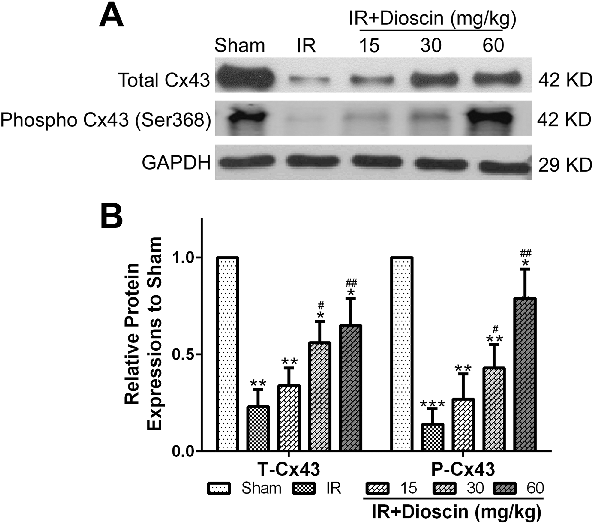
Dioscin ameliorated myocardial IR-induced ventricular arrhythmias by increasing Cx43 expression in rats. A, Western blot was used to infarct. (B) was the statistical analysis of A. n = 7 for each group. Data were presented as mean ± SD. *P < .05, **P < .01, and ***P < .001 compared to sham group. # P < .05 and ## P < .01 compared to IR group. IR indicates ischemia reperfusion.
Discussion
It has been validated that myocardial IR injury often caused by various clinical setting, including coronary bypass operation, thrombolytic threat, cardio-pulmonary resuscitation, and heart transplantation. 5 Myocardial IR injury, including postischemic arrhythmia, cardiomyocyte death, myocardial stunning, and a no-reflow phenomenon, 21 is associated with high disability and mortality in patients. Therefore, there are imperative needs to develop powerful new therapies for the treatment of myocardial IR injury.
At present, dioscin has attracted more and more interest during the treatment of various diseases, including melanoma, 22 nonalcoholic fatty liver disease, 23 obesity, 24 and organ IR injury. 13 –15 However, little is known about its effects on myocardial IR injury. In this study, animal experiments were designed to explore the protective roles of dioscin pretreatment in myocardial IR rat model in vivo. It is generally believed that ventricular arrhythmia is one of the most common causes of death during the process of myocardial IR injury. So, arrhythmias score was evaluated in this study. Furthermore, the infarct size, a gold standard when assessing the in vivo effectiveness of various cardioprotective methods, 25 was also examined in rats in different groups.
Here, 30-minute myocardial ischemia and subsequent 120-minute reperfusion of LAD led to significant deterioration of heart function parameters (including HR, MAP, and RPP) and significant increase in arrhythmia score, and infarct size in the IR group compared to the sham group. Therefore, these data demonstrated the successful establishment of myocardial IR rats. However, dioscin administration before surgery ameliorated myocardial IR injury in a dose-dependent manner as shown by evident improvement in heart function, significant decrease in arrhythmia score, infarct size and serum CKMB and cTnI levels, and obvious enhancement of T-Cx43 and P-Cx43 expression.
CKMB, the MB isoenzyme of creatine kinase, has high sensitivity for myocardial ischemia. 25 CTnI, a cardiac-specific protein, is usually undetectable in serum under healthy condition and will release into the circulation when cell membrane integrity is destroyed. 26 Apple et al found that damage of heart muscle can lead to an obvious increase in CKMB and cTnI level. 27 Hence, CKMB and cTnI are often used as biochemical markers to diagnose and monitor myocardial damage induced by IR. Moreover, Hedström et al proved that peak value of CKMB can accurately evaluate infarct size after myocardial reperfusion in patients. 28 Antman et al also demonstrated that there was a positive relationship between the cTnI level and myocardial infarct size. 26 Here, ELISA was used to analyze the serum CKMB and cTnI level in rats during myocardial IR process. The results showed that myocardial IR surgery significantly increase serum CKMB and cTnI level compared to the sham group, which was consistent with previous reports. However, dioscin preconditioning markedly suppressed IR-induced increase in serum CKMB and cTnI level. Moreover, the data showed that the trends of serum CKMB and cTnI increase were similar to those of myocardial infarct sizes in 4 treatment groups. Ischemia reperfusion + 60 mg/kg dioscin group had the smallest increase in CKMB and cTnI level and infarct size, while the IR group had the biggest increase in CKMB and cTnI level and infarct size.
Connexin 43, also known as gap junction alpha-1 protein, is a transmembrane protein that is a component of gap junctions in ventricular cardiomyocytes. 29 It has been recognized that Cx43 has an important role in synchronizing the beat rhythm. 30 Downregulation of Cx43 expression and activation impaired electrical impulse propagation resulted in ventricular arrhythmias during IR. 31,32 Moreover, increasing evidence suggested that Cx43 had a key role in cardioprotection. 29,33 Therefore, we speculated that the possible mechanism for the antiarrhythmias role of dioscin might be associated with the improvement in Cx43 expression and activation.
To confirm the abovementioned hypothesis, we used Western blot to analyze the total and phosphorylated levels of Cx43 in rats of all 5 groups. Western bolt data showed that IR surgery decreased the total and phosphorylated levels of Cx43, while the administration of dioscin resulted in a dramatical increase in the secretion of T-Cx43 and P-Cx43. Hence, the abovementioned results suggested that dioscin ameliorated myocardial IR-induced ventricular arrhythmias via increasing the expression and activation of Cx43. In addition, Reperfusion Injury Salvage Kinase and Survivor Activating Factor Enhancement cascades are 2 key pathways involved in cardioprotection, 34 and further experiments should be done to see whether either of these are involved in the cardioprotective activity of dioscin.
In summary, we found that pretreatment with dioscin had protective effects in the myocardial IR rat model, as shown by the alleviation of myocardial dysfunction, significant decrease in arrhythmia score and infarct size, dramatical drop in serum CKMB and cTnI level, and obvious increase in Cx43 expression and phosphorylation. Therefore, our study gave support to consider dioscin preconditioning as a promising therapeutic strategy for myocardial IR injury. However, more preclinical and clinical researches should be well conducted in the future to supply deeper investigation and get more necessary knowledge of the activities of dioscin on the treatment of myocardial IR injury.
Conclusion
Dioscin preconditioning may protect against myocardial IR injury in a dose-dependent fashion, which suggested that dioscin was a promising therapeutic agent for myocardial IR injury.
Supplemental Material
Supplementary_Materials - The Protective Effects of Preconditioning With Dioscin on Myocardial Ischemia/Reperfusion-Induced Ventricular Arrhythmias by Increasing Connexin 43 Expression in Rats
Supplementary_Materials for The Protective Effects of Preconditioning With Dioscin on Myocardial Ischemia/Reperfusion-Induced Ventricular Arrhythmias by Increasing Connexin 43 Expression in Rats by Jin Cheng, Chuang Sun, Jingyu Zhang, Qing Zou, Qimeng Hao and Yugang Xue in Journal of Cardiovascular Pharmacology and Therapeutics
Footnotes
Author Contributions
J Cheng contributed to design, acquisition, analysis, and interpretation; gave final approval; agrees to be accountable for all aspects of work ensuring integrity and accuracy; C Sun and Q Zou contributed to acquisition, gave final approval, agrees to be accountable for all aspects of work ensuring integrity and accuracy; JY Zhang contributed to interpretation, gave final approval, agrees to be accountable for all aspects of work ensuring integrity and accuracy; QM Hao contributed to analysis, gave final approval, agrees to be accountable for all aspects of work ensuring integrity and accuracy; YG Xue contributed to conception and design, drafted manuscript, critically revised manuscript, gave final approval, agrees to be accountable for all aspects of work ensuring integrity and accuracy.
Declaration of Conflicting Interests
The author(s) declared no potential conflicts of interest with respect to the research, authorship, and/or publication of this article.
Funding
The author(s) received no financial support for the research, authorship, and/or publication of this article.
Supplemental Material
Supplemental material for this article is available online.
References
Supplementary Material
Please find the following supplemental material available below.
For Open Access articles published under a Creative Commons License, all supplemental material carries the same license as the article it is associated with.
For non-Open Access articles published, all supplemental material carries a non-exclusive license, and permission requests for re-use of supplemental material or any part of supplemental material shall be sent directly to the copyright owner as specified in the copyright notice associated with the article.