
Abstract
Since breast cancer is the most common cancer in women, it is recommended that women over the age of 40 get annual mammogram screenings. Regular mammograms allow for early breast cancer detection, and in turn, proper treatment and improved patient prognosis. However, the stress and discomfort associated with the mammogram procedure deter many women from routine screening. Most previous work attempting to characterize mammogram-related pain utilizes subjective, questionnaire-based methods. A more objective approach is needed to fully understand the stress and pain experienced by patients during the mammogram procedure. In recent years, bio signals such as surface electromyography (sEMG) have been used to quantify various physiological states including stress and pain. Advocating women’s health and early breast cancer detection, the Human-User Interaction Lab is the first research lab to use sEMG to quantitatively measure the stress and pain experienced by patients during mammography compressions.
Objectives
One in eight females will develop breast cancer in their lifetime. Since breast cancer is the most common cancer in women, it is recommended that women over the age of 40 get annual mammogram screenings. There are 264,000 women who receive a breast cancer diagnosis in the United States each year (World Health Organization [WHO], n.d.). Regular mammograms allow for early breast cancer detection, and in turn, proper treatment, and improved patient prognosis. However, the stress and discomfort associated with the mammogram procedure deter many women from routine screening (Elwood et al., 1998; Gosein et al., 2014; Keefe et al., 1994).
Most previous work attempting to characterize mammogram-related pain utilizes subjective, questionnaire-based methods (Kornguth et al., 1996; Moshina et al., 2020; Nielsen et al., 1991). A more objective approach is needed to fully understand the stress and pain experienced by patients during the mammogram procedure. In recent years, bio signals such as surface electromyography (sEMG) have been used to quantify various physiological states including stress and pain (Cascella et al., 2023; Pourmohammadi & Maleki, 2020; Wijsman et al., 2010). Advocating women’s health and early breast cancer detection, the Human-User Interaction Lab is the first research lab to use sEMG to quantitatively measure the stress and pain experienced by patients during mammography compressions (Gielo-Perczak et al., 2024). The results of this study will provide a more holistic understanding of the discomfort that women experience during mammograms so that appropriate improvements can be considered.
For this study, researchers developed a comprehensive sEMG analysis of various muscles to quantitatively measure patient discomfort throughout the mammogram procedure. Various indices from the collected sEMG data were derived. Each of these indices presented specific characteristics of the waveform, but together they presented an intuitive summary of the muscular response to the procedure. The metrics were then interpreted within the context of the mammogram. We compared sEMG activity between relaxed and compressed (stressed) states as well as between paddle design types (flat and curved) (Hologic, n.d.).
Finally, we performed statistical analyses on the data to further interpret the collected results.
To the best of our knowledge, this study is the most complete mammography analysis to date, emphasizing a multimetric full body assessment and using sEMG signals.
Research Question
Can sEMG measures be effectively captured and used to better understand and address patient stress and pain experienced by women during the mammography procedure?
Specific Aim
Conduct a pilot study using sEMG to quantify neck, upper-limb and torso muscle activity during a mammogram procedure using both flat and curved paddles.
Approach
This research presents the use of sEMG as a means of measuring the stress and pain experienced by biological females during a mammogram.
Twenty-five healthy female volunteers ranging from 40 to 67 years old (avg. 50.84 ± 8 years) were recruited to participate in a simulated mammography procedure. The procedure involved the Hologic 3D Genius™ machine and the use of two different mammogram paddles (flat and curved). All procedures were approved by the Institutional Review Board (IRB) for human subject research. For each participant, sEMG Delsys wearable sensors were placed on 14 different muscles, including the left and right sternocleidomastoid, trapezius upper fibers, serratus anterior, external oblique, deltoid, infraspinatus, and teres major muscles. The placement of these sensors is illustrated in Figure 1. Researchers observed muscle activation and estimated stress throughout the procedure, from the relaxed state up to and including during breast compressions.
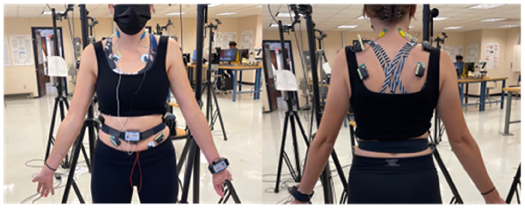
The placement of the Delsys EMGsensors on the 14 different muscles around the torso and neck of the subjects.
An initial EMG control recording (C1) was taken in a large, brightly lit room. Then, the participant was guided into the smaller room that housed the Hologic mammography system where another control recording (C2) was taken. Researchers used the same order of compressions as is used in the healthcare field, as follows: (1) right CranioCaudal view (RCC); (2) left CranioCaudal view (LCC); (3) left MedioLateral Oblique view (LMLO); (4) right MedioLateral Oblique view (RMLO). The four compressions are illustrated in Figure 2. Four compressions were first completed with the flat paddle followed by another four with the curved SmartCurve™ Hologic Paddle.
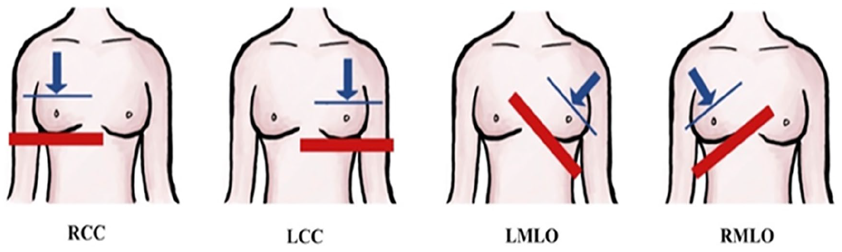
The four different positions of the breast for the simulated mammogram. Blue line/arrow indicates paddle and direction of compressive force respectively. Red line indicates imaging table on which the breast rests while being compressed.
Researchers selected tent total metrics to analyze the EMG activity during each state of the protocol. Five of the ten are time-domain metrics and the remaining ten are frequency-based metrics These metrics have been widely used to analyze EMG performance in various applications and together provide a wholistic, detailed perspective of the EMG throughout the protocol.
Findings
Analysis of the metrics derived from the sEMG data revealed that four of the muscle pairs—the deltoid, infraspinatus, teres minor, and trapezius upper fibers—frequently showed activation. The positions of the muscles, especially the first three, and their high engagement levels can be attributed to the strained torsion of the upper torso. Notably, the external obliques did not indicate frequent activation in comparison to their nominal state as might be expected in the twisted stance.
We also observed that tall women are particularly challenged with having to adjust their posture to the mammogram machine and that adjusting the height of the mammogram table to correspond to individual patient anthropometries reduced muscle activity.
Further, neither the flat nor the curved paddle consistently provoked a significantly lower or higher EMG response. This finding suggests that the proposed study methodology and sEMG application may need further refinement to detect minor variations in machine design. However, the findings did suggest a possible benefit to alternating paddle types (flat and curved) with the different mammogram table positions (right and left MedioLateral Oblique).
Finally, study participants reported lower pain scores for the simulated mammogram versus their previous “real-world” experiences. The standard mammogram compression force ranges from 100 to 140 N whereas the range of forces implemented in the study ranged between 18.25 and 71.60 N. Thus, given that participants reported pain in the simulated mammogram (albeit lower than that of actual mammograms) we can conclude that the current standard compression breast force should be significantly lowered to reduce mammogram pain and discomfort.
Takeaways
The results of this study support the potential use of the proposed methodology and sEMG analysis for identifying and quantifying muscular stress that occurs during mammograms. These sEMG responses can be used to classify the pain experienced during imaging, most notably that which is localized in the upper torso and neck and can be applied as the measure of the discomfort women experience during a mammogram procedure.
By selectively addressing those aspects of the procedure which induce muscle responses, improvements can be made to the procedure to reduce the pain and stress that is commonly associated with mammography. Based on the study findings, the standard compression forces applied during a mammogram should be significantly lowered, mammogram table heights should be adjusted for different anthropometries, and the two types of paddles (flat and curved) should be alternated during a mammogram test. Each of these proposed changes can help lower muscle activity and thus reduce pain and stress.
A more complete multimodal analysis incorporating additional signals may help elucidate the impact of specific design features and allow for additional conclusions related to stress and pain.
Footnotes
Acknowledgements
The authors would like to acknowledge Hologic Inc for their generous support in providing the Hologic 3D Genius Mammography system and related equipment. Additionally, the authors would like to acknowledge Delsys Inc for their sensor support. Finally, The DeLuca Foundation grant for their financial support.
Declaration of Conflicting Interests
The author(s) declared no potential conflicts of interest with respect to the research, authorship, and/or publication of this article.
Funding
The author(s) disclosed receipt of the following financial support for the research, authorship, and/or publication of this article: This study was founded by Hologic Inc. Sponsor did not have any involvement in the design, collection, analysis, interpretation of data, and decision to submit for publication.
Ethics Declarations
This study was approved by the University of Connecticut Institutional Review Board (IRB #H20-0146).