
Abstract
Background:
Previous studies reported the ligament attachments with regard to anatomic landmarks. However, these absolute distances may differ in size and anatomic variability. Purpose:To develop radiologic methods for assessing the tunnel placements of anatomic lateral ankle ligament reconstruction with a single common fibular tunnel and provide quantitative data on radiography and 3-dimensional computed tomography (3D CT).
Methods:
Sixteen ankle specimens were used to identify the attachment centers of the anterior talofibular ligament and calcaneofibular ligament. Subsequently, bone tunnels were created at these sites, with their positions evaluated via lateral radiographs and CT scans. Reference lines such as the anterior distal fibular line from the inferior tip of the fibula to the anterior tubercle on the fibula and the anterior lateral malleolar line from the apex of the lateral talar process to the anterolateral corner of the trochlea on the talus were employed for accurate tunnel positioning. For the calcaneal tunnel, the posterior subtalar line and its orthogonal line form the basis for a structured square measurement frame. The tunnel entries were orthogonally projected onto these references, enabling a percentage-based description of their locations. The interobserver and intraobserver reliability of the radiographic measurements were assessed using intraclass correlation coefficients (ICCs).
Results:
The fibular tunnel was projected at 35.9% on lateral radiography and 35.0% via 3D CT along the anterior distal fibular line. The talar tunnel was recorded at 62.4% on radiography and 63.5% on 3D CT along the anterior lateral malleolar line. There were no significant differences in the length of the posterior subtalar line (34.6 vs 31.4 mm, P = .140), distance a to the calcaneal tunnel (16.1 vs 14.7 mm, P = .100), and distance b to the calcaneal tunnel (27.9 vs 25.1 mm, P = .233) between the lateral roentgenogram and 3D CT. The calcaneal tunnel was observed at 80.6% on lateral radiography and 79.7% on 3D CT along the posterior subtalar line and at 46.4% on radiography and 46.3% on 3D CT along the orthogonal line. No significant differences were observed in the locations of the fibular, talar, and calcaneal tunnels between lateral radiography and 3D CT. Good interobserver agreement and intraobserver reproducibility were achieved, as indicated by ICCs.
Conclusion:
This study describes reproducible radiographic measuring techniques and provides quantitative data for intraoperative positioning and postoperative assessment of tunnel locations for anatomic lateral ankle ligament reconstruction. Both lateral radiography and 3D CT were effective modalities for evaluating the bone tunnels.
Clinical Relevance:
By offering reproducible measurement strategies and critical quantitative data on tunnel positioning for anatomic lateral ankle ligament reconstruction, this research significantly aids in optimizing intraoperative tunnel placement and postoperative evaluation, particularly for procedures involving a single common fibular tunnel and limited exposure methodologies.
This is a visual representation of the abstract.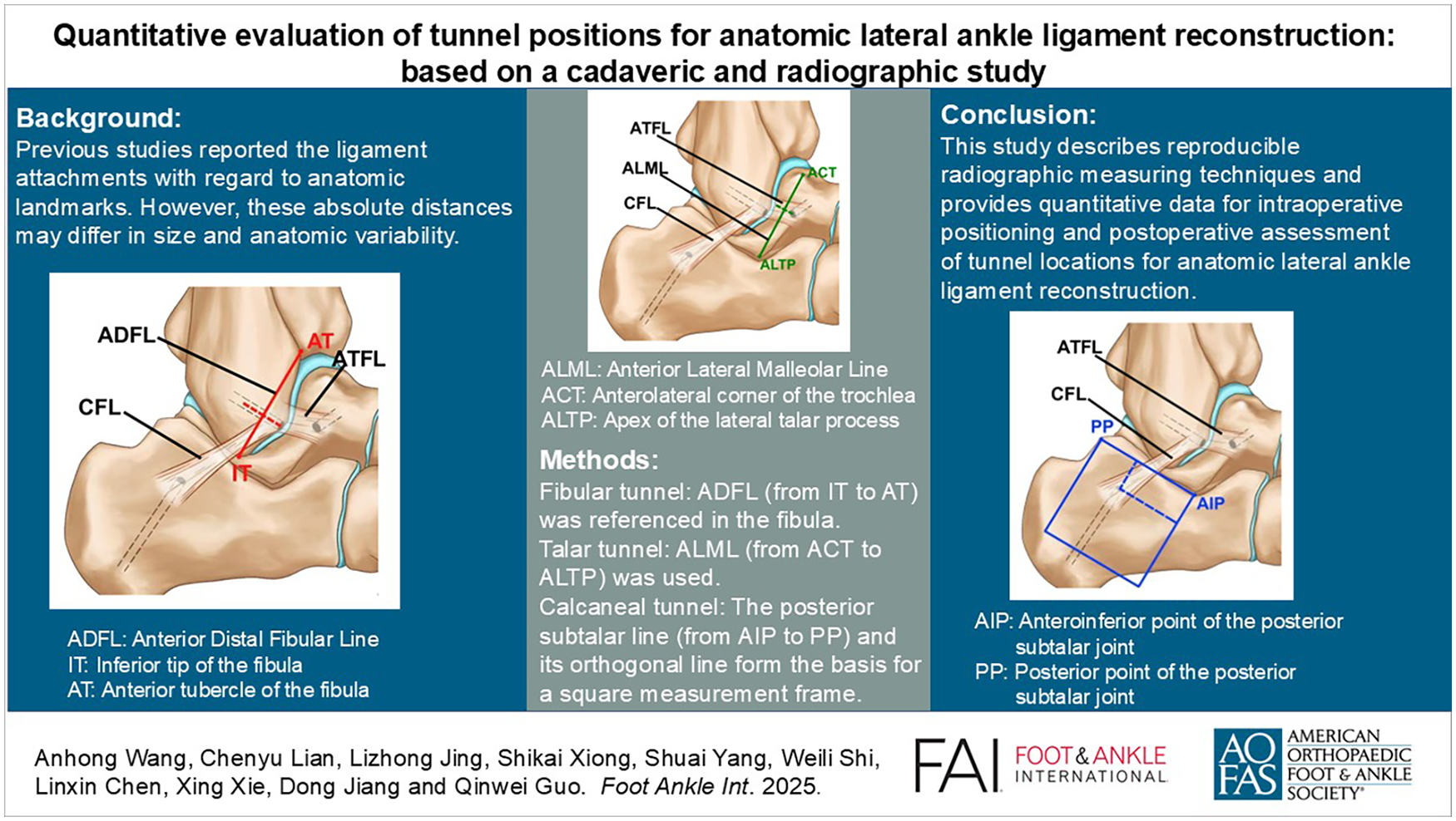
Keywords
Get full access to this article
View all access options for this article.
References
Supplementary Material
Please find the following supplemental material available below.
For Open Access articles published under a Creative Commons License, all supplemental material carries the same license as the article it is associated with.
For non-Open Access articles published, all supplemental material carries a non-exclusive license, and permission requests for re-use of supplemental material or any part of supplemental material shall be sent directly to the copyright owner as specified in the copyright notice associated with the article.