
Abstract
Rabbits are popular pets in the urban environment of Hong Kong, ranking third behind cats and dogs. Here we describe the frequency of neoplastic and non-neoplastic lesions in biopsies from pet rabbits submitted to the CityU Veterinary Diagnostic Laboratory between 2019 and 2022, comprising 247 tissue samples from 243 rabbits collected by veterinarians in 19 veterinary clinics. Among the 243 rabbits, there were 128 females (65 spayed), 114 males (54 castrated); sex information was not provided for 1 rabbit. The rabbit breeds included 45 Lionhead, 35 Dwarf, 14 Lop, 11 Dwarf Lop, 5 French Lop, 3 Angora, 2 Dutch, 2 Holland Lop, and 1 each of Netherland Dwarf, Velveteen, Mini Lop, and New Zealand White. The mean ages of rabbits with neoplastic and non-neoplastic lesions were 7.1 and 5.7 y, respectively. The most common neoplastic lesions were adenocarcinoma (26.4%), trichoblastoma (21.4%), sarcoma (9.4%), and thymoma (8.2%). The most common non-neoplastic lesion was uterine cystic endometrial hyperplasia (14.8%), followed by dermal abscess formation in the ventral abdomen or skin of the head (12.5%). Although a broad spectrum of other lesions was described, our findings in biopsies from pet rabbits in Hong Kong are consistent with those in other jurisdictions.
Domestic pets are popular in Hong Kong despite the limited living space, which averages 13.6 m2 per person in a dense urban environment. 15 The number of households keeping exotic pets, including reptiles, amphibians, rodents, rabbits, ferrets, and birds, continues to increase. 21 The rabbits kept in Hong Kong are domestic pets maintained indoors primarily; there is no rabbit farming in the city. Both the American Veterinary Medical Association 2 and the Hong Kong Government 1 have defined obligations for rabbit owners, expecting them to provide veterinary care for their rabbits similar to that for dogs and cats. Recommendations for selection of suitable pet rabbits, husbandry, and medical care are well documented. 55
Myiasis was the most common cause of death in a 2020 survey of domestic pet rabbits in England; overgrowth of claws was the most common presenting problem, with neoplasia being a less significant problem. 37 Neoplastic lesions are typically described in older pet rabbits. 57 A range of neoplastic and non-neoplastic lesions has been described in domestic pet rabbits in Europe.3,9 The prevalence of neoplasia in autopsied pet rabbits in a retrospective study was 14.4% 6 ; in rabbits >6-y-old, the prevalence was 75%. 8 Cutaneous tumors have been identified as the most commonly reported lesion in biopsy samples3,53,57; other studies utilizing autopsy cases have reported uterine adenocarcinoma, lymphoma, thymoma, and mammary gland tumors as more frequent.6,54 The most common non-neoplastic uterine lesion reported was cystic endometrial hyperplasia (CEH). 31
In Hong Kong, there has been a significant increase in the number of imported exotic pets (at least 5.2 million) between 2015 and 2021, including rabbits. 28 Studies on the incidence of neoplastic and non-neoplastic lesions in pet rabbits, especially in Hong Kong, is scarce. Our aim in our retrospective study was to describe the frequency of neoplastic and non-neoplastic lesions in biopsy samples from pet rabbits submitted to the CityU Veterinary Diagnostic Laboratory (CVDL; Hong Kong SAR, China) in Hong Kong from 2019 to 2022.
Materials and methods
Case selection
We retrospectively searched the CVDL laboratory database for cases involving biopsy samples of tissues from pet rabbits submitted for histologic examination by veterinarians in Hong Kong between January 2019 and December 2022. Samples collected from laboratory rabbits were not included in the study. The data collected included age, breed, sex, neutering status, lesion location, affected organ or tissue, and histologic diagnosis.
Histologic examination
Tissue samples were fixed in 10% neutral-buffered formalin at the veterinary clinic where surgery was performed and then sent to CVDL. After fixation, an American College of Veterinary Pathologists (ACVP)-certified veterinary pathologist examined all of the samples macroscopically, and representative sections were cut into blocks for routine processing and H&E staining. Selected slides were stained with Gram and/or Giemsa stains for bacteria, periodic acid–Schiff (PAS) and/or Grocott methenamine silver (GMS) for fungi, or Ziehl–Neelsen (ZN) for mycobacteria, as needed. All stained histopathology slides were reviewed by ACVP-certified veterinary pathologists.
The neoplastic and non-neoplastic lesions were grouped into the following categories: digestive, hematolymphoid, musculoskeletal, reproductive, respiratory, skin and soft tissues (including mammary glands), special senses, and urinary.
Data analysis
Extracted cases were entered into an Excel (v.2021; Microsoft) spreadsheet, and checked for duplicates and missing diagnoses. Lesions were categorized into neoplastic and non-neoplastic, then grouped based on the affected organs. The count and frequency, along with the 95% CI, were calculated for each lesion type. Data analysis and visualization were performed using STATA 18 (StataCorp).
Results
Animals
Between 2019 and 2022, the CVDL received 247 tissue biopsy samples from 243 pet rabbits for histologic examination, collected by veterinarians at 19 veterinary clinics across Hong Kong. Among the 243 rabbits, there were 128 females (65 spayed), 114 males (54 castrated); sex information was not provided for 1 rabbit. The rabbit breeds included 45 Lionhead, 35 Dwarf, 14 Lop, 11 Dwarf Lop, 5 French Lop, 3 Angora, 2 Dutch, 2 Holland Lop, and 1 each of Netherland Dwarf, Velveteen, Mini Lop, and New Zealand White. However, breed information was not provided for 112 rabbits. The rabbits were 0.5–14-y-old.
Lesion categorization
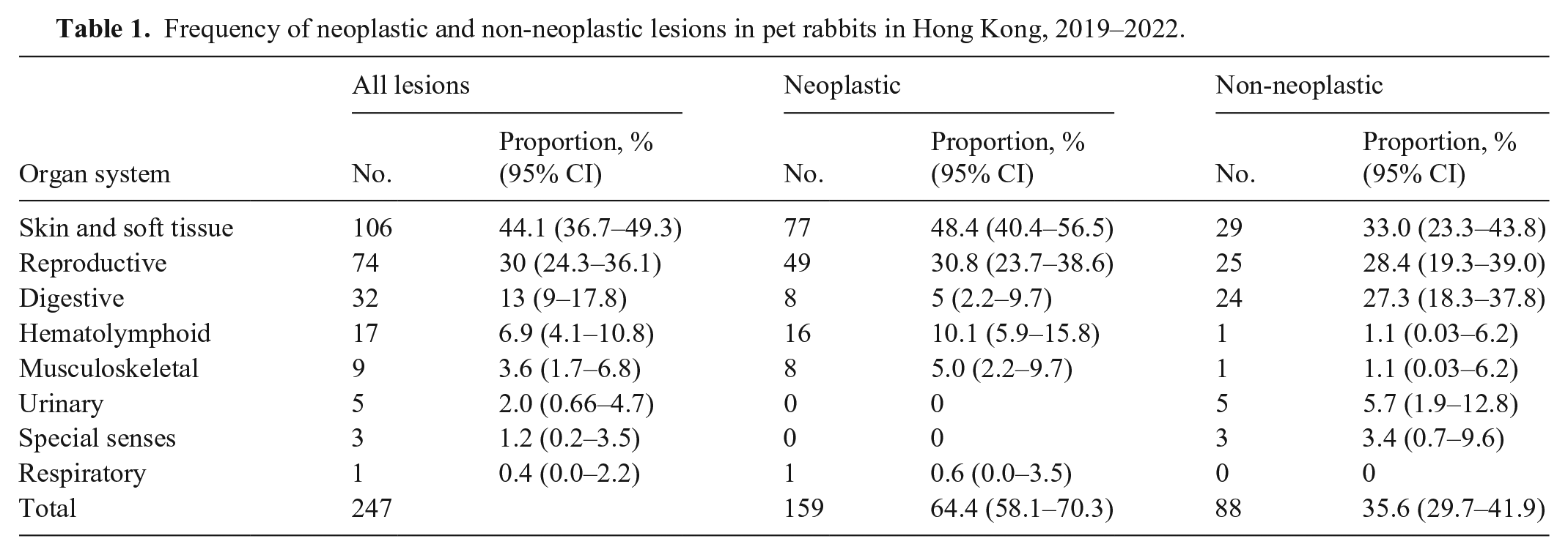
Skin and soft-tissue neoplastic and non-neoplastic lesions were the most frequent biopsies submitted, with 106 of 247 (44.1%) including 77 (48.4%) neoplastic lesions and 29 (33.0%) non-neoplastic lesions (Table 1). Mammary tumors were considered skin and soft-tissue lesions. The next most commonly submitted tissues were from the reproductive tract, followed by the digestive system (including oral tissue, salivary gland, peritoneum, and liver), then the hematolymphoid, musculoskeletal, and urinary systems; the least frequent submissions were from the organs of special senses (eyes and ears), and the respiratory tract.
Frequency of neoplastic and non-neoplastic lesions in pet rabbits in Hong Kong, 2019–2022.
Adenocarcinomas were the most frequent neoplastic lesions (42 of 159; 26.4%), and were found in the female reproductive tract (28 of 42; 66.7%), skin and soft tissue (13 of 42; 31%), and prostate gland (1 of 42; 2.4%). The next most common tumor type was trichoblastoma (34 of 159; 21.4%); various other tumor types occurred less frequently. Among common non-neoplastic lesions, uterine cystic endometrial hyperplasia (13 of 88; 14.8%) was the most prevalent; other lesions included abscesses, hepatic torsion, and dermatitis (Table 2).
Number, age, and sex of pet rabbits in Hong Kong with the most common neoplastic and non-neoplastic lesions, 2019–2022.
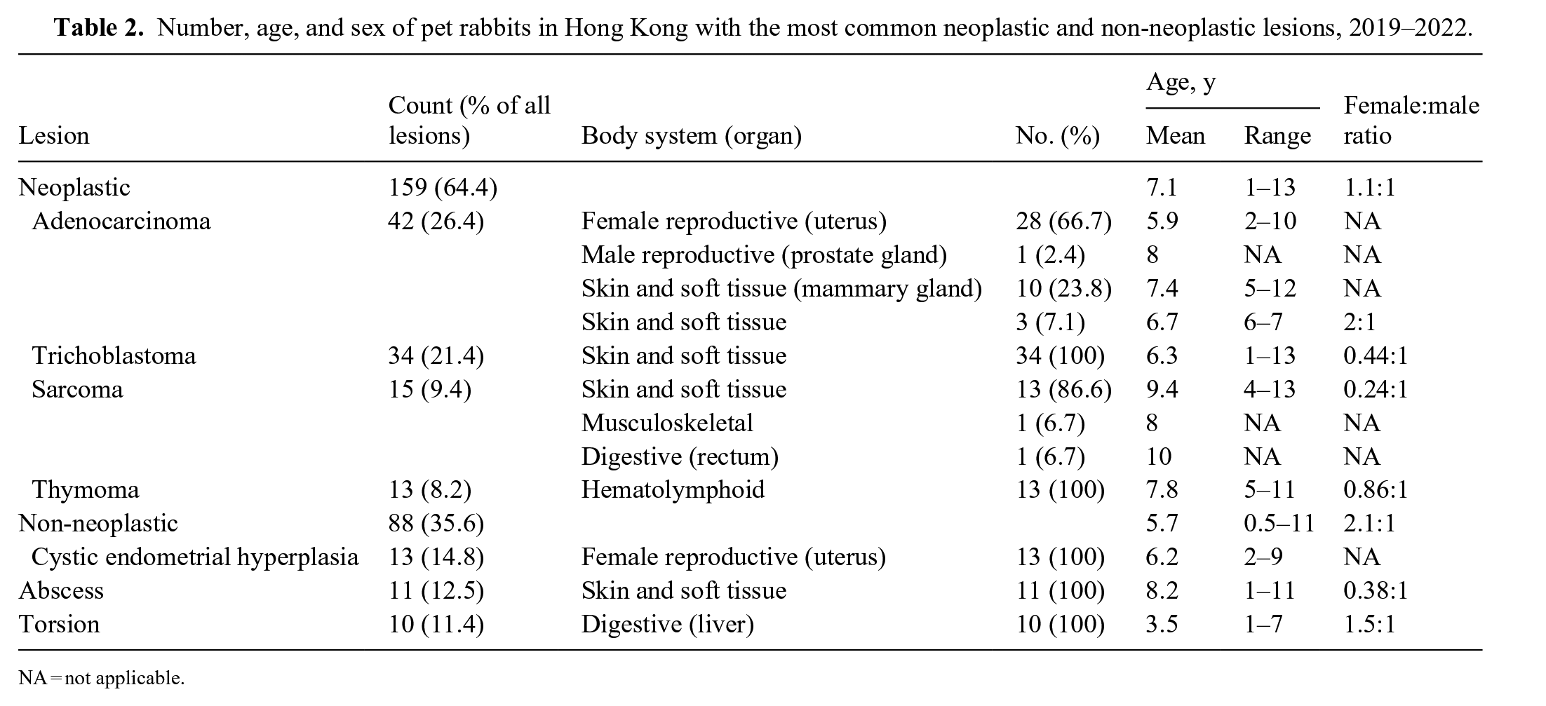
NA = not applicable.
Notably, both neoplastic and non-neoplastic lesions were more prevalent in females, with female-to-male ratios of 1.1:1 for neoplastic diseases and 2.1:1 for non-neoplastic diseases (Table 2). Neoplastic disease was more common in older rabbits (mean 7.1 y; range 1–13 y); non-neoplastic disease occurred more often in younger rabbits (mean 5.7 y, range 0.5–11 y; Fig. 1).
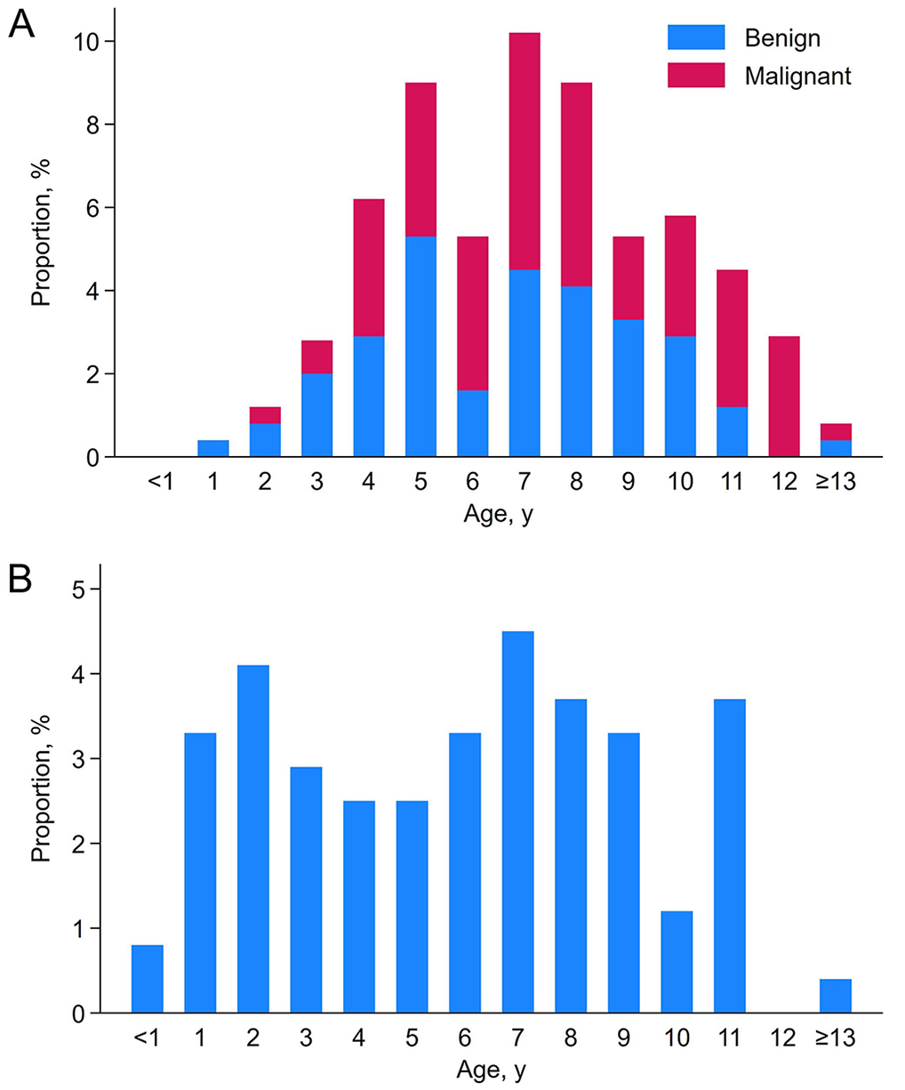
Age distribution of lesions in pet rabbits in Hong Kong, 2019–2022.
Skin and soft-tissue lesions
Trichoblastoma (Fig. 2A) was the predominant neoplastic skin and soft-tissue lesion in rabbits (34 of 77 skin and soft-tissue tumors; 44%), with 16 on the trunk, 5 on the limbs, 5 on the head and neck, and 1 on the tail; location was not recorded in 7 rabbits. The breeds affected most commonly included 7 Lionhead, 5 Dwarf, 4 Lop, and 2 Dwarf Lop rabbits. Age range was 1–13 y, with a major prevalence between 4 and 10 y; males (22 of 34) were overrepresented.
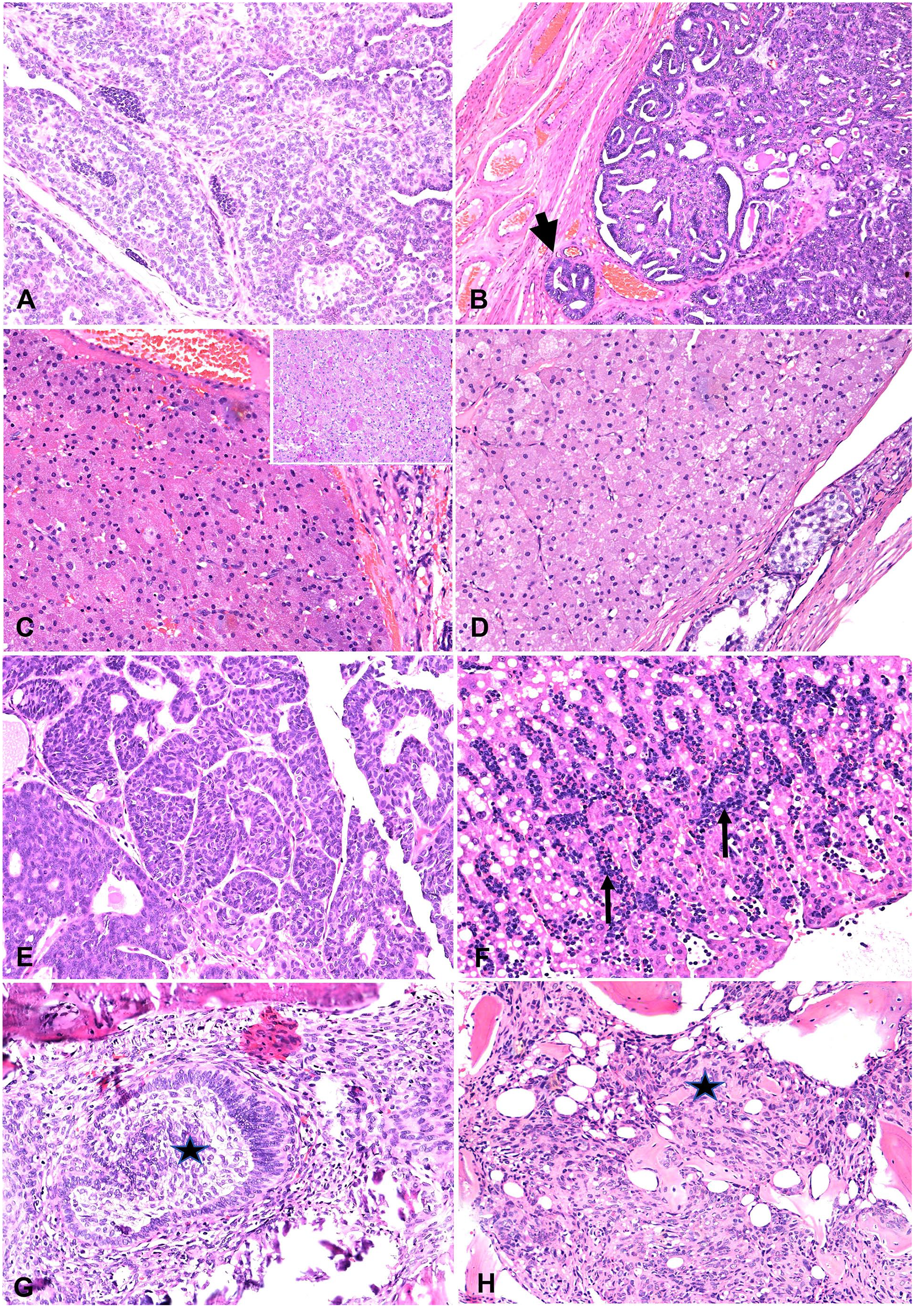
Neoplastic lesions in rabbits.
The second most frequent skin and soft-tissue tumor was sarcoma, which included 7 fibrosarcomas, 3 myxosarcomas, 2 peripheral nerve sheath tumors, and 1 giant cell sarcoma. Primarily, sarcomas were located in the limbs (8 of 13). The other locations included 2 on the head and 3 on the trunk. The breeds affected included 1 each of Dwarf lop, Dutch, Lionhead, and Dwarf, although breed was not recorded in 9 of 13 submissions. Ages were 4–12 y, with 7–12 y being the most common age range (10 of 13). Sarcomas did not have a sex predilection.
Mammary adenocarcinomas (10 of 77) were ranked as the third most common skin and soft-tissue tumor type. These adenocarcinomas were identified in 9 females (3 spayed), rabbits 5–12-y-old (mean 7.3 y). Among them, 4 Dwarf, 2 Lionhead, 1 Angora, and 2 of unknown breeds. Additionally, 1 castrated 8-y-old male rabbit, of an unknown breed, had an adenocarcinoma in the left caudal mammary gland.
Collagenous nevus was the fourth most common neoplasm in rabbit skin (4 of 77; 5%) and was most often identified in the pinna (n = 3). Squamous cell carcinoma (SCC) was identified in 3 rabbits, all located on their limbs, and ages were 8–11 y. Lipomas were observed in 2 rabbits; a histiocytic tumor, papilloma, melanoma, liposarcoma, fibrolipoma, apocrine adenoma, and apocrine adenocarcinoma occurred in 1 rabbit each.
The most common non-neoplastic skin and soft-tissue lesions included abscesses (11 of 29; 37.9%), dermatitis (9 cases), steatitis (2 cases), cellulitis (2 cases), and 1 infundibular cyst. Bacteria were identified by histologic examination in 4 abscesses, including 2 with gram-positive cocci; further bacterial identification was not pursued for these submissions. Abscesses were most frequently found on the abdomen, inguinum, and mammary chain (n = 7); 4 abscesses were located on the head. Dermatitis predominantly affected the head and neck skin of young to middle-aged rabbits (age range <1–7 y). Bacteria were identified by Gram stains on histologic examination in only 1 of the 9 dermatitis cases; further bacterial identification was not pursued. Two dermatitis cases were classified as eosinophilic dermatitis due to hypersensitivity.
Reproductive tract lesions
Reproductive tract lesions were the second most frequent. Of 74 intact rabbits 2–11-y-old (mean 6.1 y), 52 (70.3%) were presented to a veterinarian because of female reproductive tract disease. None of the rabbits in our cohort had vaginal or vulval disease. The affected breeds included 14 Lionhead, 9 Dwarf, 4 Lop, 3 Dwarf Lop, 2 Dutch, and 1 Angora, with breeds not recorded for 19 rabbits.
Thirty-one intact female rabbits (59.6%) had uterine or oviduct neoplasia, ranging in age from 2–10 y (mean 6 y), and 21 (40.4%) had non-neoplastic disease, ranging in age from 2–9 y (mean 5.5 y), which was mainly CEH. Uterine adenocarcinoma (Fig. 2B) was the most common neoplasm observed in the reproductive tract of 28 rabbits (90%); metastatic disease was observed in 3 rabbits, and was confirmed by thoracic radiography at the time of presentation. Two rabbits were diagnosed with uterine leiomyoma, one with uterine leiomyosarcoma, and one with an ovarian adenoma. CEH was often observed in rabbits in conjunction with another uterine lesion, including 13 with uterine adenocarcinoma, 1 with uterine leiomyoma, 1 with an ovarian adenoma, and 1 with pyometra. Four rabbits were confirmed to have pyometra and were younger at 3–5-y-old. Gram-stained bacteria were not observed within the inflammatory exudate of any of the rabbits. Only one rabbit had non-uterine disease, which was confirmed as an oviduct adenoma growing as an expansile, encapsulated lesion within the ovary but without any lesions in the uterus.
Lesions in the testes were identified in 21 rabbits and included 17 cases of single testicular neoplasms, 3 cases of orchitis, and 1 case of hypoplasia. Diagnosed testicular tumors included 8 granular cell tumors (Fig. 2C), 5 interstitial cell tumors (Fig. 2D), 3 seminomas, and 1 Sertoli cell tumor. Notably, one testicle contained both a seminoma and a granular cell tumor. A prostatic adenocarcinoma was identified in an 8-y-old castrated male rabbit (Fig. 2E). Of the 21 rabbits, 16 had single, unilateral testes submitted; 5 had both testes submitted. Information on the castrated testes was provided for 14 rabbits, which include 8 left testes (6 neoplastic, 2 non-neoplastic) and 6 right testes (all neoplastic). The affected rabbits were 1–11-y-old, with an average of 7 y. The affected breeds included 5 Lionhead, and 1 each of Dwarf, Netherland Dwarf, and Dwarf Lop; breed information was not available in 14 submissions.
Digestive system lesions
Lesions were noted in the digestive system of 32 of 247 rabbits. The affected breeds included 7 Lionhead, 4 French Lop, 3 Dwarf, 3 Lop, and 1 each of Dutch, Dwarf Lop, and New Zealand White; breed information was not recorded in 12 submissions. Among these digestive lesions, 14 were located in the liver; the remaining 18 were found in the oral cavity, salivary glands, stomach, intestine, rectum, and peritoneum. The lesions included 10 cases of liver lobe torsion, 7 types of neoplasia, 3 rectal polyps, and a range of inflammatory lesions. These lesions were observed in rabbits 1–11-y-old.
Hepatic non-neoplastic lesions accounted for 13 of 32 digestive system lesions (40.6%). Among the affected rabbits, there were 3 Lionhead, 2 French Lop, 2 Lop, and 2 Dwarf rabbits. Breed information was not available for 5 submissions. Within the liver, the most common lesion observed was hepatic torsion (10 of 13; 76.9%) accompanied by necrosis and thrombosis. Other liver lesions included cholangiohepatitis (3 of 13; 23%) and hepatocellular necrosis (1 of 13; 7.7%). The affected rabbits were 1–14 y-old, and there was no sex predilection. The location of the hepatic torsion was only recorded in one case, which was the caudate liver lobe. Additionally, a single case of neoplasia (lymphoma; Fig. 2F) was recorded.
Lesions within the oral cavity included an odontogenic fibroma (Fig. 2G) in a 5-y-old spayed female rabbit, an ameloblastoma in a 10-y-old female rabbit, and gingivitis in an 8-y-old spayed female rabbit. Breed information was not available for any of these rabbits.
Lesions in the salivary glands included 2 cases of sialadenitis: 1 case in a 1-y-old male French Lop and 1 case in 9-y-old, castrated male rabbit of unknown breed. Furthermore, a single stomach lesion from a 4-y-old male Lionhead rabbit was characterized by mucosal hyperplasia.
Among the 3 cecum and colon lesions submitted, all were inflammatory and non-neoplastic. These included 2 cases of typhlitis, one of which was associated with intralesional embedded plant material in a 3-y-old castrated male Lionhead rabbit, and the other exhibited diffuse typhlocolitis associated with bacteria in an 8-y-old spayed female New Zealand White rabbit. The third case had a diverticulum within the colonic wall of an 11-y-old spayed female Lionhead rabbit, which became inflamed and ruptured, leading to peritonitis.
Most of the digestive tract lesions involved the rectum; 8 diagnoses including 3 polyps, 2 rectal adenomas, and 1 each of leiomyosarcoma, fibrosarcoma, and proctitis. All of the polyps were observed in spayed females, aged 5-, 9- and 10-y-old. The affected breeds included 1 each of Lionhead, Dwarf, Dutch, Lop, French Lop, and Dwarf Lop; breed was not recorded in 2 submissions. The adenomas occurred in a 3-y-old spayed female rabbit and a 10-y-old castrated male rabbit. Proctitis occurred in a 1-y-old rabbit (sex not recorded); the fibrosarcoma occurred in a 10-y-old castrated male rabbit, and a leiomyosarcoma was found in another 10-y-old castrated male rabbit.
Two cases were found in the peritoneum: 1 intra-abdominal liposarcoma in an unknown-aged male rabbit of unknown breed, and 1 case of steatitis in a 3-y-old spayed female rabbit of unknown breed.
Hematolymphoid system
The main lesions within the hematolymphoid system included 13 thymomas located in the mediastinum. Affected rabbits were 5–11-y-old (mean 8 y), and both sexes were similarly affected. Among the breeds affected were 3 Lionhead, and 1 each of Holland Lop and Velveteen rabbits; breed was not recorded in 8 submissions. The thymomas were histologically subtyped as 11 lymphocyte-predominant types, 1 mixed lymphoepithelial type, and 1 predominant epithelial type.
Two cases of lymphoma were found. The first case involved the liver of a 3-y-old spayed female Lionhead rabbit. The second case involved diffuse lymph node enlargement in an adult castrated male rabbit of unknown breed. The lymph nodes contained large round cells with a mitotic rate of 3 per hpf.
Additionally, a splenic biopsy was submitted from a 4-y-old castrated male rabbit of unknown breed, with a history of severe anemia. Although reactive histiocytosis, hemosiderosis, and extramedullary hematopoiesis were identified, the exact cause of these conditions was undetermined.
Musculoskeletal system
Nine musculoskeletal lesions were recorded, all involving bone; among these, 5 tumors including 4 osteosarcomas (Fig. 2H). The osteosarcomas were located in the hock (2 cases), the radius (1 case), and the right foreleg (1 case). Additionally, 3 myxosarcomas were observed. The only recorded inflammatory lesion was classified as osteomyelitis and synovitis in a 1-y-old male rabbit. The affected breed was exclusively Dwarf Lop; breed information was not provided in 5 of 9 submissions.
Urinary system
Two cases of cystitis were recorded; 1 in a 1-y-old castrated male rabbit and the other in an 8-y-old spayed female rabbit. Additionally, a single renal infarct was reported in an 11-y-old castrated male rabbit; 2 cases of hydronephrosis were observed.
Special senses
Less common diagnoses included ocular lesions in 3 rabbits: 1 Lionhead, 1 Dwarf, and 1 breed unknown. In 2 cases, eyes were enucleated because of uveitis, cataract formation, or hypopyon. Encephalitozoon cuniculi was found on Gram staining of both cases of phacoclastic uveitis and scleritis. In the third case, chronic epiphora and medial canthus erosion in a 10-y-old female rabbit were associated with Harderian gland inflammation.
Respiratory system
A nasal adenocarcinoma was diagnosed in a 7-y-old castrated male Holland Lop rabbit.
Discussion
Neoplastic skin and soft-tissue lesions, particularly trichoblastoma, were the most common reason for biopsy in Hong Kong pet rabbits, followed by reproductive tract neoplasia, non-neoplastic skin disease, and non-neoplastic digestive disease. We found 13 different skin tumors, with trichoblastoma the most common, accounting for more than a quarter of all neoplastic lesions. Previous studies reported trichoblastoma as the most frequent cutaneous neoplasm in rabbits24,39,57; lesions are typically found on the trunk, head, neck, and tail; moreover, there are no signs of malignancy and there is no sex predilection. Interestingly, most of the affected rabbits in our study were male, although the reason for this sex predominance remains unclear.
Sarcoma was identified in 15 rabbits and was the second most common soft-tissue neoplasm in our study. Some of these neoplasms were further classified into subtypes such as fibrosarcoma, myxosarcoma, peripheral nerve sheath tumor, and giant cell sarcoma. This differs from the findings of a 2020 retrospective study in which sarcomas and other mesenchymal non-viral tumors were less common. 57 Unlike the 2020 study in Pennsylvania, USA, which reported a male predisposition for mesenchymal tumors, 57 we found no sex predilection for sarcomas in our case series. Furthermore, we found no cases of viral-induced neoplasia (including Shope fibroma, Shope papilloma, or myxomatosis) as reported in the 2020 study. 57 Papillomas have been reported on the skin of rabbit’s ears, 3 and the ear skin is a common location for Shope papilloma virus infections associated with arthropod bites.11,57 However, natural Shope papillomavirus infections seem to be restricted to the United States.
Mammary neoplasia is increasingly recognized in pet rabbits 7 and has been reviewed in several studies.46,47 The reported age ranges and means of affected rabbits are 2–14 y (mean 5.5), 4 2–8 y (mean 4.9), 46 0.75–14 y (mean 5.2), 47 and 5–9 y (mean 7) 49 ; these ages are similar to those in our study (5–12 y), but the mean age in our cases was higher at 7.1 y. Another feature of pet ownership in Hong Kong that we noted is a tendency not to neuter pets, with mammary and reproductive tumors being frequent reasons for biopsy. We identified one case of mammary adenocarcinoma in a male rabbit, which is considered to be a rare occurrence. Most mammary tumors are carcinomas and are often subclassified, 3 although a range of benign tumors has been reported. In our series, 10 of the 14 tumors were adenocarcinomas; only 1 mammary adenoma was described. Collagenous nevus, which is a benign neoplasm commonly described in cutaneous neoplasms in rabbits, was identified in the skin of 4 rabbits.
Inflammation, in the form of abscess, dermatitis, steatitis, or cellulitis, included most of the non-neoplastic cutaneous lesions. Bacterial organisms were identified on histologic examination in 5 of these cases. Cutaneous abscesses are commonly reported as resulting from traumatic wounds or septic events following infection of the respiratory, urinary, or reproductive tract.19,56 Although further bacterial identification was not pursued in our series, common bacterial causes of abscesses in rabbits include Staphylococcus aureus, Pasteurella multocida, and Pseudomonas aeruginosa. 19
Uterine disease was prevalent in female rabbits, with uterine adenocarcinoma the most common lesion in 28 of 243 (11.5%) rabbits. This finding is consistent with previous reports,5,8,26,61 although it surpasses the 6.8% (n = 1,238) 3 and falls below the 16.7% (n = 849) 17 reported in other studies. Ovarian disease has been reported in rabbits but at a fairly low incidence,7,8,10,61 and we found only one ovarian adenoma in our study. The mean age of the female rabbits with uterine lesions in our study was 4.9 y, which is consistent with the mean age of 5.6 y reported in another study. 2 We found CEH in 38% of the female rabbits, similar to previous reports.8,26,61
Pyometra is an uncommon finding in young rabbits and was observed in 4 cases in our study. This finding aligns with the findings of other studies suggesting that pyometra is most commonly reported in breeding does or commercial rabbit farms,18,43,48 rather than in pet rabbits where it is typically reported as a single case23,58 with fewer larger studies. 8 Other uterine lesions, including smooth muscle tumors, were rarely diagnosed, consistent with previous reports.3,26,61 Among the 18 testicular neoplasms that we found, most were granular cell tumors, which is similar to the findings of previous reports.6,22
The most common lesions in the digestive system were noted in the liver, although lesions were also present in the gingiva, salivary glands, stomach, cecum, colon, and rectum. Oral lesions are relatively common in rabbits, and a wide range of oral-specific tumors have been reported, including SCC, ameloblastoma, fibrosarcoma, osteosarcoma, cementoma, complex odontoma, giant cell epulis, chondrosarcoma, and malignant melanoma. 35 We found one case each of an odontogenic fibroma, ameloblastomas, and gingivitis.
The cecum and colon lesions found were all inflammatory; most of the lesions were rectal in origin. Digestive diseases in rabbits are often related to diet, particularly gastrointestinal stasis leading to various clinical presentations. 38 Rectal polyps are commonly found in many rabbit studies, and lesions are suggested to develop in response to inflammation rather than neoplasia. 4
Two salivary lesions were confirmed to be suppurative inflammation. Histologic examination plays a crucial role in the evaluation of salivary gland disease to differentiate sialadenitis from sialectasis and sialocoeles. 13
Liver lobe torsion was the most common non-neoplastic hepatic lesion found in rabbits in our study. Hepatic torsion is rarely reported in most species, but its detection is increasing in pet rabbits.16,44,63 Liver lobe torsion alone represented more than one-third of all digestive system lesions in our case series. Hepatic ischemic necrosis of the affected lobe was the most common lesion in our series, which is consistent with the findings of other reports.16,52,63 In one case in our series, the caudate liver lobe was the affected lobe, which is in line with a previous report that identified a predilection for this location in rabbits. 52 As described in previous reports, there was no sex predilection, and no breed predisposition was identified in our series, unlike earlier reports suggesting the overrepresentation of Mini Lops. 16
Interstitial cell tumors in rabbits have been designated as granular cell tumors due to the presence of PAS-positive cytoplasmic granules, which are otherwise identical to interstitial cell tumors in other species.6,62 PAS staining was applied to the interstitial cell tumors in our series, and if the neoplastic cells contained PAS-positive cytoplasmic granules, they were considered granular cell tumors. One study raised the question of whether a granular cell tumor is truly distinct from an interstitial cell tumor, or simply reflects a species-specific staining pattern. 4 Transmission electron microscopy (TEM), cytology, and immunohistochemical (IHC) staining for S100 and periaxin can be used to confirm a diagnosis of granular cell tumor, but they are infrequently used in routine diagnostic cases.6,22,40 PAS staining remains the most cost-effective histologic tool for diagnostic evaluation.
No other neoplasms of the male genital tract were found in our series. A testicular teratoma has been reported, 33 and testicular neoplasia was reported in 7 testicular biopsies from 37 cryptorchid testes in one study. 6 Of the 2 cryptorchid testes in our case series, a 9-y-old rabbit had a Sertoli cell tumor, and a 1-y-old rabbit had a hypoplastic testis.
In our study, one case of prostatic adenocarcinoma was found, and a previous report described a case in which a prostate lesion was similarly attached to the bladder. 45
Lesions were recorded in the hematolymphoid system of 17 rabbits and were dominated by thymomas in the mediastinum of 13 rabbits. Thymoma is the most commonly reported mediastinal neoplasm in rabbits,6,27 and whereas most are benign, malignant forms have been described. 59
Steatitis, which is a rare lesion but is reported as a consequence of liver lobe torsion, was recorded in one rabbit, as well as a single intra-abdominal liposarcoma, in our study. Liposarcoma has been reported in the forelimb, 60 head, thorax, and abdomen. 57
Most of the bone lesions were tumors, including 4 osteosarcomas and 1 myxosarcoma. Additionally, there was a single case of osteomyelitis and synovitis in a one-y-old rabbit. Osteosarcoma has been regularly reported in rabbits 54 in various locations, such as the femur, 14 humerus, 25 nasal cavity, 34 and oral cavity. 35 In our study, all of the osteosarcomas were in the limbs, including 2 in the hock, 1 in the radius, and 1 in the right foreleg (exact location not provided). Osteosarcomas are considered to be potentially metastatic and can occur in rabbits as young as 1-y-old, but are most common in rabbits >5-y-old. 32 The mean age of the rabbits with osteosarcoma in our study was 8.7 y (range 5–12 y). A single case of myxosarcoma infiltrating bone was recorded in a 9-y-old rabbit. Histologic examination revealed a mucin-rich, mesenchymal tumor involving soft tissues and skeletal muscle infiltrating the tibia and causing bone lysis. Myxosarcomas in rabbits are usually associated with a viral etiology involving connective tissue rather than invading bone. 12
Lesions in the urinary tract included cystitis, renal infarction, and hydronephrosis. Kidney and bladder lesions are relatively common in rabbits. 50 In some studies, 25% of healthy rabbits and 33% of unhealthy rabbits had evidence of kidney disease. 20
Eye lesions included phacoclastic uveitis consistent with E. cuniculi infection in 2 rabbits, identified by the gram-positive organisms within the lens or adjacent to the ruptured lens. Gram and modified trichrome stains are effective at enhancing spore detection. 42 The other ocular sample had chronic inflammation in the Harderian gland, characterized by lymphocyte and plasma cell infiltrates. This finding is typically considered an incidental or background lesion in rabbits. 51
Only 2 cases of lymphoma were described in our series, one in the liver and one in a lymph node. In the liver, neoplastic round cells were observed, and lymphoma was considered the most likely diagnosis. However, due to limitations in our laboratory in which rabbit antibodies are employed for IHC staining, we could not confirm the diagnosis conclusively, leaving this diagnosis provisional. Hepatic lymphoma is rarely reported in the literature, indicating its infrequent occurrence. 3 The second case of lymphoma was described as diffuse lymph node enlargement with the presence of large round cells, again suggesting lymphoma as the most likely diagnosis. Among lymphoma types in rabbits, large B-cell diffuse and epitheliotropic lymphoma are the most commonly described types of lymphoma 41 ; intestinal lymphoma has been reported in a small number of cases. 30
Only one respiratory lesion (nasal adenocarcinoma) was identified in our study. Instances of nasal adenocarcinomas have been documented, 29 with some cases even responding positively to treatment. 36
The primary limitation of our study lies in its retrospective nature and the fact that all of the included data were sourced from a single laboratory. This could make the findings unrepresentative of the pet rabbit population in Hong Kong. Furthermore, the frequency of lesion types is influenced by the number of submitted tissue samples. In our study, the most frequently submitted tissues in surgical biopsies were skin, soft tissue, and reproductive tract, which may have led to an overrepresentation of tumors in these organs, in contrast with autopsy cases. Nevertheless, surgical biopsies can provide valuable information on various tumor types in specific organ systems and the median age of affected animals.
Footnotes
Acknowledgements
We thank our contributing veterinarians for providing biopsies for analysis. We also thank the histology technologists for processing the samples.
Declaration of conflicting interest
The authors declare no potential conflicts of interest with respect to the research, authorship, and/or publication of this article.
Funding
The authors received no financial support for the research, authorship, and/or publication of this article.