
Abstract
Published information about fish botulism is scant. We review here the current literature on fish botulism. Freshwater fish are susceptible to botulism. Only anecdotal evidence exists about possible botulism cases in saltwater fish. With only a few exceptions, the etiology of all cases of fish botulism reported is Clostridium botulinum type E, although fish are sensitive to, and may carry, various C. botulinum types. Clinical signs of botulism in fish include loss of equilibrium and motion, abducted opercula, open mouths, dark pigmentation, and head up/tail down orientation in which attempts to swim result in breaching the surface of the water. Dark pigmentation is thought to be associated with acetylcholine imbalance in botulinum neurotoxin (BoNT)-affected fish. Rarely, but similar to the situation in other animal species, fish can recover from botulism. Fish botulism can cause secondary outbreaks of the disease in birds, as botulism-affected fish stand out from normal fish, and are selectively preyed upon by fish-eating birds, which thus become intoxicated by the BoNT present in sick fish. The source of BoNT in fish has not been definitively confirmed. Fish may ingest C. botulinum spores that then germinate in their digestive tract, but the possibility that fish ingest preformed BoNT from the environment (e.g., dead fish, shellfish, insects) cannot be ruled out. The presumptive diagnosis of botulism in fish is established based on clinical signs, and as in other species, confirmation should be based on detection of BoNT in intestinal content, liver, and/or serum of affected fish.
Keywords
Botulism is caused by botulinum neurotoxins (BoNTs) produced by BoNT-producing clostridia, including Clostridium botulinum, C. butyricum, C. baratii, and C. argentinense. Most cases of botulism are, however, associated with C. botulinum, which is, as are all of the clostridia mentioned above, a gram-positive, anaerobic, sporulated rod. BoNTs are the most powerful toxins known. Botulism is characterized by flaccid paralysis, and it has been described in numerous mammalian, avian, and fish species. C. botulinum can be found in the environment and the intestinal content of many clinically normal animals.18,20,31
Most fish botulism cases are produced by C. botulinum type E, and the disease has been historically called “bankrupt disease” in reference to the high mortality and associated economic loss that it causes.15,27,55,56 The environmental impact associated with large die-offs in natural environments is also significant (Fig. 1). Type E botulism was first reported in rainbow trout in Denmark, 27 and several additional outbreaks were reported subsequently in other species of farmed fish.5,27 In addition to its impact on the fish industry, botulism in feral fish has been associated with large-scale outbreaks of botulism-associated mortality in piscivorous birds.3,14 Although it has always been assumed that these birds become intoxicated by eating fish carrying C. botulinum, 19 the mechanism of exposure of birds to BoNT type E (BoNT/E) is unknown.
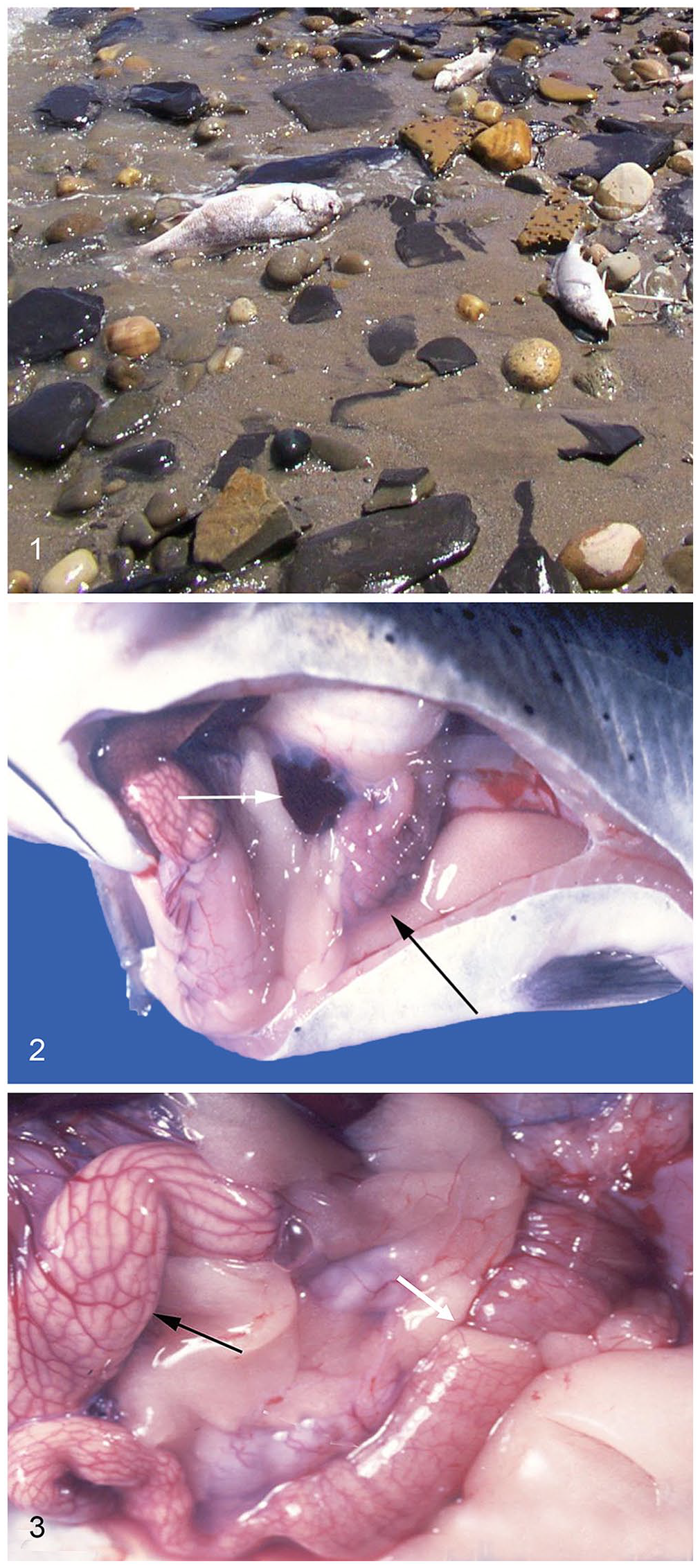
Several dead fish on a beach; part of an outbreak of Clostridium botulinum type E spontaneous intoxication affecting several species of fish in one of the Great Lakes, USA. Photo courtesy of Dr. L. Khoo.
Etiology
C. botulinum can be classified according to the antigenic specificity of the toxin produced or according to DNA relatedness.1,21 If classified according to toxin type, C. botulinum can be grouped into toxin types A–G. 21 When classified according to DNA relatedness, C. botulinum is divided into groups I–IV. 34
In North America, C. botulinum type E is mostly found in marine and fresh water, 51 soil, and fish. 2 Human and avian diseases caused by C. botulinum type E have been associated with the consumption of fish and marine mammals.10,23,50 C. botulinum type E spores are frequently found in the intestine of healthy fish.18,20,31 Different types of C. botulinum have been found in fish at prevalences of 2–21%.18,20,31 For instance, in a study of the prevalence of C. botulinum in wild fish performed in India, the C. botulinum types found on the surface of the fish included types D (8%), C (6%), A (6%), and B (2%). In the intestine of those fish, types A (4%) and C (2%) of C. botulinum were found. 30 Random amplification of polymorphic DNA (RAPD) and pulsed-field gel electrophoresis (PFGE) have been used to determine genetic relatedness among C. botulinum type E strains.25,28,32 In one of these studies, 25 strains were isolated from trout in Finnish farms. All of these studies found that there is extensive genetic variation in C. botulinum type E strains. 25
All BoNT subtypes block the release of acetylcholine at the neuromuscular junction and lead to flaccid paralysis. Different types are more prevalent in certain species; for instance, BoNT types A, B, E, and, more rarely F, are mainly responsible for human botulism, whereas toxin types C and D and their mosaic variants are mostly involved in mammalian and avian animal botulism. 34 This is not, however, a strict rule, and exceptions occur. Although C. botulinum type E has been associated frequently with fish and fish-eating birds, several cases of human disease have also been reported. 24 The number of type E outbreaks in humans depends on the country and on dietary habits. 22 Type E outbreaks were also detected in poultry on farms in France in the late 1990s 33 ; the source of contamination was not identified, but this outbreak shows that type E botulism outbreaks can occur in species other than fish, piscivorous birds, and humans.
Most cases of fish botulism reported to date have been produced by C. botulism type E. 34 However, although there is no information on cases of fish botulism produced by other types of C. botulinum, it is possible that they have occurred and have gone undetected.
Although most cases of fish botulism are produced by C. botulinum type E, 38 fish are sensitive to the other botulinum toxin types and can carry various C. botulinum types. For instance, C. botulinum types A–D were found in a large survey of fish and water sediments in the Indian subcontinent; C. botulinum types C and D were the predominant types found in both fish and the aquatic environment. 30 Types A and D were also found in cured fish in another survey in India. 31 In a survey of fish and water performed in France, C. botulinum type B was the predominant type found, followed by types A and E. 18
Pathogenesis
The pathogenesis of botulism in fish has not been studied at the molecular level as it has been in other species. However, due to similarities in the neuromuscular system of fish compared to other vertebrates, 52 and the fact that experimental exposure to BoNTs causes similar clinical signs, including flaccid paralysis, 7 it is assumed that the pathogenesis in fish is similar to that in mammalian and avian species.
Experimental infections have demonstrated that several fish species are particularly susceptible to BoNT/E. For instance, in coho salmon inoculated by the oral route, type E toxin was lethal at a dose equivalent to 90 mouse intraperitoneal minimal lethal doses. 17 The median lethal dose in channel catfish fingerlings was 13.7 pg/fish when injected intracoelomically. 7 Zebra fish have variable sensitivity to different BoNTs. The 96-h median immobilizing doses of BoNT/A, BoNT/C, BoNT/E, and BoNT/F for adult male Tübingen strain zebrafish were 16.3, 125, 4.7, and 0.61 pg/fish, respectively. 9
Two mechanisms of intoxication by BoNTs have been proposed for fish. It is thought that fish acquire C. botulinum or its spores orally by being exposed to decayed plant or organic matter (i.e., from other fish that died of botulism) in lakes and seabeds. Most fish species are also carriers of C. botulinum type E in their intestine or intestinal contents, which poses a risk of foodborne zoonotic disease. 18 Once in the intestine, spores germinate into vegetative forms and start producing BoNTs, which are then absorbed into the blood and disseminate to the rest of the organism. The second mechanism involves ingestion of preformed toxins from the contaminated environment. In both cases, the BoNTs heavy chain targets specific receptors at the end of the motor neurons in the neuromuscular junction. Subsequently, BoNTs become internalized via receptor-mediated endocytosis in either clathrin-coated molecules or recycling synaptic vesicles. Once these vesicles are acidified, the light chain is translocated into the cytosol. The BoNT light chain cleaves the synaptic fusions forming SNARE (soluble N-ethylmaleimide-sensitive-factor attachment protein receptor) proteins; specifically, BoNT/E cleaves synaptobrevin and synaptosomal-associated protein. Consequently, the formation of the fusion complex is disrupted and acetylcholine cannot be released to the synaptic cleft at the neuromuscular junctions, thereby causing flaccid paralysis.34,43
Epidemiology
Most cases of botulism reported in fish have been produced by C. botulinum type E. In humans, cases of type E botulism are most frequently associated with the consumption of fish or tissues from marine mammals.13,24,26 C. botulinum type E is found in marine and freshwater soil, 25 and also in the intestinal tract of clinically normal fish.20,30
C. botulinum type E has been isolated from several different freshwater and marine fish species including herring, salmon, trout, whitefish, and mullet. Human cases of type E foodborne botulism are most commonly seen in association with consumption of fermented or pickled fish, and based on this, it is thought that the fermentation and pickling process allows the development of anaerobic conditions that promote germination of spores and toxin formation. 24
Sporadic reports of botulism in fish are documented in the scientific literature. In commercial fish production, several large-scale mortality events in salmonid hatcheries have been attributed to type E botulism.5,15,16,27 Outbreaks are most common in juvenile salmonids (including chinook and coho salmon, and steelhead and rainbow trout) raised in earthen bottom ponds. Toxins were detected in sediments, dead fish, and, to a lesser degree, invertebrates found within the ponds.5,16,27 Dead fish are a major source of BoNTs in these epizootics, and cannibalism plays a significant role in the spread of this toxin to other fish. Outbreaks are typically managed by rapidly removing dead fish and transitioning the remaining fish to concrete ponds. 16 Similarly, visceral toxicosis of catfish (VTC) is a form of botulism described in channel catfish. VTC was first recognized in the late 1990s in commercially raised catfish in the southeastern United States.29,54 It predominantly affects food or brood-size fish, and occurs largely in the spring and fall. 29
Sporadic natural outbreaks of type E fish botulism have been documented in freshwater lakes.19,21 The bulk of the published literature regarding mortality events associated with type E botulism has focused on wildlife mortality (specifically waterfowl and shore birds) in the Great Lakes of North America. In these outbreaks, both toxigenic and nontoxigenic strains of C. botulinum were isolated from the intestinal tract of fish consumed by affected birds. 21 In one study, 21 type E toxin was only detected in a small portion of sampled fish; however, several non–toxin-producing C. botulinum isolates were PCR positive for the BoNT/E gene, indicating their potential for toxin production. Other authors have demonstrated that C. botulinum type E was detectable in both healthy and moribund feral fish. 20 A survey of fish from the Great Lakes in the United States revealed that the percentage of fish with detectable C. botulinum type E varied among sampling locations, suggesting that the distribution of toxin-producing strains is not homogeneous in the same habitat. 2
Moribund fish clinically affected by botulism often exhibit changes in behavior (e.g., head bobbing at the surface and inability to maintain a normal position within the water column), which may predispose them to consumption by piscivorous birds that only eat live fish.20,55,56 Experimental studies suggest that susceptibility to type E botulism varies between fish species.55,56 Onset of clinical signs (e.g., hyperpigmentation, behavioral changes) and mortality among evaluated species (round goby, walleye, rainbow trout, yellow trout) vary considerably. For example, at the same dose, mortality ranged from 25% in yellow perch to 92% in round goby. 56
Zoonotic risk of fish botulism
Foodborne type E botulism linked to the consumption of fish has been reported, and it was thus suggested that botulism can be considered a zoonosis. 45 However, the term zoonosis does not apply to these cases of human disease because zoonosis means direct transmission from diseased animals to humans. The cases of human botulism mentioned above resulted from consumption of contaminated food of animal origin, but not from transmission from sick animals (Michel R. Popoff, pers. comm., 2023).
Cases of human botulism type E have been associated with consumption of salted dried ham in France36,44 and Argentina. 48 In those cases, it was suggested that sea salt used for the preparation of ham could have been the source of contamination. However, although C. botulinum type E has been found in water, human activities such as swimming and fishing during botulism outbreaks are usually not considered a risk to human health.
Other cases of type E human botulism have been reported associated with consumption of fish or other seafood.22,35,41,46,53 An outbreak of human type B botulism was reported in France associated with consumption of homemade preserved sardines.11,37 In that outbreak, C. botulinum was found in the fish but not in the marinade, suggesting that the origin of the outbreak was the fish, not the other components of the preparation. 37 C. botulinum type B is highly prevalent in fish from coastal France, 19 and this was suggested to be the origin of the contamination of the sardines. 37
Clinical signs
Natural disease
Wild fish with natural botulism are described as being hypersensitive and nervous in the early stages of disease. This progresses to paralysis of fins, loss of equilibrium, swimming on one side, loss of color, gaping of gills, and elevated mortality.5,15,16 In captivity-raised fish, additional reported clinical signs include fish being pushed into screens or to low-flow areas. Muscle fasciculations, quivering barbels, and occasionally eversion of the stomach and exophthalmia have been reported in affected catfish. 29
Experimental disease
Clinical signs in experimental infections are similar to those described above for natural disease, although some subtle variations in clinical signs between fish species were noted. For instance, in fish experimentally infected with C. botulinum type E, clinical signs included melanosis or discoloration, abnormal swimming patterns, loss of equilibrium or loss of fin control, vertical orientation, opercular abduction, variations in respiration rate, and mortality.55,56
Thus far, there does not appear to be a consensus as to the best method of administration of BoNT/E to reproduce botulism in fish. Some authors administered the toxin orally55,56 in an attempt to replicate what occurs in natural infections, whereas other studies have attempted introducing the toxin via intracoelomic injection.7,8
Pathology
Natural disease
In salmonids that died of botulism, extended gill covers and curved bodies were observed. In these animals, the caudal 10–15 mm of the intestinal tract contained very viscous amber fecal material suggestive of constipation, as described in humans and other animals with botulism. 16
The pathology of VTC was described in detail. 29 Grossly, chyle-rich ascites (Figs. 2, 3), intestinal intussusceptions (Fig. 3), eversion of the stomach into the oral cavity, pale proximal intestine with congested blood vessels (Fig. 3), splenic congestion, and a reticular pattern were observed in the liver. Histologically, there was cerebral, splenic, and hepatic congestion, coupled with splenic lymphoid depletion and perivascular edema, vascular dilation and edema in the gastrointestinal tract, and perivascular edema in the anterior and posterior kidneys.
Experimental disease
In rainbow trout experimentally dosed per os with different doses of BoNT/E, the animals showed dorsal recumbence and opercular abduction,43,44 similar to descriptions in the first experiment of oral exposure to BoNT in fish. 48 The same experimental setting induced progressive dark pigmentation that started as a faint 1–2-cm wide black band just caudal to the pectoral fins and progressively evolved caudally until almost the entire body became black.55,56 Similar results were reported in a separate study with round gobies experimentally exposed to repeated low doses of BoNT/E.55,56 Walleye were in dorsal recumbency with adducted opercular plates and ventrally arched bodies. Yellow perch had abducted opercular plates and darkening of pigmented areas that also progressed to the rest of the body. Loss of color and gaping of the gills was noticed in a field experiment, in which rainbow trout were inoculated with BoNT/E. 5 The mechanism associated with loss of color in fish with botulism is not fully understood. It has been suggested that the chromatophores in fish are innervated, and both the parasympathetic and sympathetic innervation of chromatophores can affect the expansion or contraction of these cells and the degree of pigmentation. 40 It is therefore possible that the changes in pigmentation in BoNT/E-affected fish are related to loss of control over these nerves.
Channel catfish fingerlings developed lesions similar to VTC after experimental intracoelomic inoculation with different doses of BoNT/E, including terminally opened mouths with abduction of the opercula, exophthalmos, ascites, splenic congestion, and intussusception and blanching of the intestine. 7 Zebrafish inoculated intracoelomically with serum of VTC-affected catfish or with BoNT/E had renal tubular necrosis histologically. 8 The gross and microscopic changes observed in some cases of fish botulism seem to be nonspecific and are probably of little diagnostic significance. Something similar occurs in avian and mammalian species in which nonspecific gross findings can be seen occasionally (e.g., nuchal ligament edema in horses). 34
Diagnosis
A presumptive diagnosis of botulism in any animal species is based on clinical signs. However, although highly suggestive, clinical signs alone are not diagnostic and laboratory confirmation is required. As in other species, a botulism outbreak in fish is suspected when there is a rapid increase in mortality associated with flaccid paralysis. Absence of significant gross and microscopic lesions during postmortem examination is supportive of a diagnosis of botulism, although some of the lesions described above in channel catfish with VTC may suggest a diagnosis of this disease. Culture and/or detection of C. botulinum in animal or feed samples is also supportive but does not confirm a diagnosis of botulism because the organism can be found in the environment and the intestinal content of healthy animals, including fish.
Diagnostic confirmation requires laboratory detection and identification of the neurotoxin type in animal or feed samples. Despite significant recent developments in methods for the detection of BoNT, mouse bioassay (MBA) remains the gold standard for toxin detection, and it is still the method used most commonly by veterinary diagnostic laboratories. The sensitivity of the MBA is, however, relatively low, and false-negative results are common. Another problem with the MBA is that this test cannot differentiate mosaic types. Finally, the MBA method is time-consuming, laborious, expensive, and presents ethical issues associated with the use of live animals. Very few laboratories throughout the world are equipped to perform the assay. Several alternative laboratory tests to detect BoNTs have been developed, including ELISAs, enzymatic methods based upon BoNT cleavage of synthetic peptides followed by differential detection of the specific product by spectrometry, or cell-based assay.4,6,12,39,42,43,47,49 These techniques, although promising, have, however, not been validated for the diagnosis of fish botulism.
The reported cases of VTC were initially confirmed using a catfish bioassay and mass spectrometric–based endopeptidase methodology, 21 and more recently by bioassay in adult zebrafish. 8 The latter is considered a reliable and practical alternative to rapidly detect BoNT/E in sera from VTC-affected catfish. The preferred samples for MBA in mammals are, in decreased order of sensitivity: gastrointestinal content, liver, and serum, although, if feed suspected to be associated with an outbreak is available, this is the ideal sample to test. However, little information is available in the literature about the sensitivity of testing such samples for the diagnosis of fish botulism.
Because the classic presentation of botulism in wild fish is just en-masse fish mortality, there are several differential diagnoses that should be considered, which vary based on fish species. For example, infection by Flavobacterium psychrophilum or Myxobolus cerebralis would be appropriate differential diagnoses in salmonids. Usually in these large mortality events, various viral, bacterial, and parasitic agents are detected. One of the most significant features of fish botulism is that multiple fish species are affected. Additionally, conditions such as hypoxia and other water quality–related issues also must be considered; these also affect a wide variety of species. In an aquaculture setting, however, the absence of other pathogens and significant gross and microscopic lesions should be considered suggestive of botulism.
Discussion
Subclinical carriage of C. botulinum type E occurs frequently in fish. As analyses of wildlife fish are usually performed on postmortem samples collected from carcasses, it is difficult to determine if detected BoNT is the cause of death or if it has been produced after death.
The lack of published information about botulism in fish is probably in part associated with the fact that very few laboratories in the world perform routine diagnostic work for this disease and that the current gold standard, MBA, is expensive, laborious, and has low sensitivity. It is therefore possible that at least some cases of botulism in fish go undiagnosed. Another fact that may compound the problem is that the clinical signs and postmortem lesions of botulism are not specific, and a presumptive diagnosis of the disease is not established in many cases. Finally, when mortality occurs in wild fish populations, it is possible that clinical signs are not observed and samples for postmortem examination and ancillary testing are not available.
But even when samples from affected fish are available for laboratory testing, the low sensitivity of the gold standard MBA conspires against a successful diagnostic outcome. As with other species, the low sensitivity of MBA is probably associated with the higher sensitivity of fish to botulism compared to mice. Several other tests for the detection of BoNT, including mass spectrometry, have been used for diagnosis of botulism in humans. 37 Some of these techniques are being validated for use in veterinary medicine and hopefully will be made available to diagnostic laboratories in the not-too-distant future.
To date, only C. botulinum type E has been found to be responsible for cases of botulism in fish. This situation is not unique as other animal species are particularly affected by certain types of C. botulinum (e.g., waterfowl are mostly affected by C. botulinum type C followed by type D, cattle by mosaic types C/D and D/C, and type D and C, and so on).
The reason for this species preference for some types of C. botulinum is not known and it can be associated with animal and/or environmental factors. For instance, C. botulinum type E seems to be more prevalent in fresh water, and only anecdotal evidence is available about possible cases of botulism in saltwater fish. This may reflect an environmental difference in the pathogenesis of fish botulism, which is supported by the fact that salmonids are affected by botulism, but these species have both freshwater and marine life stages, and botulism has only been reported in freshwater- or hatchery-raised salmonids. Additional information is needed to understand the pathogenesis of botulism in fish.
Footnotes
Acknowledgements
We thank Ms. Seana Fitisemanu for her work on the references for this paper.
Declaration of conflicting interests
The authors declared no potential conflicts of interest with respect to the research, authorship, and/or publication of this article.
Funding
The authors received no financial support for the research, authorship, and/or publication of this article.