
Abstract
CNS tumor diagnosis in dogs often relies on immunohistochemistry (IHC) given similar histologic features among tumors. Most CNS tissue samples encountered by diagnostic pathologists are collected during autopsy, and postmortem specimens can be susceptible to autolysis and prolonged formalin fixation, both of which have the potential to influence IHC results and interpretation. Here we evaluated the effects of experimentally controlled autolysis induced by delayed tissue fixation (sections of brain held for 2, 4, 8, 12, 24, 48, and 72 h in 0.9% NaCl at either room temperature or 37°C prior to fixation) as well as the effects of prolonged formalin fixation times (1 wk, 1 mo, 2 mo) on a panel of 8 IHC markers (CNPase, GFAP, Iba1, OLIG2, PGP9.5, MAP2, NeuN, synaptophysin) relevant to brain tumor diagnosis. Prolonged fixation of up to 2 mo had no detrimental effect on any immunomarker except NeuN, which had reduced immunolabeling intensity. Delayed fixation led to autolytic changes as expected, on a gradient of severity corresponding to increased time in saline prior to fixation. Several immunomarkers should be used with caution (CNPase, OLIG2) or avoided entirely (MAP2, NeuN) in markedly autolyzed brain and brain tumor tissues. Our results suggest that autolysis has minimal effect on most immunomarkers, but that advanced autolysis may cause a loss of specificity for GFAP, MAP2, and PGP9.5, a loss of intensity of CNPase and OLIG2, and loss of labeling with MAP2 and NeuN. Prolonged fixation affected only NeuN, with mildly decreased intensity.
CNS tumors in dogs can have overlapping histologic features, which can create diagnostic challenges for the pathologist who may be limited to evaluating H&E-stained tissue sections.1,7,9,12,13 For this reason, the application of immunohistochemistry (IHC) is often necessary to arrive at a definitive diagnosis. Although veterinary pathologists with neuropathology subspecialty expertise and interest are increasingly seeing biopsy specimens from intracranial tumors submitted for histopathology, most specimens that generalist diagnostic pathologists will encounter are collected during autopsy. In contrast to small biopsy specimens, autopsy-collected brain tissues containing tumors are often subjected to much longer periods of formalin fixation, which can be prolonged in some instances. Prolonged fixation presents a dilemma given that, although there is abundant anecdotal lore about the use of IHC on autolyzed or over-fixed CNS tissues, there is scant published scientifically rigorous evaluation of the effects of these variables on specific IHC markers of relevance to canine brain tumor diagnosis. Much of the published information about the validity of IHC as applied to autolyzed postmortem brain in animals refers to its use in detecting infectious agents rather than endogenous tumor or tissue antigens. Similarly, there is little published information available regarding the effect of time in fixative on IHC markers relevant to brain tumor diagnosis in animals.
In the largest and most comprehensive contemporary study on the effects of prolonged fixation on IHC in canine and feline tissues, 21 glial fibrillary acidic protein (GFAP), neurofilament (NF), and protein gene product 9.5 (PGP9.5) were included in the panel of evaluated immunomarkers and assessed for labeling intensity by 3 pathologists at various fixation times (1 and 3 d, then weekly until wk 7). In that investigation, the authors did not explicitly state which tissues were used to generate the data; hence, the relevance of these results to brain tissue remains unknown.
We assessed the effect of experimentally controlled autolysis and formalin fixation time on a set of 8 IHC markers routinely used in the diagnosis of canine brain tumors. Our hope is that this information will help guide diagnostic pathologists performing IHC on brain tumors obtained during autopsy or retrospective studies using archived material, which may have various degrees of tissue changes associated with postmortem autolysis and formalin fixation time.
Materials and methods
Tissue preparation
For experimental investigations into the effects of autolysis and formalin fixation time on a selected panel of IHC markers, sections of a normal canine brain were collected immediately post-euthanasia with owner consent from a geriatric mixed-breed dog undergoing elective euthanasia for non–CNS-related terminal disease. A single animal was deemed sufficient because brain tissue would be expected to be extremely similar across individuals and the size of the dog permitted the acquisition of all experimental samples. Approximately 1-cm wide × 1-cm long × 5-mm thick symmetrical sections of cerebrum and cerebellum were placed either immediately into 10% neutral-buffered formalin (NBF; “control”) or into containers containing sterile 0.9% saline that were subsequently held at either room temperature (RT) or in a standard humidified 5% CO2 tissue-culture incubator at 37°C for 2, 4, 8, 12, 24, 48, or 72 h prior to subsequent immersion fixation in NBF for 48 h, followed by routine tissue processing. Separately, sections of cerebrum and cerebellum were placed immediately into NBF and allowed to fix for either 2 wk, 1 mo, or 2 mo. For all tissues in fixative, there was a 1:20 tissue:fixative ratio by volume. The cerebrum and cerebellum were chosen for sampling to have adequate amounts of relatively uniform tissue available for all experimental times. Given that all samples were subjected to experimental conditions ex situ, the specific location of the samples within the cerebral cortex was not deemed relevant nor recorded.
Histomorphology and immunohistochemistry
At the end of each experimental period, samples were processed routinely, embedded in paraffin wax, sectioned at 4-µm, and subjected to H&E staining per routine protocols on an automated stainer (Autostainer XL; Leica). To minimize variation in IHC procedures across all samples, representative areas comprising both gray and white matter, free from crush artifact, were identified on H&E, and then 3-mm diameter cores from these areas of the paraffin block were removed with a disposable skin punch biopsy tool and re-embedded into tissue microarrays of 7–9 punches, which were then sectioned at 4-µm and subjected to IHC for 2′,3′-cyclic nucleotide 3′-phosphodiesterase (CNPase), GFAP, ionized calcium-binding adaptor molecule 1 (Iba1), microtubule-associated protein 2 (MAP2), neuronal nuclear protein (NeuN), oligodendrocyte transcription factor 2 (OLIG2), PGP9.5, and synaptophysin (SYN; Table 1). All IHCs are in current use in one or more of our laboratories for diagnostic applications and have been validated using negative controls (external = omission of primary antibody or use of a non-targeting isotype control; internal = lack of labeling in structures in which it would not be expected), positive external controls (tissues or tumors with known immunolabeling), and positive internal controls (immunolabeling of expected cells/structures). Additionally, the use of all of these IHC protocols has been published in canine tissues.2,3,6,8,11,13,16–18
Immunohistochemical protocols used to assess the effects of autolysis and prolonged formalin fixation on histomorphology and IHC of normal canine brain tissue.
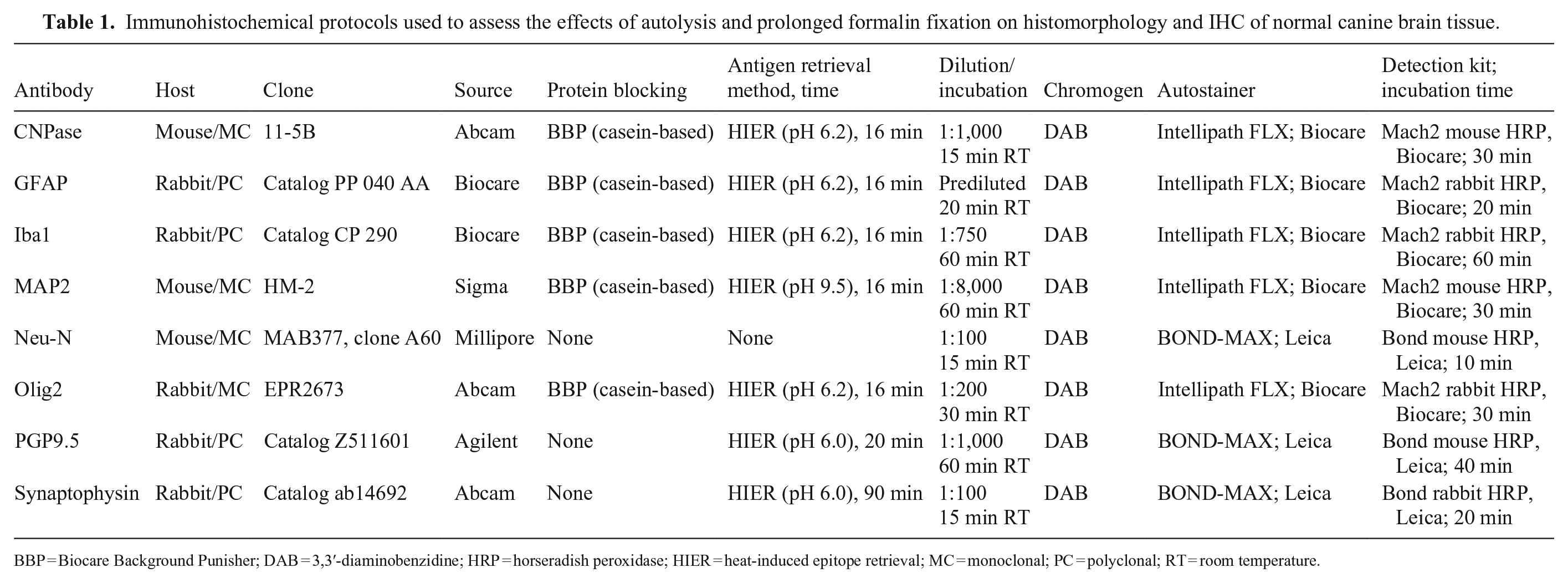
BBP = Biocare Background Punisher; DAB = 3,3′-diaminobenzidine; HRP = horseradish peroxidase; HIER = heat-induced epitope retrieval; MC = monoclonal; PC = polyclonal; RT = room temperature.
IHC specificity and intensity scoring
For scoring of immunolabeling, we scanned slides (VS200 digital slide scanner; Olympus) at 20 × magnification. One pathologist (J. Koehler) took representative photomicrographs from each experimental time and aggregated them into a slide deck in which the normal control and test image were immediately adjacent to each other to facilitate direct comparison. Using this slide deck presentation, 3 experienced pathologists with neuropathology expertise scored each experimental time for both immunolabeling specificity and intensity using a novel 4-tier classification system (Table 2).
Scoring system for evaluation of IHC specificity and sensitivity in canine brain tumors and normal brain with experimentally controlled postmortem autolysis (fixation delay) and prolonged formalin fixation.
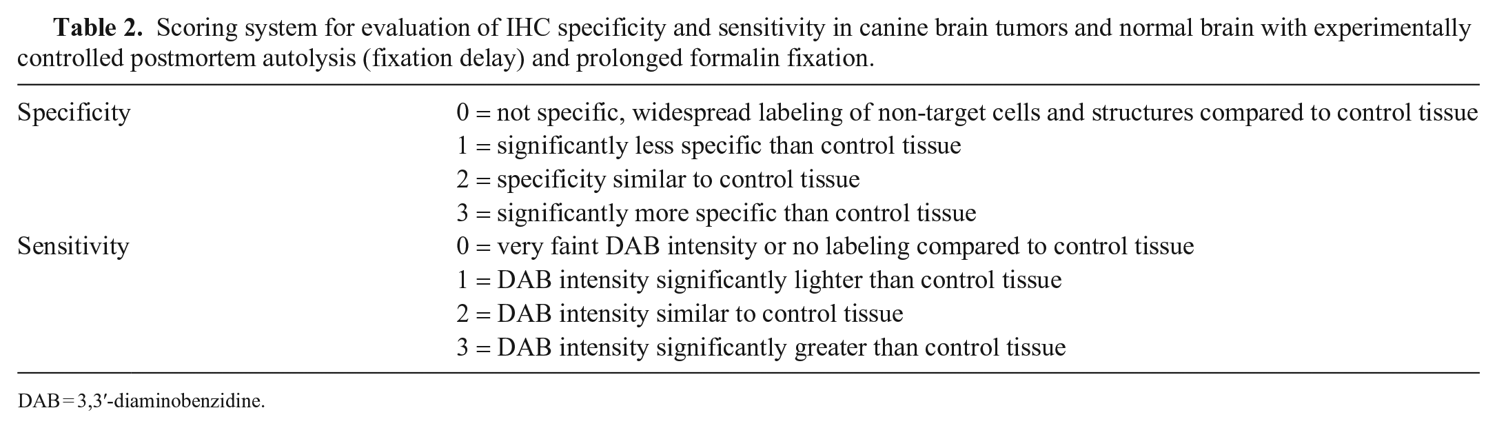
DAB = 3,3′-diaminobenzidine.
Labeling specificity was defined as the degree to which IHC maintained appropriately restricted subcellular localization relative to control tissues; labeling intensity was defined as the amount of chromogen observed using light microscopy. All 3 pathologists performed independent evaluations; cases of discrepancy were reviewed together to arrive at a consensus score (Table 3), often by evaluating the entire digitally scanned slide to ensure that the photomicrograph was not inadvertently misrepresentative. We did not perform statistical analysis because our study was primarily descriptive and was not aimed at establishing the validity of the scoring system or calculating measures of inter-observer variability.
Consensus scores for IHC labeling specificity and intensity for IHC markers evaluated in canine brain tissue with experimentally controlled autolysis (fixation delay) or prolonged fixation.
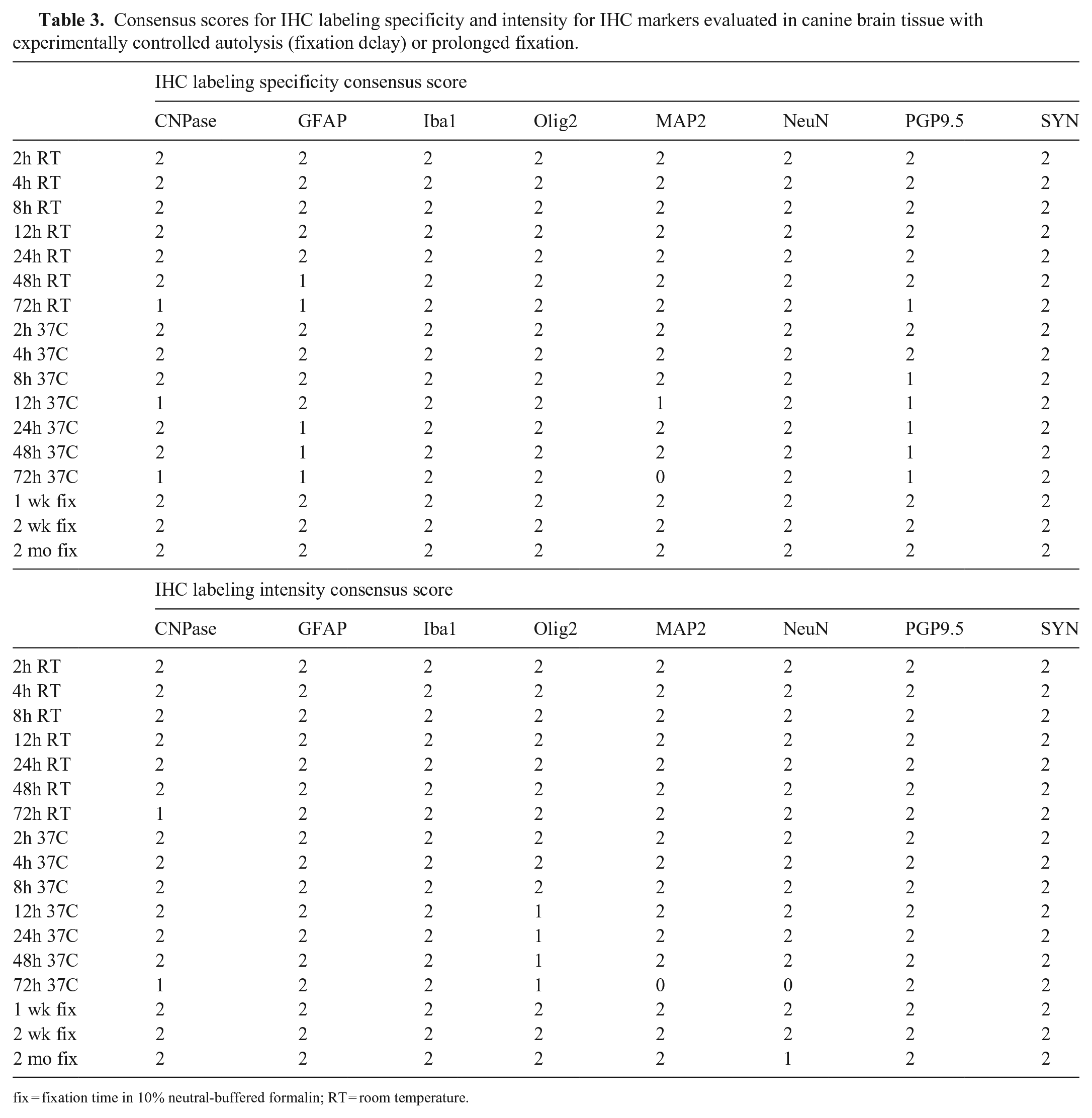
fix = fixation time in 10% neutral-buffered formalin; RT = room temperature.
Results
Histomorphology
We confirmed autolysis on H&E-stained sections via morphologic changes that manifested on an increasing gradient along with increasing times until fixation occurred (Fig. 1), consistent with reported criteria.4,5,10,14,15,19,20,22 The primary observed changes were 1) increased coarseness to fine vacuolation of the neuroparenchyma; 2) nuclear changes, including loss of chromatin detail, loss of distinction of the nuclear membrane, and pyknosis; 3) cytoplasmic shrinkage, loss of neuronal Nissl substance, and artifactual pericellular clear space around glial cells and neurons; and 4) increased overall amphophilic staining of the neuroparenchyma. The most striking and easily recognized of these changes were parenchymal vacuolation, nuclear pyknosis, and pericellular clear space (especially noticeable around oligodendroglia and small neurons of the cerebellar granular layer). Importantly, as time elapsed, differentiation of cell types became increasingly impossible as a result of pyknosis and other aforementioned nuclear changes.
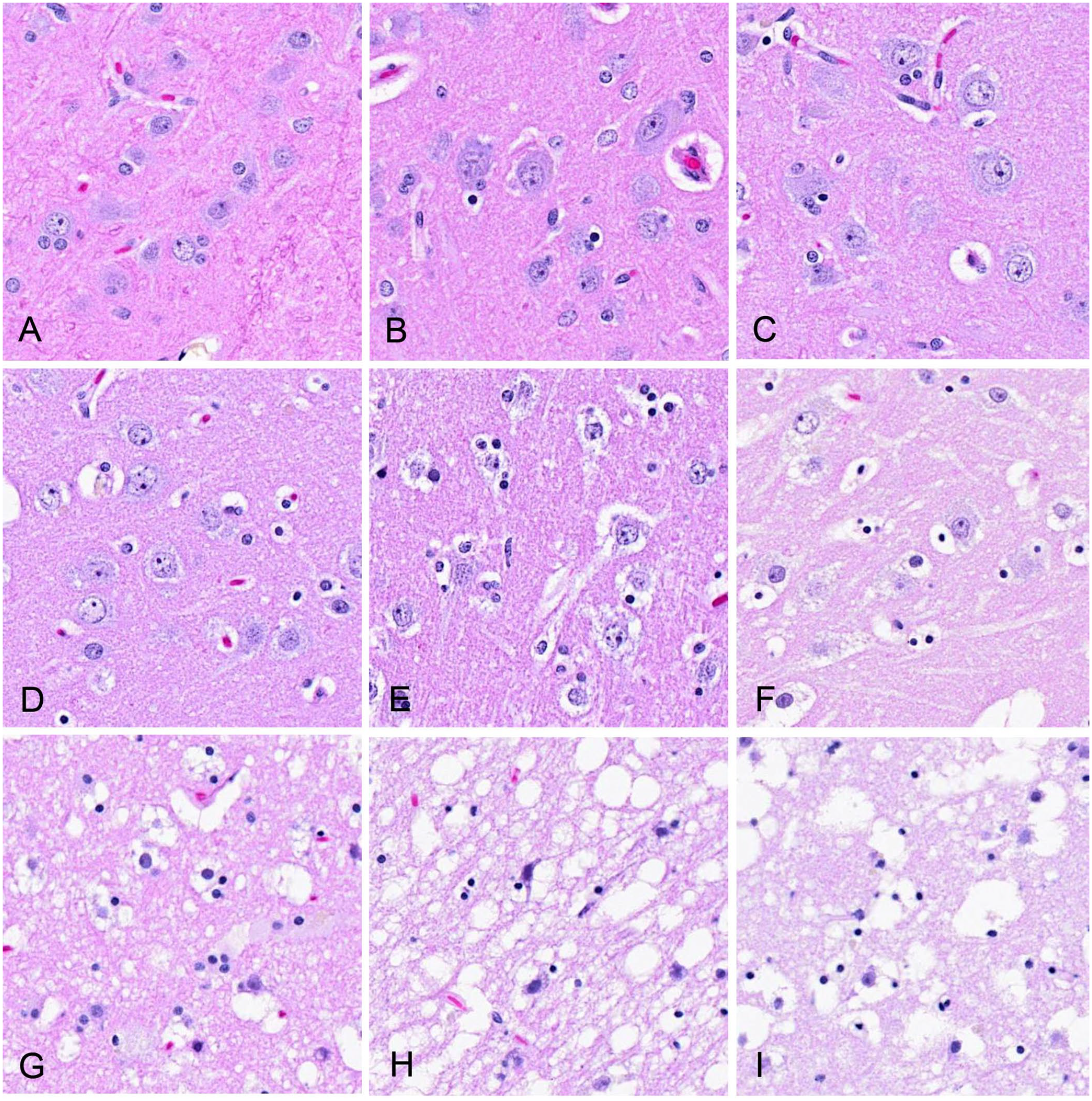
Effect of experimentally induced autolysis via delayed formalin fixation on histomorphology in H&E-stained sections of canine brain.
IHC specificity and intensity scoring
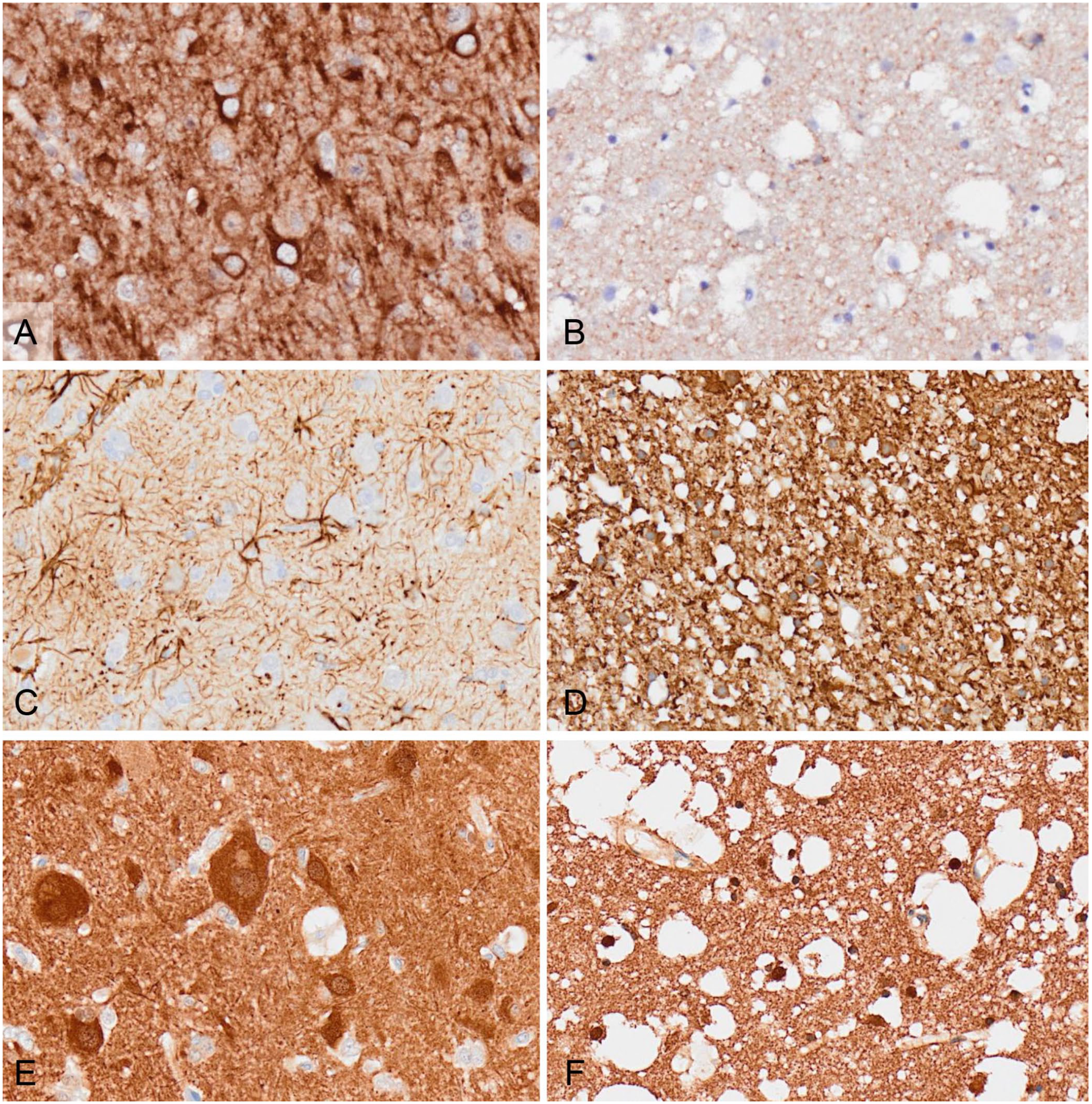
In terms of the specificity of labeling, most immunomarkers were minimally affected by the degree of autolysis, with some markers having slightly less-specific labeling at certain times (generally the longest delayed fixation times; Table 3, Figs. 2, 3). OLIG2 immunolabeling became less confined to the nucleus with increasing autolysis, leading to a slight “bleeding” appearance to the expected nuclear localization but with the bulk of labeling still restricted to the nucleus. Two immunomarkers (CNPase and MAP2) had results for the 12-h 37°C time that were discordant with the previous and subsequent times; hence, we interpreted these as spurious results that may have been related to the handling of that particular tissue or the specific anatomic location of the section. None of the immunomarkers had a loss of labeling specificity with prolonged fixation.
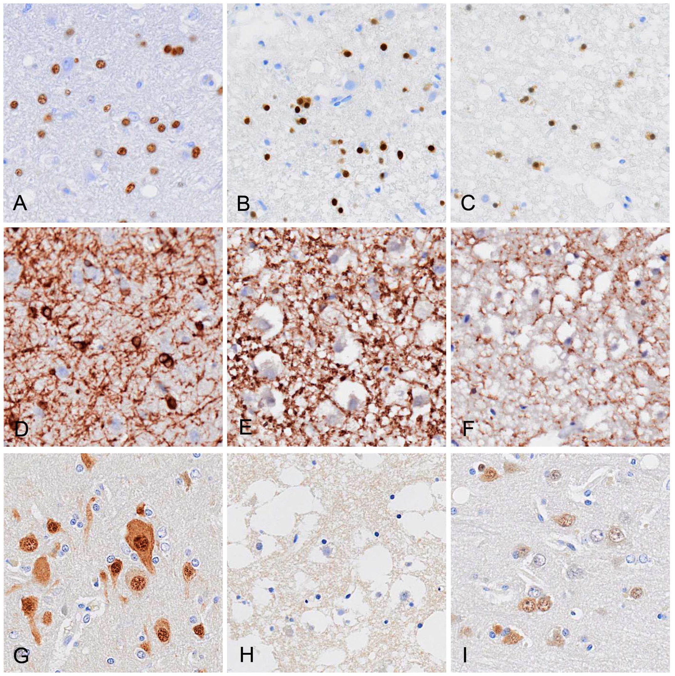
Effect of experimentally induced autolysis via delayed formalin fixation or prolonged formalin fixation on IHC specificity and intensity in canine brain.
Effect of delayed fixation on IHC specificity and intensity in canine brain.
In terms of the intensity of labeling, CNPase immunolabeling was decreased at 72-h at both RT and 37°C. OLIG2 immunolabeling was decreased at all times after 8-h in the 37°C group only. MAP2 and NeuN had no decrease in immunolabeling, except at 72-h 37°C, at which there was no discernable labeling, likely as a result of near-total collapse of neuronal structural integrity. For GFAP, PGP9.5, SYN, and Iba1, there was no loss of immunolabeling intensity at any time. The only immunomarker that was affected by fixation times was NeuN, which had a moderately decreased labeling intensity after 2 mo of fixation.
Discussion
A search of PubMed, CAB Direct, Web of Science, Scopus, and Google showed no publications using experimental fixation delay and prolonged fixation at specific time points to evaluate histomorphology along with a panel of IHC markers commonly used in the diagnosis of canine brain tumors. Our results suggest that autolysis has a minimal effect on most immunomarkers, but that advanced autolysis may cause a loss of specificity for GFAP, MAP2, and PGP9.5, making them more difficult to interpret. CNPase and OLIG2 immunolabeling were slightly decreased in intensity in samples with marked autolysis but without a loss of specificity. Severely autolytic samples had total loss of the neuronal immunomarkers MAP2 and NeuN, suggesting that these IHCs are not suitable for highly autolyzed samples.
Based on our findings, mild-to-moderate autolysis (such as that seen routinely in animals that die and are placed fairly quickly into a cooler to undergo autopsy within 24 h) is unlikely to have any significant detrimental effect on the use of any of the immunomarkers that we tested. Likewise, apart from NeuN, which appears sensitive to prolonged fixation, fixation of up to 2 mo in NBF should not preclude the use of any of these IHC markers in the diagnosis of autopsy-obtained canine brain tumors or CNS tissue. Given that only one IHC protocol per marker was tested, it is possible that results could be influenced by an individual laboratory’s formalin solutions, IHC protocols, equipment, or reagents.
Footnotes
Acknowledgements
We thank Dr. Mutsumi Yamazaki, formerly of Azabu University, Japan, for her technical assistance with experimental procedures, and Dr. Brad Matz (Auburn University, Department of Clinical Sciences) for donor identification and coordination.
Declaration of conflicting interests
The authors declared no potential conflicts of interest with respect to the research, authorship, and/or publication of this article.
Funding
The authors received no financial support for the research, authorship, and/or publication of this article.