
Abstract
Clostridial infections in goats have been associated frequently with enteric diseases or gas gangrene but very rarely with the reproductive system. We describe here 12 cases of fatal postpartum gangrenous metritis in does associated with infection by several clostridial species. Clinically, these cases were characterized by rapid onset of hyperthermia followed by death after kidding. On postmortem examination, the uteri appeared to be necrotic and were hemorrhagic and edematous. Microscopically, the uteri had diffuse coagulative necrosis, edema, hemorrhage, and fibrinous thrombi with intralesional gram-positive rods. Clostridium perfringens was isolated from 7 of 9 uterine samples cultured, and C. perfringens, C. septicum, C. novyi, or C. chauvoei were demonstrated by immunohistochemistry (IHC) in the 5 cases examined. IHC for Paeniclostridium sordellii was negative in all 5 cases. PCR performed on 3 of the C. perfringens isolates was positive for alpha toxin and perfringolysin, identifying these isolates as type A. Clostridial infection should be considered in cases of postpartum gangrenous metritis of does.
Keywords
A large number of infectious agents can cause metritis in goats, including, among others, Coxiella burnetii, Chlamydia spp., Yersinia pseudotuberculosis, and Mycoplasma spp.2,12,22 However, the literature on clostridial metritis in this species is very limited. Postpartum hemorrhagic or gangrenous metritis and sepsis have been described in Angora goats, associated with Clostridium septicum, C. novyi, and/or C. chauvoei.5,30 A series of 9 cases of periparturient metritis associated with Paeniclostridium sordellii has been reported in goats. 13
Clostridia are mostly gram-positive, sporulating anaerobic rods that are associated with several major diseases in goats. C. perfringens type A, C. septicum, and C. novyi are associated with gas gangrene,9,11,14 C. perfringens types B and C with hemorrhagic enteritis,10,29 C. perfringens type D with enterotoxaemia,17,28 and C. novyi with necrotic hepatitis. 14 We present here a series of 12 cases of Clostridium spp.–associated gangrenous metritis in does originating from 8 dairy goat farms in France.
Material and methods
Twelve uterine samples from postpartum does were submitted to the Pathology Service of the University Animal Hospital, Oniris, Nantes, France, between 2017 and 2021. They originated from 8 high-producing dairy goat farms located in western France. The clinical history was similar in all cases and was characterized by early postpartum metritis, with high lethality, affecting a more-or-less similar proportion of primiparous and multiparous does. No abortions, dystocia, or gross placental lesions were observed, and most kids were born alive and clinically healthy. According to the submitting veterinarians, 1.2–4.3% of the dams in each of the 8 farms were affected. The affected does developed clinical signs 24–48 h after kidding, consisting of marked hyperthermia, depression, and, inconsistently, foul-smelling vaginal discharge. After a diagnosis was established in these cases, treatment with injectable penicillin at parturition was established in all flocks, and no more cases were observed.
Twelve field autopsies were performed individually by 2 of the authors of this paper (Cyrille Tesson, Jérôme Despres) within 2–8 h of death. All carcasses were in a mild-to-moderate state of postmortem decomposition. Samples of uterus were collected in all cases, fixed by immersion in 10% neutral-buffered formalin for a minimum of 72 h, and processed routinely to produce 4-μm thick sections, which were stained with H&E and Gram stains.
Sections of 5 of the 12 uterine samples were also processed by immunohistochemistry (IHC) for C. perfringens, C. septicum, C. chauvoei, P. sordellii, and C. novyi, using rabbit polyclonal antibodies against each of these microorganisms, as described previously. 1 Tissues from goats or cattle known to be infected by any of the clostridial species mentioned above were used as positive controls. Uterine sections processed with normal rabbit serum instead of anti-clostridial polyclonal antibodies were used as negative controls.
Additional uterine samples from 9 of the 12 does were collected aseptically and inoculated onto brain-heart infusion plus Drigalski agar (Bio-Rad) and incubated aerobically at 37°C for 24–48 h. Aliquots of these samples were also incubated onto blood colistin–nalidixic acid agar (Bio-Rad) and incubated anaerobically at 37°C for 24–48 h. Additionally, samples of uterus from these 9 animals were also incubated onto tryptone–sulfite–cycloserine selective agar for C. perfringens (Bio-Rad) and incubated anaerobically at 45°C for 24–48 h. If black colonies were observed, they were subcultured onto blood agar and incubated anaerobically at 37°C for 24 h. All isolates were identified by conventional biochemical techniques using the API identification system (API 20 E; BioMérieux).
Toxinotyping was carried out on 3 isolated strains of C. perfringens by PCR to amplify the genes encoding the following toxins: alpha (CPA), beta, epsilon, iota A, iota B, beta 2 (CPB2), delta, enterotoxin, TpeL toxin, necrotic enteritis toxin B–like, and perfringolysin (PFO), as described elsewhere.20,21 DNA was extracted from clostridial strains (InstaGene; Bio-Rad) according to the manufacturer’s recommendations. 19
Results
On postmortem examination, all does were in good nutritional condition and had similar gross lesions. There was edema and congestion of the entire thickness of the uterine wall, and the serosa was diffusely and severely congested, with well-delimited extensive areas of dark-red-to-blue discoloration and petechiae (Figs. 1, 2). The uterine mucosa was diffusely hemorrhagic, appeared to be necrotic, and was covered by a fibrinous pseudomembrane (Fig. 3). Most other abdominal organs were congested, and a few does had moderate and diffuse black discoloration of the ruminal mucosa. No other significant gross abnormalities were observed in the other organs of any of the animals. No animals had uterine perforation and secondary peritonitis.
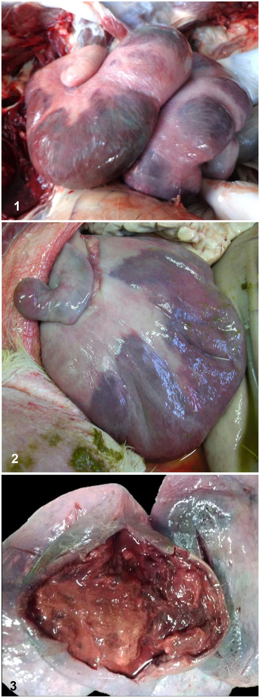
Uteri of goat does with gangrenous metritis.
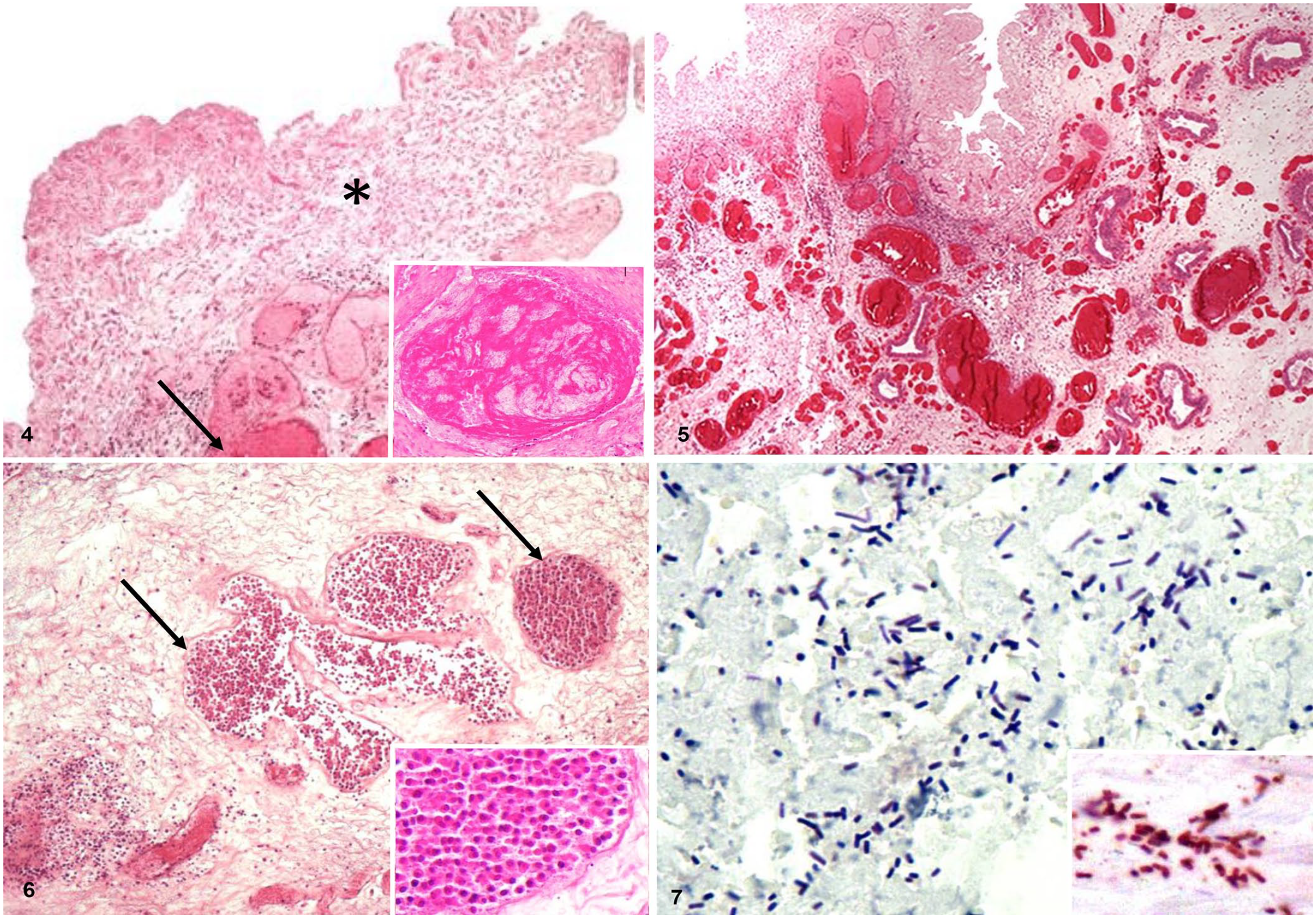
The microscopic uterine lesions were similar in all 12 does (Table 1). Histologic lesions, graded according to their severity and extent of uterine wall involvement, consisted of superficial (5 of 12) to extensive transmural (7 of 12) coagulative necrosis. The superficial lesions were characterized by necrosis of the superficial epithelium and sometimes lamina propria (Fig. 4); transmural lesions also affected muscle layers and serosae. Necrotic areas were hypereosinophilic and contained extravasated erythrocytes, strands of fibrin, and a small number of neutrophils, viable or degenerate. The neutrophils were mostly present at the margins of the areas of coagulative necrosis; there was minimal infiltration in the center of these areas. Circulatory disturbances were prominent and were characterized by marked edema, congestion, hemorrhage (Fig. 5), and inconsistent fibrinous exudate. These circulatory disturbances were often transmural. Leukostasis and thrombosis were observed in blood vessels (Figs. 4, 6). Variable numbers of gram-positive rods, single or in clusters and rarely sporulated, were present on the surface of epithelium, throughout the necrotic mucosa, and occasionally within the lamina propria and submucosa (Fig. 7). Some of the C. perfringens isolated from the uterus may have been postmortem invaders; however, the fact that they were seen associated mostly with inflammatory infiltrate suggests otherwise.
Histologic lesions observed in uterus of 12 goat does with gangrenous metritis.
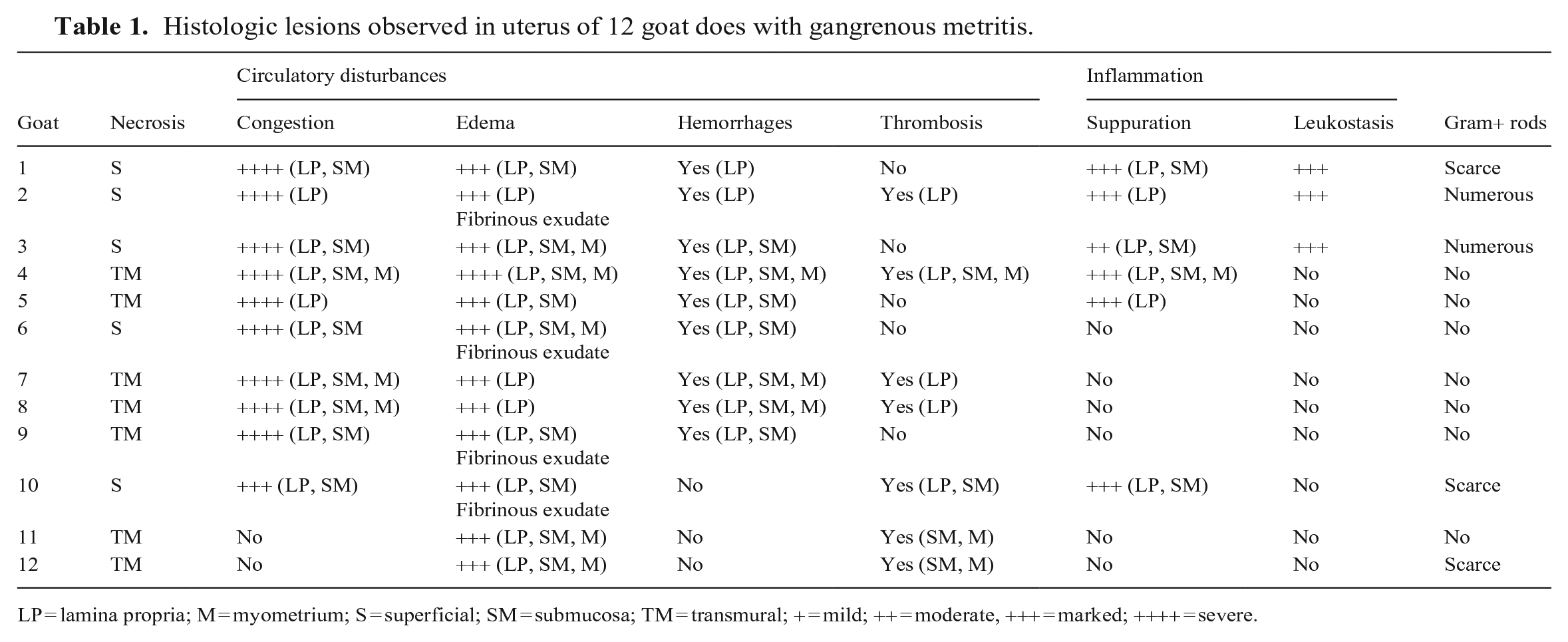
LP = lamina propria; M = myometrium; S = superficial; SM = submucosa; TM = transmural; + = mild; ++ = moderate, +++ = marked; ++++ = severe.
Uteri of goat does with gangrenous metritis.
Escherichia coli was isolated from 8 of 9 uteri (cases 3, 4, 6–8, 10–12), C. perfringens from 7 of 9 uteri (3, 4, 6, 8, 10–12), and P. sordellii from 4 of 9 uteri (7, 10–12).
The rods seen in H&E- and Gram-stained uterine sections stained positively with antibodies to C. chauvoei from 2 of 5 uteri (cases 2, 6), C. septicum from 3 of 5 uteri (1, 2, 6), C. perfringens from 4 of 5 uteri (1, 2, 4, 6), or C. novyi from 2 of 5 uteri (4, 6), but not with P. sordellii antibodies.
PCR for toxin genes was performed in the 3 isolates from cases 3, 6, and 10. The genes encoding CPA and PFO were detected in the 3 isolates; PCR for all other toxin genes was negative, except for a positive CPB2 gene in case 6.
Discussion
We established diagnoses of clostridial metritis based on clinical and postmortem findings, coupled with demonstration of the involved clostridial species by culture and/or IHC. Metritis in does is usually associated with retained placenta, retained kids, or trauma and infection of the uterus in the course of dystocia. Among the most common infectious causes of metritis in does following abortion are C. burnetii, Chlamydia abortus, Y. pseudotuberculosis, and Mycoplasma spp.2,12,22 In C. burnetii, C. abortus, Y. pseudotuberculosis, and Mycoplasma spp. infections, lesions are usually restricted to the placenta. In C. burnetii infection, the placenta is thickened and leathery, with multifocal-to-confluent areas of mineralization. In C. abortus infection, the lesions are characterized by vasculitis without thrombosis. Necrotizing metritis with circulatory disturbances (e.g., hemorrhages, edema, thrombosis) is strongly suggestive of, although not specific for, clostridial uterine infections. We ruled out other etiologies in our cases based on epidemiologic (no reported abortions) and clinical data as well as gross (no reported placental lesions) and microscopic lesions.
Histologically, uterine sections had superficial-to-transmural massive coagulative necrosis, hemorrhages, thrombosis, and edema associated with the presence of gram-positive rods, and paucity of leukocyte infiltration, the latter a hallmark of C. perfringens histotoxic infections. These changes are similar to those reported in cases of gas gangrene by C. perfringens in humans and mice 4 and of postpartum vulvovaginitis caused by C. septicum in cattle. 15 Although it is possible that lesions occurred in other organs, unfortunately, tissues from organs other than the uteri were not collected during the field autopsies. We cannot, therefore, rule out that lesions occurred in extra-uterine tissues.
The involvement of Clostridium spp. in uterine infections of ruminants is poorly documented. Clostridium spp. were isolated from 14 of 76 postpartum uterine samples of cows with acute puerperal metritis. 18 Gas gangrene caused by C. septicum has been reported in cattle in the postpartum period in the perineal, perivulvar, and perivaginal areas. 15 Gangrenous metritis with vaginal discharge and perineal and/or vulvar swelling was reported in Angora goats in South Africa, affecting does kidding twins in outdoor pens. The disease was caused by C. septicum, C. novyi, or C. chauvoei, and it was thought to be associated with lacerations of the genital tract during birth (https://www.angoras.co.za/article/clostridium-septicum-baarmoedersponssiekte).5,30 A series of 9 cases of postpartum metritis has been reported in does in association with P. sordellii infection. 13
In 9 of our cases, we isolated Escherichia coli (8 of 9), C. perfringens (7 of 9), and P. sordellii (4 of 9); C. perfringens, C. septicum, C. chauvoei, and C. novyi were detected in some of the cases by IHC. These results indicate that our metritis cases were polymicrobial. Because C. perfringens was found in 5 of the 12 uterine samples, this microorganism seems to have an important role in the pathogenesis of the infection.
PCR analysis of the 3 isolates tested detected the genes encoding CPA and PFO. In cases of C. perfringens–mediated gas gangrene in humans, it has been shown that both toxins act synergistically to mediate vascular impairment and vascular leukostasis, leading to a paucity of leukocytes at the site of infection.7,8,24,26,31 CPA leads to great vascular impairment by increasing vascular permeability and injuring microvasculature, inducing thrombosis, and activating the inflammatory cascade.6,25,27 Although the significant role of CPA in human gas gangrene is recognized and well-studied, the same does not hold true for gas gangrene of other mammals; the in vivo role of CPA remains speculative. 16
Although knowledge about the pathogenesis of gas gangrene in animals is limited, it is believed that vegetative or spore forms of at least one Clostridium spp. can cause the disease in animals. 16 These bacteria can enter the body through damaged skin or mucous membranes, as a consequence of surgical wounds, vaccination, parturition, shearing, marking, docking, bleeding, and other actions. 16 Cases have been reported of septic shock in women associated with post-abortion metritis caused by C. perfringens and P. sordellii. 3 In these cases, it was hypothesized that these bacteria moved from the gastrointestinal tract, as either spores or vegetative bacteria, into the genital tract (via the perineum), where they were introduced into anatomic niches hospitable for infection. 3 We cannot exclude from our study the possibility of bacterial infections that ascended via the vagina and cervix following parturition and entered the uterine wall through micro-lesions.
Under the influence of progesterone, the uterus (including during pregnancy) is more susceptible to many nonspecific bacterial infections and, conversely, is remarkably resistant to microbial invasion when progesterone levels are low. 22 Much of the diminished uterine resistance to microbial invasion during diestrus and pregnancy is the result of luminal secretion of progesterone-induced immunosuppressants, which inhibit lymphocytic reaction.
All of the affected does in our study had been pregnant, and gestational progesterone may have predisposed them to clostridial infection. It is not clear, however, why the infection occurred in these cases and not in the great majority of does that survive pregnancy and parturition without suffering clostridial infections. Increased susceptibility of the uterus to infections might be predisposed by mineral deficiencies. 13 We did not explore this possibility, and the source of infection and the route of transmission were not determined, but we hypothesize that micro-lacerations associated with the birth process could have favored the entry of clostridia.
The Infectious Diseases Society of America practice guidelines for the management of Clostridium spp. skin and soft-tissue infections recommends high doses of intravenous penicillin. 23 After the occurrence of these cases of gangrenous metritis in the affected farms of our study, does that were ready-to-kid were systematically treated with penicillin just before parturition, and no new cases were reported. It is assumed that this treatment prevented new cases to occur. To reduce the routine use of antibiotics, vaccination against clostridial diseases should be considered.
Footnotes
Acknowledgements
We thank Emmanuelle Blandin and Anne-Sophie Noël for their valuable technical assistance in bacteriology.
Declaration of conflicting interests
The authors declared no potential conflicts of interest with respect to the research, authorship, and/or publication of this article.
Funding
The authors received no financial support for the research, authorship, and/or publication of this article.