
Abstract
Two rock hyraxes (Procavia capensis), from the Chattanooga Zoo, were submitted separately for autopsy at the University of Tennessee Veterinary Medical Center. The first was a 4-y-old intact female that died without premonitory signs and the second was a 10-y-old intact male that was euthanized because of severe renal disease. Microscopically, the lungs of both hyraxes had multifocal-to-coalescing, <1-mm diameter aggregates of epithelioid macrophages separated by streams of fibrous tissue. Macrophages contained intracytoplasmic, clear, acicular, birefringent crystals. Transmission electron microscopy and energy-dispersive x-ray spectroscopy findings on the lung samples were consistent with silica crystal deposition. The hyraxes had been housed together on commercially sourced play sand composed of 99–99.5% quartz, a crystalline silica polymorph. The microscopic findings, transmission electron microscopy, and energy-dispersive x-ray spectroscopy of the intrahistiocytic crystals, in addition to the history of exposure to crystalline silica, were consistent with pulmonary silicosis. Pulmonary silicosis has not been reported previously in rock hyraxes, to our knowledge.
Rock hyraxes (Procavia capensis) are small social mammals that live throughout semiarid and arid areas of Africa, specifically in locations with cliffs and rock formations.2,14 Rock hyraxes can also be found invading residential areas in South Africa where they are considered a pest species. 20 Although rock hyraxes phenotypically resemble rodents, they are phylogenetically more closely related to elephants and manatees. 14 These animals can be found in zoologic settings or research settings throughout the United States; however, there are few published husbandry recommendations, with no known published recommendations for substrates. 17 There are various reports on rock hyrax diseases, including a retrospective case series of 103 rock hyraxes, and individual case reports describing the following diseases: hemosiderosis, pancreatic islet fibrosis, diabetes mellitus, herpesviral stomatitis, dilated cardiomyopathy, metastatic mineralization, systemic toxoplasmosis, disseminated tuberculosis, and demodicosis.7,9,10,12–14,19 Pulmonary silicosis has not been reported previously in rock hyraxes, to our knowledge.
A 4-y-old, intact female rock hyrax from the Chattanooga Zoo (Chattanooga, TN, USA) was submitted to the University of Tennessee (Knoxville, TN, USA) for postmortem examination after being found dead; no premonitory signs were reported. The gross examination revealed hemorrhagic enterocolitis suggestive of a Clostridium spp. infection. A culture of the intestines yielded colonies of Clostridium perfringens; however, toxin typing was not performed. Microscopically, there was necrohemorrhagic enterocolitis with gram-positive rods coating the necrotic mucosal surface, supportive of clostridial enteritis. Six months following the death of the female rock hyrax, her breeding partner, a 10-y-old intact male, was euthanized because of chronic progressive kidney and dental disease and was also submitted for postmortem examination at the University of Tennessee. Microscopic examination confirmed severe chronic lymphoplasmacytic tubulointerstitial nephritis and necrosuppurative mandibular osteomyelitis and gingivitis with a tooth fracture. Additional tissues examined microscopically from both cases included heart, liver, lung, kidney, and brain. All tissues were fixed in 10% neutral-buffered formalin and underwent routine histologic processing.
Microscopic examination of the lungs from both rock hyraxes identified similar changes. Affecting 30–50% of the parenchyma were ~1-mm diameter nodules composed of epithelioid macrophages separated by variable amounts of collagen (Fig. 1). Macrophages had intracytoplasmic clear, acicular, birefringent crystals. Ziehl–Neelsen and Fite–Faraco acid-fast stains and Gomori–Grocott methenamine silver (GMS) stains were performed, and no microorganisms were identified. The crystals stained pink, orange, yellow, and green with the acid-fast stains (Fig. 2), and green with GMS. Transmission electron microscopy was performed on lung from both animals. Pulmonary tissue was retrieved from 10% neutral-buffered formalin, post-fixed in 1% osmium tetroxide, and serially dehydrated with graded acetones. The tissue was embedded in epoxy (EMbed 812; Electron Microscopy Sciences), and sections were cut at 1-mm thickness and stained with toluidine blue to confirm the presence of crystals in the processed tissues. These areas were thin-sectioned (100 nm; UMC7 ultra-microtome; Leica), placed on 200-mesh copper grids, and stained with uranyl acetate and lead citrate. The grids were examined with a transmission electron microscope (Libra 200MC; Zeiss), and digital images were acquired (US1000 X camera; Gatan). Within the cytoplasm of macrophages were variably sized electron-dense, crystalline-to-amorphous inclusions (Fig. 3). Energy-dispersive x-ray spectroscopy (E-Flash detector; Bruker), with an accelerating voltage of 20 keV and collection time of 60 s, was performed on the electron-dense crystalline material, and revealed tall peaks of silicon and oxygen, characteristic of silica (Fig. 4), and shallow peaks of aluminum, potassium, copper, chloride, and iron.
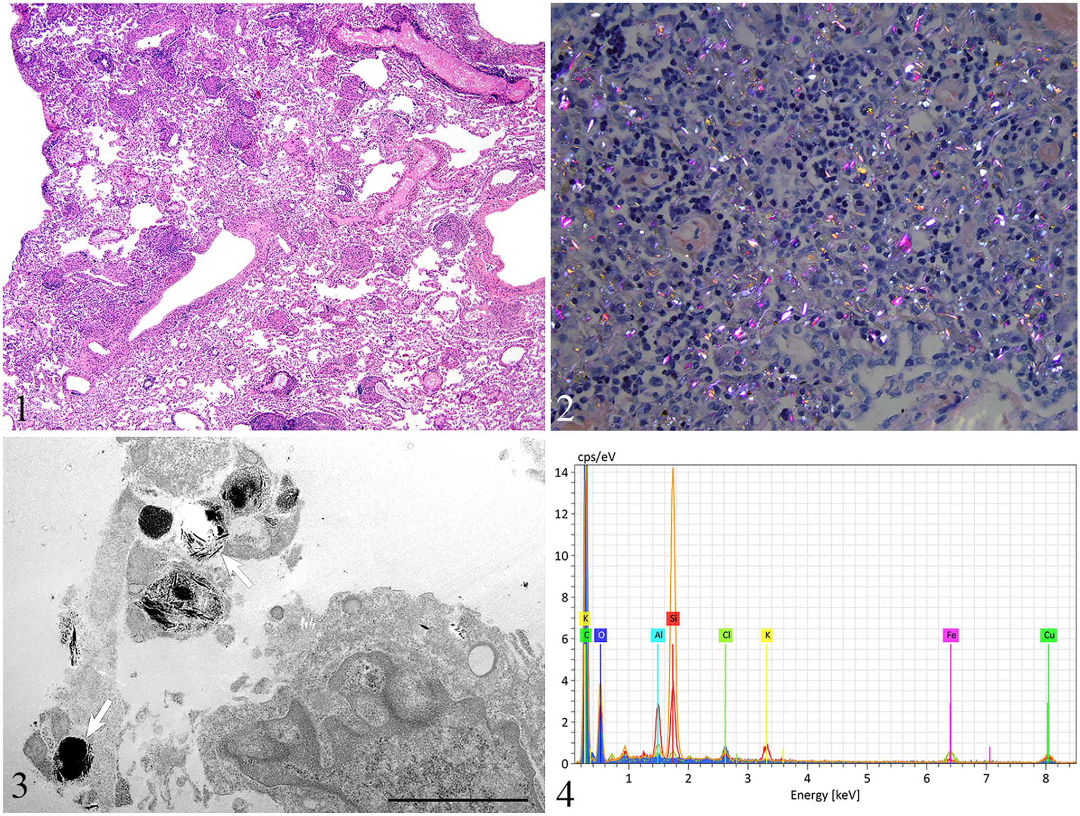
Pulmonary silicosis in 2 rock hyraxes.
The light microscopic, transmission electron microscopic, and energy-dispersive x-ray spectroscopy findings were consistent with a diagnosis of pulmonary silicosis in both rock hyraxes. To determine the cause of the pulmonary silicosis, we investigated the potential shared environment and dietary risk factors. The hyraxes were co-housed in an indoor enclosure with a substrate of play sand and ledges of various heights. The diet consisted of timothy hay, timothy hay cubes, rabbit pellets (Oxbow), and a mix of greens, excluding spinach, and squash; however, we found no peer-reviewed resources regarding appropriate diet and husbandry of rock hyraxes. 17 The only known source of silica available to the 2 rock hyraxes was their substrate, play sand, which is composed of 99–99.5% quartz, a polymorph of silica. A differential diagnosis considered for our cases was asbestos exposure; this cannot be ruled out completely given that asbestos has the same composition via spectroscopy and looks similar with light microscopy. Asbestos was considered less likely given the lack of known exposure to asbestos within the facility, as well as the absence of similar lesions in other species of animals within the facility.
We searched PubMed under the terms: pulmonary silicosis, silicosis, silica, silicates, pneumoconiosis, and silicate pneumoconiosis. Silica occurs as a variety of polymorphs, which have the same chemical components but a different crystal structure; these include crystalline silica (quartz, cristobalite, tridymite, coesite) and fibrous silicates (asbestos). When inhaled, any of these polymorphs can contribute to the development of pulmonary silicosis, an interstitial lung disease caused by inhalation of dust particles, also known as silicate pneumoconiosis. 11 In human medicine, acute and chronic forms of pulmonary silicosis are described, with chronic forms including simple and complicated manifestations.4,15 All forms have been considered occupational health hazards. 11 The acute form involves intense high-dose exposure to inhaled silicates and has not been described to occur naturally in animals.4,15 The chronic simple and complicated forms of silicosis develop over years of continuous low-dose exposure to silicates.4,11,15 In humans, simple pulmonary silicosis is characterized by silica-laden macrophages within a fine network of reticulin and collagen <1-cm diameter.4,11 As exposure increases and nodules continue to enlarge to >1-cm diameter, the disease progresses to complicated pulmonary silicosis in which clinical signs develop from compromised lung function. 11
In veterinary medicine, only the chronic forms of pulmonary silicosis have been described.1,5,6,16,18 The distinction between simple and complicated pulmonary silicosis based on the size of nodules has not been explored, but pulmonary silicosis has been described in many species. The most notable naturally occurring reports are from horses in the Monterey-Carmel Peninsula of California where the soil is composed of cristobalite, one of the silica polymorphs.1,3 The extensive pulmonary involvement in these horses is associated with respiratory distress and exercise intolerance. The equine disease appears most compatible with complicated silicosis as described in people, even though a specific size criterion is not defined for equine pulmonary nodules. 11 An associated syndrome in horses with pulmonary silicosis is osteoporosis; the mechanism and pathogenesis for this development remains unclear. 1
Pulmonary silicosis without associated clinical signs has been reported in a wide range of other veterinary species; these are often described as incidental postmortem findings. A survey for pulmonary silicosis in 100 specimens from San Diego’s zoologic collection included 11 semiarid and arid zoologic mammalian species: koalas, kangaroos, okapis, goats, antelopes, camels, oryxes, giraffes, saiga, bobcats, and rabbits, and 8 avian species. 5 Fifty percent of cases had only crystal-laden macrophages within alveoli or lymphatics, 20% had granulomas with intracytoplasmic crystalline material (dust granuloma) and a few collagen fibers, 15% had mild fibrosis, and only 5% of animals had severe fibrosis with surrounding emphysema. 5 The most affected mammalian species were older koalas and kangaroos, and the most affected avian species was the kiwi, which had the most severe lesions of all of the animals. 5 The large retrospective study found no lesions within camels 5 ; however, a study examining camels living in Somalia found dust granulomas with few areas of fibrosis. 8
Although no clinical signs were observed in our rock hyraxes, which is consistent with simple pulmonary silicosis, the extent of pulmonary involvement was more severe than reported in most other semiarid and arid zoologic species. Further evaluation of substrates for zoologic rock hyraxes is necessary to determine the most appropriate bedding for this species, and options other than a quartz-based substrate should be considered.
Footnotes
Declaration of conflicting interests
The authors declared no potential conflicts of interest with respect to the research, authorship, and/or publication of this article.
Funding
The authors received no financial support for the research, authorship, and/or publication of this article.