
Abstract
Multilobular tumor of bone (MLTB) is an infrequent, slow-growing, bone neoplasm formed predominantly on the head. These tumors can behave as malignant neoplasms clinically and pathologically and can metastasize occasionally. No cases of MLTB in rodents have been reported, to our knowledge. We describe a novel case of an MLTB in a guinea pig. An adult guinea pig had an exophytic mass fixed on the frontal bone, maxilla, and nasal bone. On radiography, the mass had a spherical contour and variable density and was formed on the surface of the cranial bones. The mass was excised surgically. The cut surface was light-yellow to milky-white and had a granular texture with fine fibrous septa. Histologically, the neoplasm had a multilobular pattern, which consisted of many islands of bone and/or cartilage matrix surrounded by small cells and separated by fibrous septa, which closely resembles the equivalent neoplasm in dogs.
Multilobular tumor of bone (MLTB) is an infrequent, slow-growing, bone neoplasm predominantly formed on the head, including the mandible and maxilla.4,5,11,15 Uncommon sites reported include the orbit, 4 tympanic bullae, 4 zygomatic arch, 6 hard palate, 1 rib, 16 axilla, 7 and baculum. 17 This tumor can invade adjacent tissue and cause neurologic signs in cases that exhibit intracranial growth,9,12 and can metastasize occasionally.8,15 These neoplasms have been given various names (e.g., multilobular osteochondrosarcoma, chondroma rodens) based on their morphologic similarity to human tumors or their biologic features.8,16 MLTB is uncommon in the veterinary field and has been reported mainly in canine cases,4,15 with one case in an African wild dog. 9 Only a few cases in cats,3,10,14 and a single case in a horse, 13 have been described. To our knowledge, no cases have been reported in rodents. Guinea pigs have a very low incidence of spontaneous neoplasms. 2 We describe herein the pathology and prognostic categorization of an MLTB in a guinea pig.
An adult guinea pig, breed and sex unknown, was an individual in a group reared in a zoological garden. An exophytic, spherical mass fixed on the left side of the frontal bone, maxilla, and nasal bone, caused mild compression of the left eyelid (Fig. 1). On radiographic examination, the mass tightly adhered to the surface of the affected bones and had a disorganized reticular density with a smooth contour (Fig. 2). There was neither obvious compression nor invasion of the surrounding tissues. The mass was excised surgically, with a minimal surgical margin. Neither recurrence nor other clinical signs were found during follow-up 8 mo after the operation.
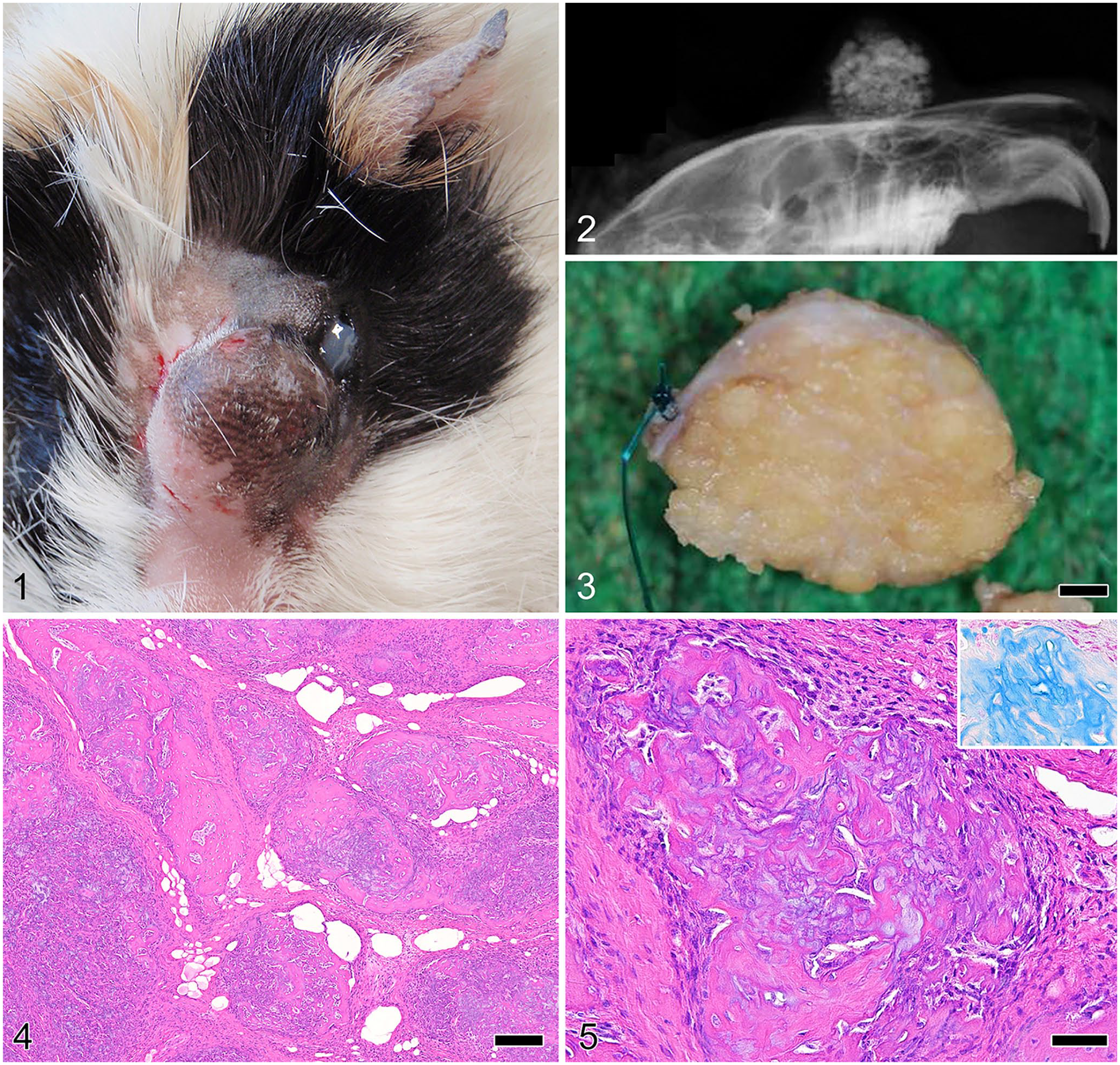
Multilobular tumor of bone on the forehead of a guinea pig.
The excised mass was encapsulated with fibrous tissue, ~ 1.5 cm diameter, and hard. The cut surface was light-yellow to milky-white and had a granular texture with fine fibrous septa (Fig. 3). The mass was fixed in 10% neutral-buffered formalin, routinely processed after demineralization (K-CX solution; Falma), sectioned at 3 µm, and sections stained with hematoxylin and eosin and with alcian blue (pH 2.5).
Microscopically, the mass had a multilobular pattern that consisted of many islands of bone and/or cartilage matrix of variable sizes and irregular shapes (Fig. 4). These islands were surrounded by small spindle-to-ovoid cells with small oval nuclei; lobules were separated by fibrous septa (Fig. 5). The quantity of bone or cartilage matrix in each lobule was variable. Some islands were similar to mature compact bone, and others consisted of a randomly mixed matrix of bone and cartilage. Proliferative small cells were immature and mesenchymal, and exhibited minimal atypia of nucleus and cytoplasm with no mitotic figures. On the section stained with alcian blue, the cartilage matrix stained blue (Fig. 5, inset). These findings were consistent with the characteristics of MLTB.
The affected site in our case is identical to that of many found in canine reports: the tumor was formed on the area from the frontal bone to the maxilla.4,15 The age of the guinea pig was not known but it appeared to be an adult. In dogs, the average age of MLTB onset is 8 y, with a range of 4–17 y.4,15
Radiographic images of MLTB characteristically show a sharply demarcated border and a granular appearance caused by mineralization, described as a “popcorn ball” appearance.4,15 On gross inspection, an MLTB is a projecting nodular mass enveloped by a fibrous membrane.4,16 Upon sectioning, the neoplasms consist of numerous gray-white to yellow gritty nodules divided by fibrous septa.8,11 The radiographic and gross morphologic features of our guinea pig case are almost identical to those in a typical canine case. A typical MLTB has a unique histologic pattern of lobules, consisting of variable amounts and maturity of bone and/or cartilage; lobules are surrounded and outlined by small mesenchymal cells and fibrous connective tissue. 16
MLTBs must be differentiated from other primary bone tumors, but they are generally straightforward to diagnose because they have the unique morphologic features (radiographic, macroscopic, and histologic) described above. Enough neoplastic tissue needs to be inspected histologically for a definitive diagnosis, given that a small portion of tumor tissue may yield a misdiagnosis as a chondrosarcoma, osteosarcoma, or other mesenchymal tumor. On computed tomography (CT) the characteristic patterns of MLTB are visualized in the bone window 6 ; the utilization of CT is also helpful for determination of the surgical margin. 15
Histologic grading for MLTB has been attempted with a 3-grade system based on assigning scores for 6 criteria: borders (pushing, pushing and invasive, invasive), size of lobules (small, medium, large), organization (well, moderate, poor), mitotic rate (1–5, 6–10, > 10 per 10 HPFs), pleomorphism of cells (monomorphic, mild, moderate, marked), and necrosis (none, present). 15 According to this system, our case is scored as grade I (low grade) because there was no evidence of apparent invasion, mitotic figures, or pleomorphic cells; the neoplasm contained bone and/or cartilage tissue differentiated in various degrees. Without recurrence and metastasis, the postoperative course was uneventful.
Footnotes
Declaration of conflicting interests
The authors declared no potential conflicts of interest with respect to the research, authorship, and/or publication of this article.
Funding
The authors received no financial support for the research, authorship, and/or publication of this article.