
Abstract
The only Sarcocystis species currently known to inhabit the fibers of skeletal and cardiac muscles in horses are S. fayeri, S. bertrami, and S. asinus. We describe herein the invasion of myofibers in a horse by S. gigantea, a sheep-specific species with low virulence in the original host. A hunter gelding was referred to a veterinary surgeon in Newmarket (UK). The anamnestic data reported that the horse had an initial history of swelling of the right forelimb with fluid on the front of the carpus and edema spreading up the forearm. Subsequently, 2 firm lumps were found on the left pectoral muscle adjacent to the axilla of the left forelimb. Histologic examination of biopsies from the lumps revealed multifocal granulomatous eosinophilic myositis associated with intact and degenerate encysted parasites, consistent with Sarcocystis spp. Based on amplification and DNA sequencing of the 18S rRNA gene obtained from formalin-fixed, paraffin-embedded tissue blocks, S. gigantea was identified. The presence of sarcocysts in equine skeletal muscles has been considered an incidental finding, and there are only sporadic associated reports of myositis. Our finding suggests that some Sarcocystis spp. have a wider intermediate host range than believed previously, and that Sarcocystis of other species (not considered horse-associated) can invade the muscle fibers of equids, leading to myositis.
Keywords
In July 2016, a 9-y-old hunter gelding was examined in the stables near Billingshurst, West Sussex, UK. The gelding was biopsied after a 6-wk history of progressive swelling on the forelimbs that extended to the chest and which was only partially and temporarily responsive to administration of nonsteroidal anti-inflammatory drugs. The horse was purchased in April 2015 having been imported from Eire a few months beforehand. He was kept in a stable but was turned out to pasture, shared with sheep, for large parts of the day.
On physical examination, the horse was alert and responsive, in good body condition (580 kg), and clinical signs recorded were within normal reference ranges. A non-painful, non–well-defined locally extensive mild swelling of the middle side of the chest up to the shoulder joint was visible and palpable, associated with 2 well-defined oval-shaped, 4 × 3 cm, symmetrical lumps on the pectoral region. The lumps were slightly firmer than normal muscle, were slightly painful, and covered by intact skin. An ultrasound of the pectoral region revealed a perfectly continuous extension between the muscle fascia and the masses, which showed increased echogenicity consistent with edematous myositis.
A complete blood cell count and serum biochemistry panel were performed. The most significant findings were moderate normocytic normochromic anemia (packed cell volume 32 L/L, reference interval [RI]: 32–45 L/L) and significant increases in muscle enzyme activities, (i.e., aspartate aminotransferase 588 U/L, RI: 175–340 U/L; creatine kinase 537 U/L, RI: 1–28 U/L). These findings indicated muscle damage. The eosinophil count was within the RI.
The horse had been treated with a broad-spectrum antibiotic and nonsteroidal and steroidal anti-inflammatory drugs. Partial response was achieved, with a slight decrease in the chest and forelimb swelling. However, the 2 masses over the pectoral region persisted, although they were subjectively less sensitive to palpation. A surgical incisional biopsy was performed under local anesthesia.
The palpable masses were well-delimited, unencapsulated, and located deep within the pectoral muscles, with diffuse hyperemia of the surrounding tissue; on cut surface the tissue was mottled, composed of red and yellow fiber bundles separated by white thickened interstitium with scattered white pinpoint lesions.
Tissue biopsies were fixed in 10% neutral-buffered formalin and submitted to the Diagnostic Laboratory of the Department of Animal Health Trust (Newmarket, UK) for histologic examination. The tissue was processed routinely, and slides stained with hematoxylin and eosin.
Histologically, disseminated oval protozoan cysts were noted in the skeletal muscles, with aggregates of epithelioid macrophages and giant cells encircling amorphous eosinophilic material and nuclear debris interpreted as degenerating and degranulating eosinophils with necrosis (Fig. 1A). Occasionally, residual degenerate protozoan cysts were embedded in this eosinophilic necrotic material (Fig. 1B). Large numbers of eosinophils were present with macrophages in granulomas; diffuse mild eosinophilic infiltration was widespread in the endomysium and perimysium. Some muscle fibers had homogeneous eosinophilic sarcoplasm with fragmentation (myonecrosis), and there was moderate multifocal endomysial fibrosis.
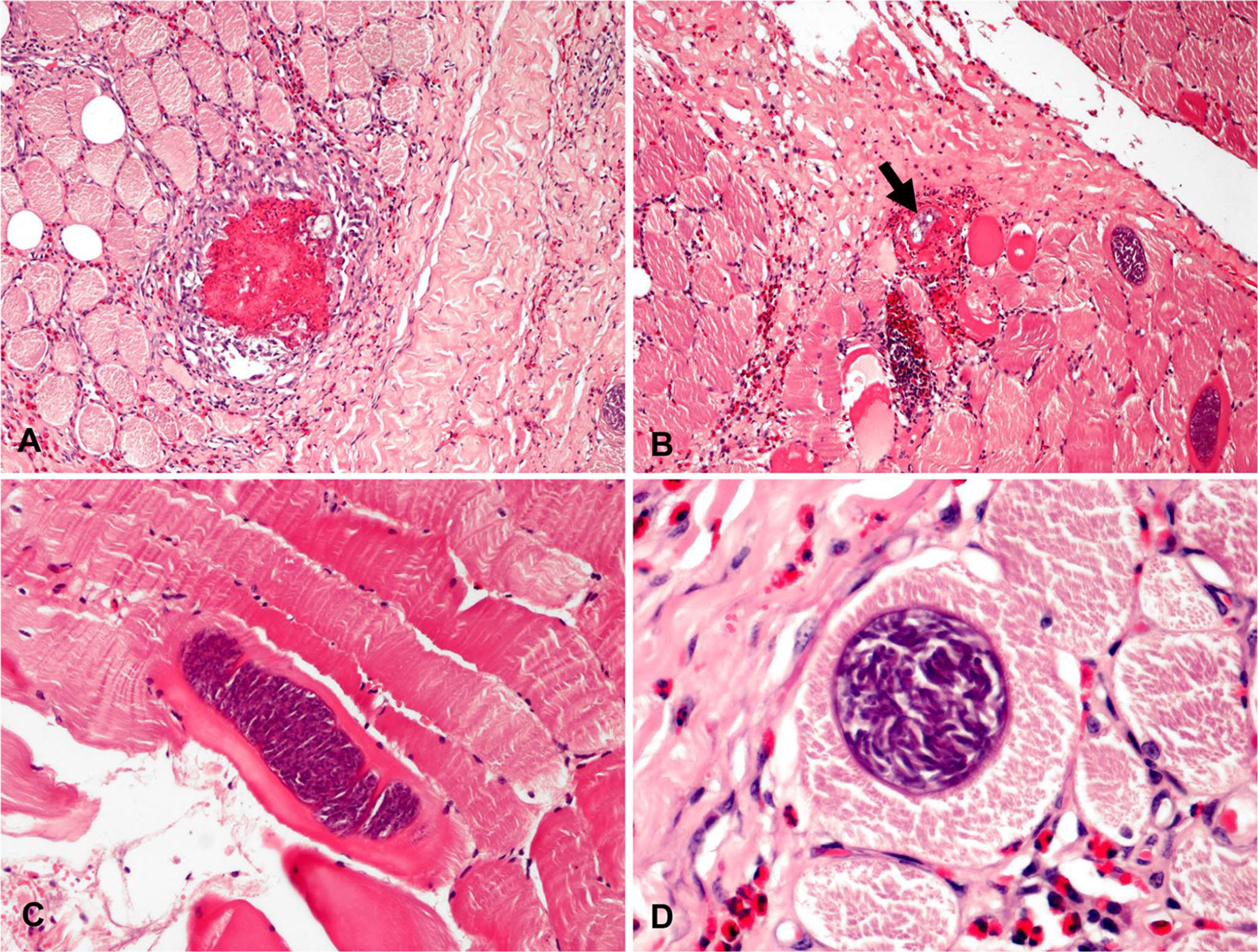
Pectoral muscle of a horse affected by granulomatous eosinophilic myositis caused by Sarcocystis gigantea.
Elongated or more circular cystic forms, depending on the plane of section and orientation of myocytes, were observed within intact and degenerate myofibers (Fig. 1C). The cystic forms of variable size (180–430 μm long, 35–130 μm wide) had a thin wall, which was radially striated by villar protrusions (Fig. 1D). The wall, including protrusions, was 2.3–3.8 μm thick. The cysts were septate and contained numerous crescent-shaped, dark-staining zoites (~ 4–6 μm long) centrally together with some rounded and pale-staining zoites at the periphery consistent with bradyzoites and metrocytes of Sarcocystis spp., respectively (Fig. 1D).
To identify the species of Sarcocystis, DNA was extracted from formalin-fixed, paraffin-embedded muscle blocks (DNeasy blood and tissue kit; Qiagen), following the manufacturer’s protocol. The complete 18S ribosomal RNA (18S rRNA) gene was amplified by PCR, with a pair of generic apicomplexan 18S rRNA-specific primers. 14 Amplifications were performed in 25-µL reaction final volumes, containing 1× PCR buffer, 0.2 mM dNTPs, 2.5 mM MgCl2, 0.4 mM of each primer, and 1 U of Taq DNA polymerase. 14 The concentration of DNA samples was measured (NanoVue spectrophotometer; GE HealthCare); ~20 ng of DNA were used in each PCR reaction. The reaction cycle consisted of an initial step of 10 min at 95°C, followed by 45 cycles of 30 s at 95°C, 30 s at 65°C, and 1.5 min at 72°C. The last cycle included an extended elongation step of 5 min at 72°C. PCR amplicons were separated by electrophoresis on 1.5% agarose gel in 1× TAE (Tris–acetate–EDTA) buffer, stained (GelRed; Biotium), and visualized under ultraviolet light. Low DNA mass ladder (Life Technologies) was used to estimate the size of the amplicons. Amplified products were purified (QIAquick PCR purification kit; Qiagen) following the manufacturer’s protocols and then sequenced (Bio-Fab Research, Rome, Italy). The sequences were compared with those available in GenBank using BLAST (http://blast.ncbi.nlm.nih.gov/Blast.cgi), and analyzed using MEGA7 (https://www.megasoftware.net/). 16 The sequence of the amplicons obtained had a similarity score of 99% with S. gigantea (accession KC209733.1) sequence from GenBank. The sequence obtained was deposited in GenBank as accession KY594259 (Fig. 2).
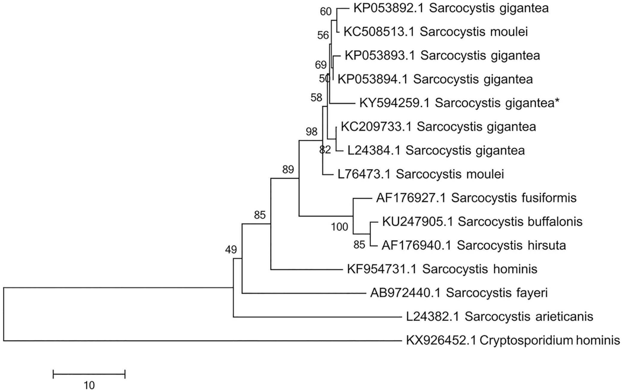
Phylogenetic tree based on 18S sequence data, compared with representatives of the genus Sarcocystis available in GenBank. The tree was constructed using the neighbor-joining method and rooted against Cryptosporidium hominis as out-group. The sequence in our case (accession KY594259) is marked with an asterisk.
To investigate the relationships among Sarcocystis isolates from other animals, sequences were phylogenetically analyzed with those available in GenBank. Selected isolates previously sequenced for S. gigantea (accessions KP053892.1, KP053894.1, KC209733.1, KP053893.1), S. moulei (L76473.1), S. fusiformis (AF176927.1), S. buffalonis (KU247905.1), S. hirsuta (AF176940.1), S. hominis (KF954731.1), S. arieticanis (L24382.1), and S. fayeri (AB972440.1) were included in the analysis. The evolutionary history was carried out using the neighbor-joining method and the Tajima–Nei model. The evolutionary distances were computed using MEGA7. 16 The bootstrap consensus trees, inferred from >1,000 replicates, were taken to represent the evolutionary history of the taxa analyzed. Cryptosporidium hominis was chosen as the out-group (KX926452.1). A bootstrap support of 50 was considered significant. The phylogenetic analysis showed that the isolate clustered in one clade, as a sister group to S. gigantea isolates (Fig. 2).
Based on histologic and molecular findings, multifocal granulomatous eosinophilic myositis caused by S. gigantea was diagnosed. Daily oral administration of toltrazuril sulfone (10 mg⁄kg; Baycox; Bayer) and trimethoprim–sulfadiazine (30 mg/kg; Norodine; Norbrook Laboratories) for 28 d was added to the therapeutic plan with recovery of the clinical signs.
The genus Sarcocystis consists of cyst-forming apicomplexan protozoa with an obligatory 2-host life cycle. 4 Sarcocysts are usually found in striated muscles of herbivores or omnivore intermediate hosts; carnivores are the definitive hosts. Three species of Sarcocystis have been described in horses: S. bertrami, S. fayeri, and S. asinus, all of which have a canine–horse cycle, but with different geographic distributions.1,12,18 To our knowledge, a clinical case of sarcocystosis has not been reported previously in equids in Europe, nor has the histologic description and biomolecular confirmation of S. gigantea natural infection been documented in the horse.
A high prevalence of Sarcocystis spp. infection (4–93%) has been reported in horses and donkeys worldwide, and its presence in skeletal muscle has long been considered as an incidental finding.1,5,9,12,21 However, Sarcocystis may be of greater pathogenic significance than previously thought, and it may be involved in neuromuscular disorders in horses.1,13,21 In addition, it is sometimes also associated with general signs (i.e., chronic weight loss, abnormal gait, mild-to-severe muscle soreness, esophageal dysphagia, acute-to-chronic and mild-to-severe anemia).2,5,7
In a case reported in 2015, 13 the infection was widespread and mostly affected skeletal muscles including the shoulder muscles, as in our case. In both cases, there was no blood eosinophilia, and a normal blood count was reported. Similarly, no eosinophilia was present in a case described in 1994. 21
From the comprehensive medical information obtained from our case, only local signs of shoulder muscle pain were observed, without any apparent impairment of the general health status of the animal. However, given that a significant increase in muscle enzyme activities was reported in blood chemistry, a general myopathic problem should not be ruled out.
Differential diagnoses for horses with eosinophilic myositis include multisystemic epitheliotropic eosinophilic syndrome and aberrant larval migration. In our case, the first hypothesis can be ruled out based on a lack of blood eosinophilia and associated lesions of the skin, gastrointestinal tract, and other organs. The second hypothesis seems unlikely given the widespread distribution of the lesions and close relationship between degenerate cysts and eosinophilic granulomas. Immune-mediated myositis in horses has been described; however, it is histologically characterized by lymphomonocytic infiltrates without eosinophils and/or the formation of discrete granulomas. 17 Most of the granulomas observed in our case may have resulted from the intramuscular presence of sarcocysts that act as chronic inflammatory stimuli.
The clinical cases of sarcocystosis in horses have been mostly described in North American equids and are associated with intramyofiber-encysted S. fayeri, which have not been isolated in Europe.1,13,21 S. bertrami is the species that is reported to infect the striated muscles of horses and donkeys in Europe, usually without clinical manifestations. 19
The sequence obtained by molecular analyses of the muscle samples containing parasitic DNA did not match those of 2 horse-specific species. A complete DNA match was detected with S. gigantea, a worldwide sheep-associated parasite that has felids as definitive hosts. 6
S. gigantea infection is considered to be a mildly pathogenic, but quite frequent, parasite of sheep.6,15 In a 1986 study, 20 macroscopic cysts were visible in 4.5% of the slaughtered sheep; however, microscopic cysts were present in up to 93% of the cases. Inflammation in these cases has very rarely, if ever, been described. 20 The sarcocysts detected microscopically in our case are similar to those described for S. gigantea in sheep. 6 However, a more detailed structural description was not possible because we were unable to perform electron microscopy and thus the specific identity was assessed molecularly.
From a histopathologic point of view, the lesions observed in our case consisted of multifocal granulomatous eosinophilic myositis associated with intact and degenerate encysted parasites. Muscle sarcocystosis associated with diffuse eosinophilic myositis with occasional granulomatous inflammation has also been described in cattle, camelids, and sheep. 3
Sarcocysts were observed only in a small proportion of granulomas in our case. This is not surprising given that some studies have demonstrated that many serial sections are often required to demonstrate sarcocysts. 11 In addition, the destructive nature of granulomatous reactions may partially or completely destroy the sarcocysts within lesions.
Some cystic forms were observed within intact myofibers and not surrounded by an inflammatory reaction. The pathogenesis of the lesions induced by cyst-forming protozoa such as Toxoplasma and Sarcocystis has been studied for decades; however, the mechanism that regulates tolerance rather than an immune and inflammatory reaction to the cyst is still poorly understood. Most cysts are silent and do not induce an inflammatory response; however, if the cyst ruptures, free bradyzoites can cause severe inflammation, mostly in the sensitized host, by hypersensitivity mechanisms. 8
Our finding supports the hypothesis that some Sarcocystis spp. have a wider intermediate host range than believed previously. Given that ultrastructural and biomolecular studies have indicated that Sarcocystis spp. rather than species-specific ones may be involved in bovine and sheep myositis, horses may represent an alternative intermediate host for S. gigantea.10,15 The infection of our horse by S. gigantea could be related to the free and regular access that the animal had to pastures shared with sheep.
Our finding is likely a one-off instance; the prevalence of S. gigantea in sheep is usually low compared with other species, with dogs as the definitive host (e.g., Sarcocystis arieticanis, S. tenella), given that cats usually have no close relationship with sheep, especially those reared in extensive systems, and the access of cats to sheep carcasses is also limited.
Footnotes
Declaration of conflicting interests
The authors declared no potential conflicts of interest with respect to the research, authorship, and/or publication of this article.
Funding
The authors received no financial support for the research, authorship, and/or publication of this article.