
Abstract
A captive, adult female Brent goose (Branta bernicla) with a history of severe feather picking by its mate, was presented with 0.5–2.5 cm skin nodules on the head and neck. Histologic examination revealed a well-delineated dermal mass that surrounded an intact feather follicle and was composed of lakes of proteinaceous fluid and fibrin with scattered foamy macrophages and multinucleate giant cells. No bacteria or fungi were identified with histology, microbial culture, or PCR. Sterile panniculitis is an infrequent finding in animals and traumatic panniculitis is rarely sterile.
Various forms of sterile panniculitis have been described in domestic animals, many are idiopathic, although a cause can be suspected or identified in others, such as vitamin E deficiency or pancreatic enzyme leakage. Traumatic panniculitis is usually the result of the introduction of foreign material or infectious agents into subcutaneous tissues. To our knowledge, sterile traumatic panniculitis has not been reported in species other than humans, cats, and dogs.3,4
An adult female Brent goose (Branta bernicla) from a captive flock was referred to the Exotic Animal Clinic of the Université de Montréal for nodular skin lesions over the head and neck. Although there was no information available regarding the onset and growth pattern of the lesions, the owner reported the bird’s feathers had been severely picked in this area by its mate during the breeding season a few months prior to presentation.
On physical examination, the animal was in good body condition, and firm, pink skin nodules of 0.5–2.5 cm diameter were present over the head and neck (Fig. 1). Lesions were either isolated or tended to coalesce, the latter finding was especially prominent in the ventral part of the neck. No pain was elicited upon palpation. The remainder of the physical examination was unremarkable. Biopsy of 2 nodules was performed under general anesthesia; 1 was fixed in 10% neutral-buffered formalin for histologic examination and 1 was submitted for microbial culture. A tissue sample was also sent for bacterial PCR testing.
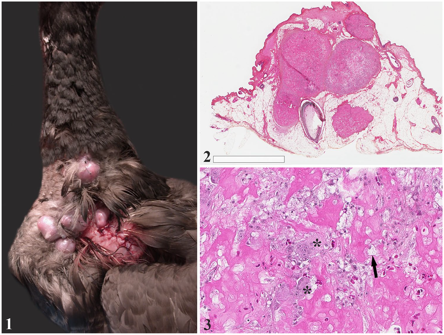
Sterile traumatic panniculitis in a Brent goose.
The dermal mass submitted for histopathology was 1.5 cm, firm, multilobulated, and white-to-yellow on section. Tissue samples for histologic examination were processed within 24 h of formalin fixation. Sections (3 µm-thick) were stained with hematoxylin–eosin–phloxine–saffron (HEPS), periodic acid–Schiff (PAS), Ziehl–Neelsen (ZN), and Fite–Faraco (FF) stains, and examined by a board-certified veterinary pathologist (M-O Benoit-Biancamano).
Under microscopic examination, a well-delineated, multilobular, granulomatous dermal mass surrounded an intact feather follicle (Fig. 2). The mass was primarily composed of large lakes of proteinaceous fluid and fibrin that contained foamy macrophages and multinucleate giant cells (Fig. 3). Foci of hemorrhage and necrosis were scattered throughout the mass. Follicular debris was not observed on the examined slides. No fungi or mycobacteria were seen with the PAS, ZN, and FF stains. Fungal and bacterial cultures yielded no growth.
A PCR for a 492-bp 16S ribosomal RNA fragment was performed to identify bacterial agents. 18 The formalin-fixed, paraffin-embedded tissue block was melted, the epidermis was carefully trimmed off the mass, and the epidermis and the mass were processed for DNA extraction. The following primers were used: F: 5’-AGAGTTTGATCMTGGCTCAG-3’ and R: 5’-GWATTACGGCGGCKGCTG-3’. PCR conditions were 50°C for 30 min then 95°C for 15 min for initial denaturation, followed by 45 cycles at 94°C for 45 s, 55°C for 45 s, and 72°C for 75 s, with a final extension at 72°C for 10 min. The PCR was positive on the skin surface, providing an internal positive control, whereas it was negative on the mass, confirming the absence of bacterial DNA in the lesion. A diagnosis of aseptic, fibrinonecrotic panniculitis was therefore made. Based on the history of feather picking in the region, it was believed to be associated with cutaneous trauma.
Our case appears to be one of sterile panniculitis at the site of previous trauma in a bird. Most described cases of sterile panniculitis in animals do not have a traumatic origin; traumatic panniculitis in domestic animals is more commonly associated with infection or foreign material. 19
Granulomatous inflammation occurs more readily and in a wider range of tissue injury in birds compared to their mammalian counterparts. Indeed, inflammation in avian species tends to be caseous or fibrinous rather than liquefactive. 10 Macrophages often enter inflamed areas at an early stage; multinucleate giant cells, which are frequent in birds, may be found 1–3 d after injury. In birds, skin granulomas are generally associated with bacterial, parasitic, or fungal infections.5,13,17 Cases of avian mycobacteriosis are rare in wild birds; in psittacines, the disease is manifested by swellings around the head and neck.13,16 Although some cases of contact dermatitis have been described in close association with feather follicles, such lesions are usually located on the plantar surface of the footpads and toes (pododermatitis) of broilers, turkeys, and Anseriformes.1,11,15
Sterile cellulitis is infrequent in avian species, and the most common form is xanthoma, which can also manifest as xanthomatosis, especially in some psittacine birds. Xanthomas are rare, benign, typically granulomatous lesions that occur in skin, subcutaneous tissues, tendons, and internal organs, and consist of nodular aggregates of lipid-filled macrophages, multinucleate giant cells, and free cholesterol clefts. Xanthomas generally occur as a result of disturbances of lipid synthesis, metabolism, and transport. 11 They have been described in humans, and domestic and wild animals.2,11,12 In our case, the histologic lesions were not compatible with xanthomas, given that the lesions in our case were primarily composed of large amounts of fibrin and serum with scattered macrophages, and there were no cholesterol crystals.
Although nutritional subcutaneous fat necrosis has been reported in birds, lesions are normally widespread in all deposits of fat.6,7,9 A granulomatous inflammatory response to exogenous lipids has been described in fowl, most commonly from oil-based adjuvants in vaccines. 8 There was no history of vaccination in our case.
Nodular changes in subcutaneous fat have been reported following trauma in humans and companion animals, although infrequently, and are considered likely underdiagnosed in cats and dogs.3,4 Obese cats and dogs are considered more likely to develop traumatic panniculitis, 3 and the high amount of subcutaneous fat in the goose in our case might have predisposed the bird to such lesions. Fat necrosis and panniculitis secondary to trauma are also a common feature of lipomas in companion animals. Damage to lipocytes in any tissue results in the liberation of lipid, which is hydrolyzed into glycerol and fatty acids. Fatty acids vigorously stimulate inflammation, especially granulomatous reactions, thereby enhancing the original inflammatory reaction.3,14 Given that the skin of birds has a high lipid content, 8 trauma from the severe picking by the male might have induced the reaction observed in this goose.
Footnotes
Declaration of conflicting interests
The authors declared no potential conflicts of interest with respect to the research, authorship, and/or publication of this article.
Funding
The authors received no financial support for the research, authorship, and/or publication of this article.