
Abstract
We report herein a choroid plexus papilloma in a beluga whale (Delphinapterus leucas). This case was positive for choroid plexus tumor marker Kir7.1 on immunohistochemistry. These results and the high conservation of Kir7.1 across species at the amino acid sequence level strongly suggest that antibodies directed against Kir7.1 not only can be employed for the diagnosis of choroid plexus tumors in cetaceans, but are also likely to be diagnostically useful in other animal species.
Keywords
Choroid plexus tumors (CPTs) are rare intraventricular papillary neoplasms derived from the choroid plexus epithelium. The majority of CPTs in humans represent benign choroid plexus papillomas. 4 In dogs, choroid plexus papillomas comprise 10% of all primary intracranial central nervous system tumors, 7 but CPTs in cetaceans have not yet been described, to our knowledge. We report herein a tumor in a beluga whale (Delphinapterus leucas), in which an antibody against human CPT marker Kir7.1 2 was successfully employed for the diagnosis of choroid plexus papilloma.
In 2007, a captive female adult beluga whale (estimated age 26 years) suddenly and unexpectedly died in her pool. The recent medical history of the whale had been unremarkable. A thorough autopsy did not show any striking lesions aside from a 10-cm in diameter, space-occupying mass within the right cerebellar hemisphere, which expanded dorsally between the cerebrum and cerebellum, and appeared to be connected to the fourth ventricle. On section, it was dark red and of soft texture (Fig. 1A). On histologic examination, a tumor composed of tightly packed papillary structures was encountered (Fig. 1B). The cuboidal to columnar tumor cells exhibited mildly pleomorphic round or elongated nuclei. Mitotic activity was low (<1 mitosis per 10 high-power fields). Blurring of the papillary growth pattern and small foci of necrosis were present focally. Using various antibodies (Table 1), tumor cells as well as normal choroid plexus tissue were immunoreactive for cytokeratins (MNF116) and showed membranous staining for CPT marker Kir7.1 2 (Fig. 1C). In contrast, various normal beluga tissues (lung, hypodermis, muscle, thyroid gland, mesenteric lymph node, kidney, adrenal gland, mammary gland, uterus, esophagus, stomach, liver, spleen, pancreas, intestine, myocardium, brain stem, cerebrum, and hypothalamus) did not show membranous Kir7.1 staining. When the Kir7.1 antiserum was replaced by nonimmune rabbit serum, staining was negative. The cerebellar cortex adjacent to the tumor showed reactive changes and stained for synaptophysin, glial fibrillary acidic protein, and S100, whereas tumor cells were negative for these markers. The Ki-67/MIB-1 proliferation index was low and accounted for 3%, also supporting a benign neoplastic process.
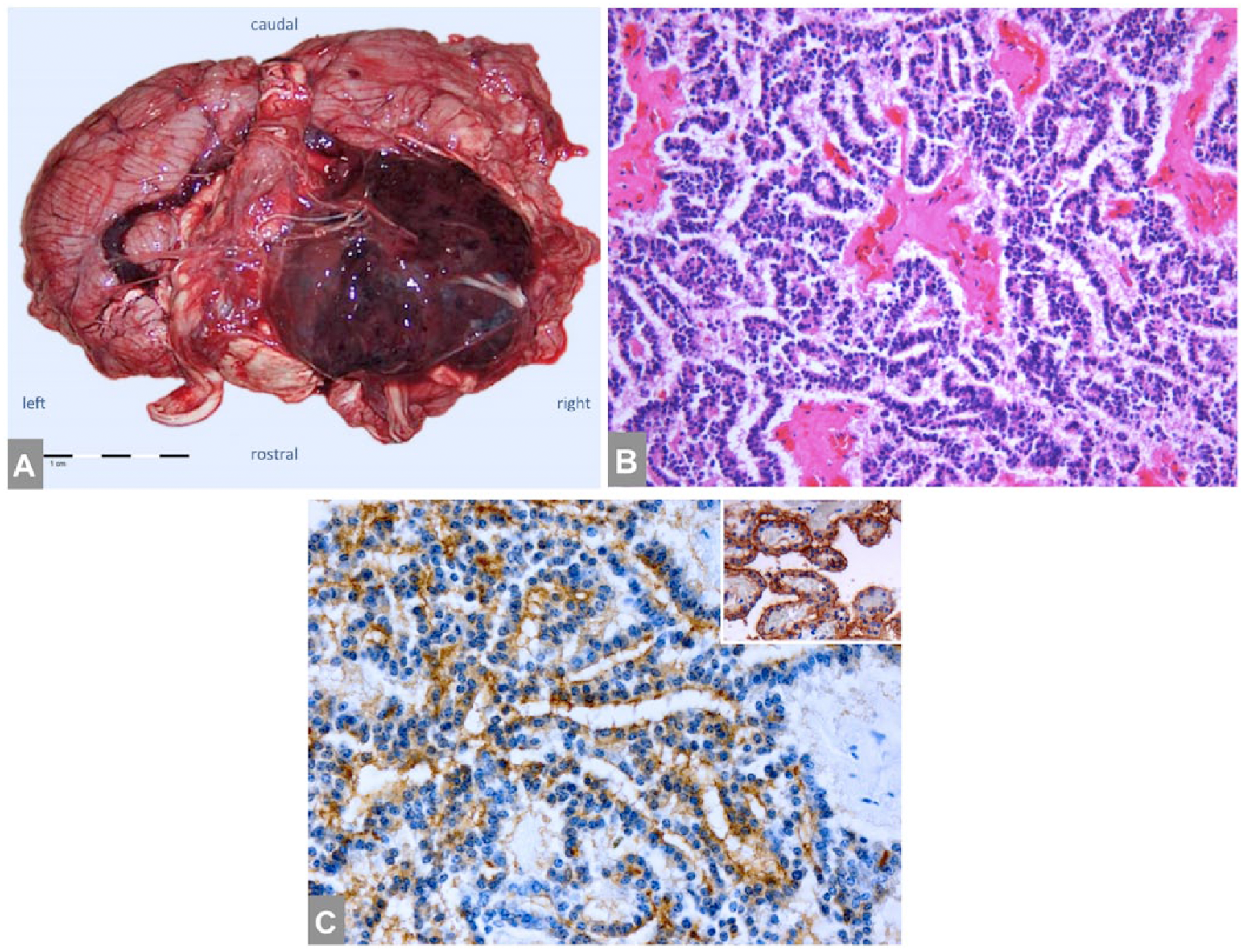
Choroid plexus papilloma in a beluga whale (Delphinapterus leucas).
Immunohistochemistry: antibodies and staining results in Delphinapterus leucas.

Dako = Dako Denmark A/S, Glostrup, Denmark.
Based on these findings, a diagnosis of choroid plexus papilloma was established. This case is only the second brain tumor reported in a beluga whale. 5 Reports on brain tumors in cetaceans are scarce. 1 The most likely explanation could be simply that the brains of cetaceans are not often examined. In the field, the examination of cetacean brains is hampered by logistical and technical problems and autolysis; there might also be conflicts between scientists who wish to examine the brain and those who want to preserve the skull for taxonomic studies. That a brain tumor had not been suspected as the cause of death in this case emphasizes the importance of routinely examining the brains of cetaceans dying in the wild or in captivity.
The diagnosis of CPTs may pose difficulties, and (metastatic) carcinoma is an important differential diagnosis. Interestingly, the only other brain tumor reported to date in D. leucas has been a poorly differentiated carcinoma of the brain stem. 5 Metastatic carcinoma and CPTs both show cytokeratin expression. In contrast, the inwardly rectifying potassium channel Kir7.1 has been shown to be specifically expressed in CPTs, but not in brain metastases 2 or primary intracranial carcinomas 6 in human patients. The observation that a polyclonal antibody directed against human Kir7.1 was successfully employed for the diagnosis of choroid plexus papilloma in D. leucas is interesting. Kir7.1 plays an important role in choroid plexus physiology 3 and is evolutionarily highly conserved. Using a Protein BLAST search (http://blast.ncbi.nlm.nih.gov/), the amino acid sequence of human Kir7.1 was found to be 96–97% identical to that of cetaceans examined (Orcinus orca, Tursiops truncatus, Lipotes vexillifer, Physeter catodon, and Balaenoptera acutorostrata scammoni).
This finding emphasizes the importance of routinely examining the brains of cetaceans dying in the wild or in captivity. Our histopathologic and immunohistochemical results and the high conservation of Kir7.1 amino acid sequence across species suggest that antibodies directed against human Kir7.1 not only can be employed for the diagnosis of CPTs in cetaceans, but might also be diagnostically useful in other animal species.
Footnotes
Acknowledgements
We thank Dr. S. Hirose (Department of Biological Sciences, Tokyo Institute of Technology, Japan) for generating and kindly providing the Kir7.1 antiserum. Ralf Mersmann provided expert assistance in the preparation of the figures.
Authors’ note
Daniel Martineau and Martin Hasselblatt contributed equally to this work.
Authors’ contributions
C Thomas contributed to acquisition, analysis, and interpretation of data, and drafted the manuscript. J Mergl, E Gehring, and W Paulus contributed to acquisition, analysis, and interpretation of data, and critically revised the manuscript. D Martineau and M Hasselblatt contributed to conception and design of the study; contributed to acquisition, analysis, and interpretation of data; and critically revised the manuscript. All authors gave final approval and agreed to be accountable for all aspects of the work in ensuring that questions relating to the accuracy or integrity of any part of the work are appropriately investigated and resolved.
Declaration of conflicting interests
The author(s) declared no potential conflicts of interest with respect to the research, authorship, and/or publication of this article.
Funding
The author(s) received no financial support for the research, authorship, and/or publication of this article.