
Abstract
A 2-year-old dappled Percheron horse had a wasting condition that did not respond to antibiotic treatments and ultimately resulted in death. Thickening of the wall of the large colon and enlargement of the mesenteric lymph nodes were observed at postmortem examination, along with the presence of pinpoint whitish foci in the liver. Microscopic examination of affected tissues revealed diffuse chronic granulomatous enterocolitis, granulomatous mesenteric lymphadenitis, and multifocal granulomatous hepatitis. The DNA extracted from paraffin-embedded intestinal and lymph node samples was analyzed using both a polymerase chain reaction (PCR) assay and PCR–restriction endonuclease analysis and demonstrated the presence of Mycobacterium bovis.
Keywords
Tuberculosis (TB) is an infectious disease caused by pathogenic acid-fast bacilli of the genus Mycobacterium belonging to the M. tuberculosis complex. Among M. tuberculosis complex members, M. bovis is the species that has demonstrated the widest host range. Although it typically infects cattle, M. bovis has also been isolated from a number of domestic and wild species, including human beings. Mycobacterium bovis is the main agent of zoonotic TB, an important disease from the public health perspective in many developing countries.2,31
Cattle are the main reservoir of M. bovis, but wildlife species can also maintain the infection in certain epidemiological settings.14,25 In addition to cattle, other livestock species that may be infected by M. bovis include goats, 4 sheep,13,16 and occasionally cats, 21 dogs, 22 and horses.4,18,20 Infection of horses with M. tuberculosis complex members has been known to occur since the beginning of the 20th century, 18 but experimental studies suggest that equids are naturally resistant, 8 and few reports of natural cases have been published.11,18 For example, among 116 horse, donkey, and pony submissions to the Animal Health and Veterinary Laboratories Agency (United Kingdom) between 1970 and 2010, there were only 2 cases of M. bovis infection. 1 In the few reports addressing TB-associated pathological findings in horses, lesions typically consist of disseminated white-to-ivory granulomas mainly affecting the digestive, respiratory, and urinary systems and lymph nodes.15,18,20 The frequent involvement of the digestive tract suggests that ingestion may be a common source of infection. 28
In Argentina, where animal TB is mostly caused by M. bovis, 32 prevalence of TB-like lesions in cattle as determined by slaughterhouse surveillance was 1.2% in 2004, 5 with disease clustering in areas of the highest density of dairy farms. 19 The current report provides a description of pathological and molecular findings associated with a case of TB caused by M. bovis in a horse in Argentina, with severe gross lesions in the intestine, mesenteric lymph nodes, and liver.
The horse was a 2-year-old dappled Percheron purchased at an auction and transported to a farm located in the province of Entre Rios, in eastern Argentina. Since being purchased, the horse had watery diarrhea. The animal continuously lost weight and was treated with a combination of trimethoprim–sulfamethoxazole and loperamide. The animal died after the second antibiotic dose approximately 1 month after introduction to the farm. At postmortem examination, the mucosa of the large colon was diffusely thickened. Numerous irregularly transversely folded mucosal rugae of the large colon created a gyriform mucosal contour (Fig. 1). Small (1–2 mm) white foci with irregular edges were present in the liver. The colonic lymph nodes were enlarged to a size of 1–2 cm in diameter, and were pale and edematous, especially in the medulla. Samples of the large intestine, liver, lung, kidney, and mesenteric lymphoid nodes were collected, refrigerated, and submitted for histological testing to the Veterinary Pathology Diagnostic Laboratory of the Facultad de Ciencias Veterinarias, Universidad Nacional de Rosario (Argentina). Tissues were fixed in 10% buffered formalin, embedded in paraffin, sectioned at 5 µm, and stained with hematoxylin and eosin. Sections of liver, colon, and lymph node were also subjected to Ziehl–Neelsen staining.
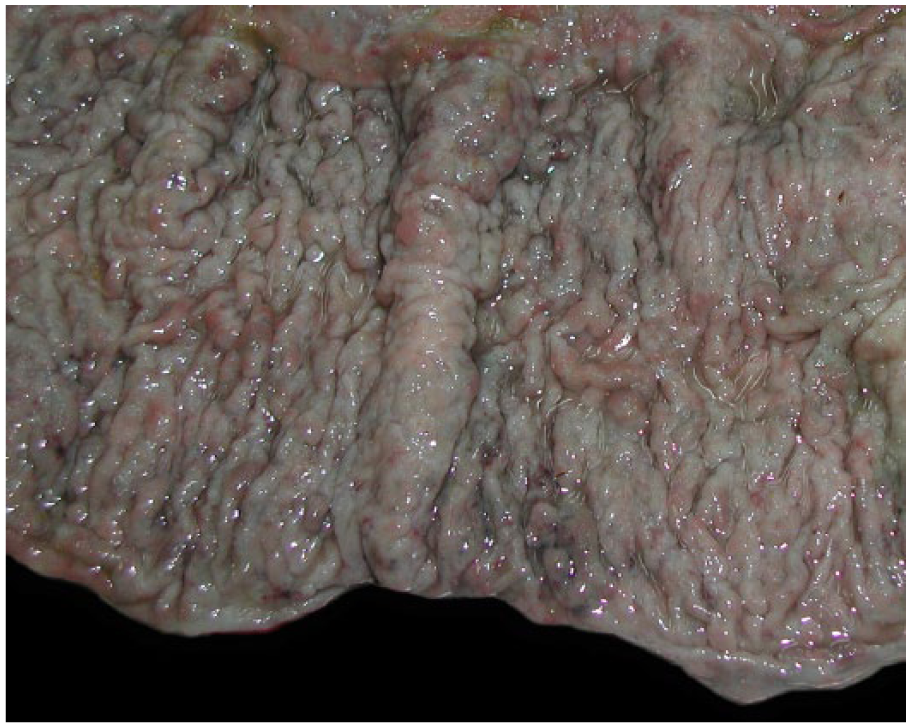
Horse; large colon with thickened gyriform mucosa.
Histologically, the colonic lesions were limited to the mucosa and submucosa. The lamina propria mucosae was widely infiltrated and expanded by epithelioid macrophages. Occasional multinucleated giant cells and aggregates of lymphocytes and plasma cells were scattered among the infiltrate. The infiltrate expanded multifocally past the muscularis mucosae into the submucosa. In these sites, clusters of lymphocytes (interpreted to be remnants of gut-associated lymphoid tissue) were usually present in the center of the histiocytic infiltration. The submucosa was markedly edematous. The mesenteric lymph nodes and liver had numerous cellular aggregates, less than 1–2 mm in diameter, composed of epithelioid macrophages and multinucleated giant cells. Ziehl–Neelsen-stained sections revealed numerous acid-fast bacteria with bacillary morphology within the cytoplasm of epithelioid macrophages and multinucleated giant cells in the large intestine, liver, and mesenteric lymph nodes. Macroscopic or microscopic lesions were absent in the submitted lung and kidney tissue.
Four blocks of paraffin-embedded tissue (including intestine, liver, and lymph nodes) were submitted to the Institute of Biotechnology of the National Agricultural Technology Institute (Hurlingham, Buenos Aires, Argentina), for identification of the acid-fast etiological agent. For DNA extraction, 3–4 paraffin-embedded tissue samples were boiled in 400 µL of Tris–ethylenediamine tetra-acetic acid buffer for 30 min, centrifuged for 10 min at 12,000 × g for removal of paraffin from the surface, and incubated with 100 µL of 10% sodium dodecyl sulfate and 20 μL of proteinase K for 45 min at 65°C. An equal volume of a chloroform–isoamyl alcohol (24:1) solution was added, and the supernatant was collected and mixed with 100 µL of a 5 M NaCl solution, and then precipitated with a 0.6 volume of isopropanol at −20°C. The product was centrifuged at 12,000 × g for 15 min, and, after discard of the supernatant, the precipitate was washed with 1 mL of 70% ethanol, and then resuspended in 40 µL of nuclease-free water. Finally, 1:20 and 1:40 dilutions of the extracted DNA were subjected to polymerase chain reaction–restriction endonuclease analysis (PCR-REA) of the hsp65 gene as previously described, 23 and to PCR detection of specific insertion sequences present in the M. tuberculosis complex (IS6110 and IS1081),26,33 the M. avium complex (IS1245), 10 and M. avium subsp. paratuberculosis (IS900). 6 The PCR assays were carried out using a commercial Taq DNA polymerase. a All steps (DNA extraction, PCR mix preparation, DNA addition to the mix, and gel electrophoresis visualization of the PCR products) were carried out in different areas to prevent cross-contamination, and negative controls were included in both the DNA extraction and PCR analysis. The hsp65 PCR-REA yielded a typical M. tuberculosis complex restriction pattern and only IS6110 and IS1081 were detected, while no reaction was detected in the negative controls, thus confirming a positive diagnosis of TB. To distinguish between M. tuberculosis and M. bovis, DNA was further analyzed using a PCR aiming at the RD7 region. 7 A single band of 635 base pairs, typical of M. bovis, 7 was observed, thus demonstrating the involvement of this bacterial species.
Infection in horses by M. tuberculosis complex members is rare. The incidence in countries with an eradication national program is considered low, 3 although cases due to M. bovis have been reported in countries with high prevalence of bovine TB, such as the United Kingdom 1 or Spain, 15 and even in infected areas located within otherwise bovine TB-free countries. 12 In the early reports of TB infection in horses, cases were characterized by multifocal granulomas in the spleen, liver, lungs, pancreas, kidney, and mesenteric, hepatic, and mediastinal lymph nodes.14,17,21 In the case reported herein, lesions were seen in the intestine, with a diffuse thickening of the large intestinal mucosa, as well as in the mesenteric lymph nodes and liver.
Differential diagnosis of granulomatous diseases in horses may include infection with other mycobacteria, mainly M. avium complex members (M. avium subsp. avium and M. avium subsp. hominissuis), which may cause disseminated granulomatous lesions not only in the intestinal tract (granulomatous enteritis and colitis) but also in other organs.18,24 As well, other possible agents include Rhodococcus equi, 9 Burkholderia mallei (more usually associated with respiratory disease), 27 strongyles, 11 fungal infections (including Blastomyces dermatitidis and Histoplasma capsulatum),17,29 and toxins. 30
In conclusion, the current report is a reminder that natural infection of horses with M. bovis occurs in horses despite their relative resistance to infections with mycobacteria. It is important to follow up the pathologic examination of suspect cases with molecular methods to unequivocally identify the causative agent due to trade and the public health importance of the diagnosis of bovine TB.
Footnotes
Acknowledgements
The authors thank Dr. Mario Marengo (practitioner, Argentina) for his clinical collaboration and Dr. Jim Collins (Director, Veterinary Diagnostic Laboratory, Minnesota) for providing valuable comments on this article.
a.
GoTaq DNA polymerase, Promega Corp., Madison, WI.
Declaration of conflicting interests
The author(s) declare no potential conflict of interest with respect to the research, authorship, and/or publication of this article.
Funding
The author(s) received no financial support for the research, authorship, and/or publication of this article.