
Abstract
Sparganosis is a zoonotic cestodiasis of human beings and animals caused by plerocercoid or second-stage larvae (sparganum) of pseudophyllidean tapeworms in host tissues. Cats are among definitive hosts in which the larva develops to adult stage in the intestines. Reports on larval infection involving various tissues and organs in cats are scarce. Rare single case reports of visceral sparganosis in cats are previously documented. The present report documents an unusual subcutaneous sparganosis in 2 Domestic Shorthair cats from southern Georgia. Veterinary clinicians should consider sparganosis as differential diagnosis for subcutaneous cyst-like masses in cats. As infected animals and animal tissues are sources of human infection, sparganosis warrants public awareness and due precaution to avoid human infection.
Keywords
Sparganosis is a rare aberrant tapeworm larval tissue infection caused by plerocercoid or second-stage larvae (sparganum) of a pseudophyllidean tapeworm. 18 Most infections are nonproliferative and are associated with the presence of a single larva of Spirometra erinaceieuropaei or Spirometra mansonoides. Proliferative sparganosis is caused by the larva of Sparganum proliferum, which is characterized by asexual replication with continuous branching and budding. The resulting progeny invade tissues,1,3,13 where they grow and repeat the process, ultimately causing death of the host. 3 All tissues may be affected except bone. 13 The spargana in proliferative sparganosis do not mature when fed to presumptive definitive hosts such as the dog and cat. 3
The sparganum is a highly paratenic larva, which if landed in the intestine of an unsuitable host, leaves the intestine, migrates to other tissues, and establishes itself as a tissue parasite.10,11 The sparganum exhibits low host specificity, develops in any class of vertebrate except fishes, and has a wide tolerance of temperature and other stresses. 11 Various species of animals including apes, pigs, dogs, cats, and human beings can serve as paratenic or second intermediate hosts in which the spargana develop.3,6,14 The adult tapeworm infects dogs, cats, and other carnivores 8 and causes little or no clinical signs. 16
Two Domestic Shorthair cats, an intact male and spayed female (both 8 years old) from south Georgia, developed subcutaneous swellings in lumbar and ventral neck areas, respectively. A subcutaneous epithelial cyst or abscess was suspected clinically. Large numbers of inflammatory cells were observed in the aspirate of copious caseous material from one of the masses. There was no significant bacterial growth on culture from the exudate and swab. The masses were surgically removed and fixed in 10% buffered formalin solution for histological evaluation at the Tifton Veterinary Diagnostic and Investigational Laboratory, College of Veterinary Medicine, The University of Georgia. The tissues were processed for routine histology, sectioned at 5 μm, stained with hematoxylin and eosin, and examined by light microscopy. Periodic acid–Schiff (PAS) reaction and Von Kossa stains were applied to demonstrate the glycogen-rich matrix and the calcified (calcareous) bodies, respectively.
Microscopically, large numbers of cross and tangential sections of plerocercoid cestode larvae (Fig. 1A) were present in variably sized cystic spaces in subcutaneous tissue in both cats. The cystic spaces and the larvae were surrounded by abundant reactive fibrocollagenous stroma. The larvae measured approximately 1–1.5 mm in diameter, had thick eosinophilic tegument, and abundant PAS-positive glycogen-rich parenchyma (Fig. 1B) underlying a single row of columnar cells subjacent to the tegument. Occasional shallow invaginations of the tegument were evident. Scattered within parenchyma were several calcified bodies (calcareous corpuscles), muscle contractile fibers, and excretory ducts (Fig. 1C, 1D). Scolices, suckers, a digestive tract, and reproductive organs were not present. Severe inflammation composed of abundant lymphocytes, plasma cells, macrophages, neutrophils, and occasional multinucleated giant cells admixed with reactive fibroblasts infiltrated the fibrocollagenous stroma.
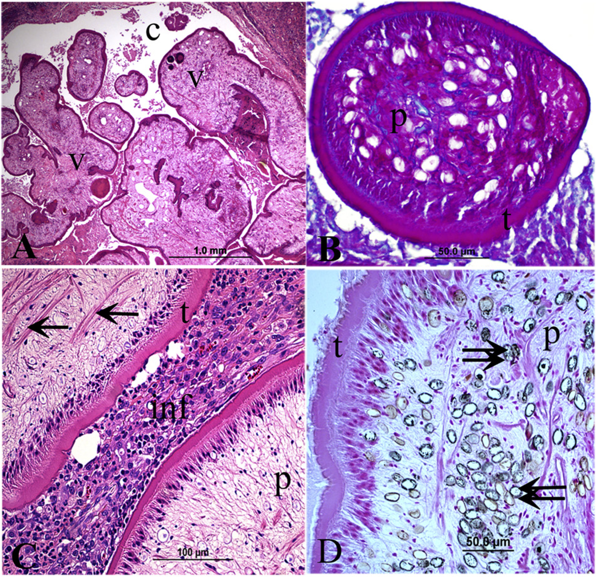
Subcutaneous tissue, cat.
The characteristic feature of cestodes is the presence of calcareous corpuscles embedded in the parenchyma, as well as complete absence of a digestive tract at all stages of development.7,21 The main groups of cestodes that would be encountered in tissue sections are the cyclophyllideans and pseudophyllideans. Adults and larvae of cyclophyllidean species possess suckers while plerocercoid larvae of pseudophyllidean cestodes do not have scolices and suckers. 7 Similar to S. proliferum, tetrathyridia are reported to occasionally reproduce asexually, but possesses characteristic invaginated scolex. Multiple histological sections should be examined to ensure that neither scolex nor suckers are present in cases of suspected plerocercoid larvae in tissues.1,7 Consistent with the larvae of S. proliferum, scolices and other head structures, a digestive tract, and reproductive organs were not observed in multiple sections of the larvae in the present cases.
Reports on proliferative sparganosis in dogs and cats are very rare. A case of proliferative sparganosis involving visceral organs (stomach, spleen, liver) was reported in a 6-year-old male Domestic Longhair cat from Florida. 3 Sparganosis involving the fundic portion of the stomach was recorded in a 7-year-old Burmese cat in 1967. The cat was originally from Cambodia and spent 3 years in Taiwan before moving to the United States in 1966. 16 Proliferative peritonitis associated with S. proliferum was also reported in a 6-year-old spayed female Labrador Retriever mix dog from Germany. 17 Proliferative sparganosis involving the subcutaneous tissue and intermuscular fascia was reported in a 21-month-old spayed female Border Collie from the United States. 6 Reports on subcutaneous sparganosis in the cat are unavailable to the authors’ knowledge. The current report documents rare cases of subcutaneous sparganosis in 2 cats. The finding suggests that sparganosis is enzootic in the region.
Cats are among final hosts in which the spargana develop to adult stage. Adult tapeworm has been reported several times in cats in the United States. 16 How cats serve as paratenic hosts and harbor tissue sparganosis is speculative. 11 Tissue sparganosis is suggested to develop in cats when cats ingest sparganum of a species not normally parasitic for cats 16 or when cats ingest crustaceans containing procercoids, which then develop into the parenteral spargana. 11 In a cat with proliferative peritoneal sparganosis, 3 it was speculated that the cat ingested infected copepods and served as an abnormal second intermediate host in which the spargana developed. How the cats in the current cases acquired the infection is unknown.
Cats are among pets with close association to human beings and hence infection in cats suggests a potential threat to human health. In human beings, sparganosis occurs more frequently in eastern Asia than in other parts of the world4,9 and is often associated with ingestion of raw or insufficiently cooked meat of various intermediate hosts such as frogs and snakes infected with the sparganum, drinking water contaminated with infected copepods, or by applying the flesh of an infected intermediate host as a poultice to the eye or an open wound.4,5,9 It usually appears as subcutaneous nodules all over the body and can involve the eye, brain, and spinal cord.15,20,22
The first human case of proliferative sparganosis due to S. proliferum was reported in 1908 in a man from Florida. 12 Most human cases of sparganosis in the United States have been reported from the southeast, although sporadic cases have also been documented in other parts of the country, 19 and almost all presenting as a subcutaneous lesion. 8 Genetic analysis should confirm whether the subcutaneous spargana in human beings and cats belong to the same or different species. Although cats occasionally develop tissue sparganosis as is in the current cases, they mainly serve as definitive hosts in which spargana develop to adult stage maintaining the enzootic cycle with a potential of environmental contamination and animal, as well as human, infection. Contaminated drinking water is suggested to be the most probable source of human infection in the United States. 19 A prevalence of 6.9% sparganosis recorded in abattoir slaughtered carcasses of feral swine in Florida suggests that consumption of raw or undercooked feral swine carcass can pose a public health risk for human sparganosis. 2
Because infected domestic animals can be sources of human sparganosis, comprehension of the epidemiology of the disease in a given area is important for public awareness and to envisage appropriate control measures to avoid animal and human infection. Anti-parasitic drugs such as praziquantel or mebendazole may be effective against sparganosis. However, complete extraction of larvae with the infected tissue is a treatment of choice. 5 Infection of animals such as dogs and cats can be controlled by preventing consumption of contaminated water or ingestion of vertebrates that could serve as second intermediate hosts. 6 Sparganosis is a zoonotic cestodiasis warranting due precaution to avoid human infection. The current report documents rare cases of subcutaneous sparganosis in cats, highlighting that veterinary clinicians, particularly in enzootic areas, should consider sparganosis as differential diagnosis in subcutaneous cyst-like masses in cats.
Footnotes
Declaration of conflicting interests
The author(s) declared no potential conflict of interest with respect to the research, authorship, and/or publication of this article.
Funding
The author(s) received no financial support for the research, authorship, and/or publication of this article.