
Abstract
Geotrichum spp. are ubiquitous, saprotrophic fungi found in soil, organic matter, and silage, as a contaminant in food products and in the digestive tracts of mammals. The current study reports a case of Geotrichum candidum infection with dermatitis in an aborted bovine fetus with skin and lung lesions. A 6-month-old aborted male Holstein Friesian fetus displayed unusual lesions on the skin of the abdomen, thorax, and head, which was excessively thickened and wrinkled. These changes corresponded to orthokeratotic hyperkeratosis, neutrophil accumulation in the stratum corneum, a pyogranulomatous inflammatory infiltrate, and superficial dermal necrosis. Moderate suppurative multifocal pneumonia was observed. Large numbers of mononuclear cells and occasional fibrin thrombi within blood vessels were found in the lungs, brain, and cerebellum. Gridley staining revealed fungal structures within the skin lesions. The mycological exam demonstrated the growth of G. candidum, and phase contrast microscopy conducted on the abomasal fluid revealed hyphae compatible with this agent. The skin lesions observed, in association with the fungus isolated, indicated that the abortion was due to G. candidum infection of the bovine fetus.
Keywords
Mycotic infections can cause placentitis and abortion in a number of animal species. 1 Fungi are common causes of bovine abortion worldwide and are associated with 1–24.9% of all bovine abortions. 1 Such infections are sporadic and rarely affect more than 1 animal in a herd; the infections occur mainly during the winter when the cows are generally fed large amounts of hay. 8 When a pregnant cow contracts a fungal infection, the conidia can penetrate lesions in the gastrointestinal or respiratory tract and spread hematogenously to reach the placenta and the fetus, where conditions are ideal for the full development of the fungus. 6 Bovine mycotic abortion generally occurs between the sixth and eighth months of gestation and is frequently followed by retention of the placenta. Hemorrhagic necrotizing placentitis is frequently observed and is generally associated with necrotic, thick yellow cotyledons. 8 In the fetus, parakeratotic dermatitis is generally evident, characterized by raised plaques and blepharitis. 6 The diagnosis of mycotic abortion requires macroscopic evaluation, histological examination, and culture testing, mainly of the placenta and the abomasal contents. 1 The fungal species that have been isolated from aborted fetuses include Aspergillus fumigatus, Aspergillus nidulans, Absidia corymbifera, and Mortierella wolfii, as well as species in the genera Rhizopus, Mucor, and Rhizomucor. 1 Geotrichum spp. are ubiquitous saprotrophic fungi that are found in soil, decomposing organic matter, 2 food products, and the digestive tracts of mammals, including human beings. 1 Infections with Geotrichum candidum have been associated with skin lesions in horses. 4 In addition, G. candidum has been isolated from the reproductive tracts of cows and buffalo with and without reproductive problems. 5 Widespread infection, cerebral abscesses, traumatic joint infection, and oral and cutaneous infections have been reported in conjunction with Geotrichum infection in immunocompromised human beings. 7 The current study reports a case of G. candidum infection associated with skin and lung lesions in an aborted bovine fetus. The case highlights the importance of G. candidum as a possible cause of abortion in cattle.
In July 2008, a male Holstein Friesian fetus was submitted to the Veterinary Pathology Sector (Setor de Patologia Veterinária; SPV) of the Federal University of Rio Grande do Sul (Universidade Federal do Rio Grande do Sul, Brazil; UFRGS) for diagnostic purposes. The fetus was necropsied, and tissue specimens from brain, lung, liver, kidney, heart, skeletal muscle, thymus, spleen, abomasum, and skin were fixed in a 10% buffered (pH 7.2) formalin solution, processed for routine histological examination, and stained with hematoxylin and eosin; selected sections were also stained with the Gridley method for fungal staining. Samples of the abomasal contents and skin were collected aseptically and analyzed by phase contrast microscopy and mycological culture. The abomasal contents and liver, skin, and lung samples were collected aseptically and submitted for aerobic and microaerobic bacterial. Brain–heart infusion medium was used to culture Campylobacter spp. Kidney smears were tested for Leptospira spp. by direct immunofluorescence test. 10 Samples of thymus, brain, and cerebellum were submitted for immunohistochemical staining for the detection of Bovine viral diarrhea virus (BVDV; Schmitz M: 2006, Caracterização patológica e imunoistoquímica da infecção pelo vírus da diarréia viral bovina [Pathological and immunohistochemical characterization of infection with bovine viral diarrhea]. Dissertação de Mestrado, Faculdade de Medicina Veterinária, Universidade Federal do Rio Grande do Sul, Porto Alegre. In Portuguese. Abstract in English), and liver tissue was used for the detection of Bovine herpesvirus 1 (BHV-1). The aborted fetus, with an estimated gestational age of 6 months, originated from a small dairy farm located in southern Brazil. The farmer reported the occurrence of additional abortions during the same period. At necropsy, abnormalities were observed mainly on the skin, which was excessively thickened and wrinkled in the thoracic, abdominal, and head regions, particularly on the eyelids, ears, and close to the nose (Fig. 1). The lungs were partially expanded.
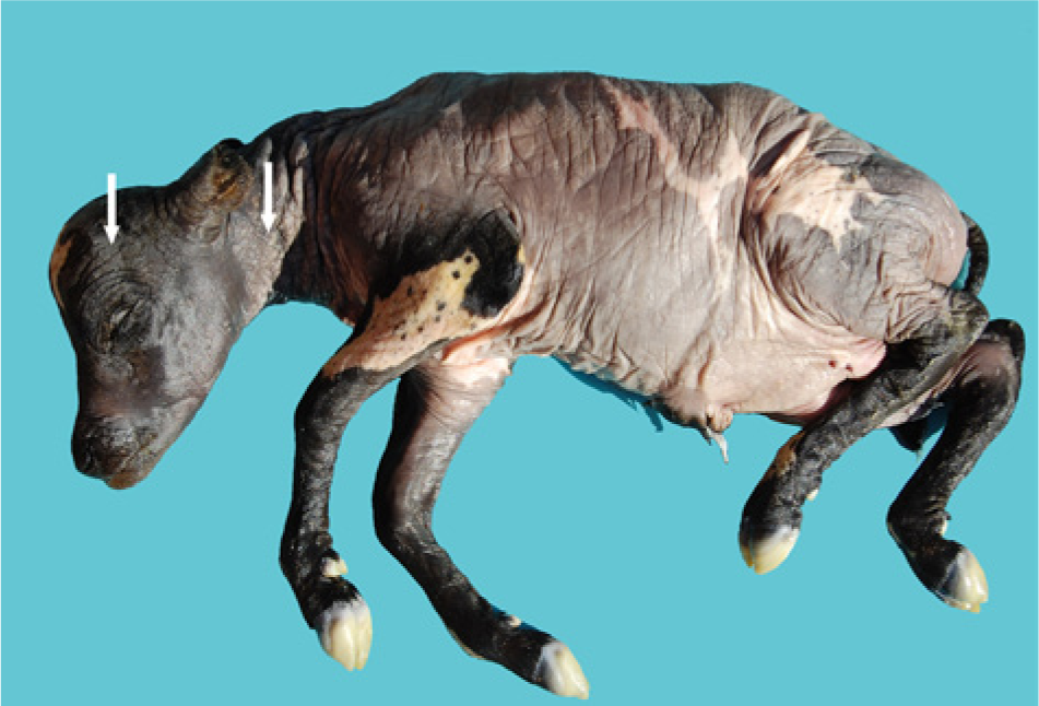
Aborted bovine fetus. Thickened and wrinkled skin bearing numerous plaques (arrows).
Histological examination of the skin revealed diffuse accentuated orthokeratotic hyperkeratosis with infiltrated neutrophils in the stratum corneum and moderate multifocal pyogranulomatous inflammatory infiltrate associated with areas of superficial dermal necrosis and intravascular fibrin thrombi (Fig. 2). Gridley staining revealed fungal structures in the foci of cutaneous necrosis (Fig. 3) that were associated with dermal thrombi. Moderate suppurative pneumonia was observed in the lungs, along with large numbers of mononuclear cells and occasional fibrin thrombi within the blood vessels, which were also present in the cerebral vessels. After 6 days of incubation, the culture plates inoculated with skin or abomasal contents exhibited growth of pure G. candidum, characterized by the development of white-to-cream–colored smooth colonies (4–5 cm in diameter) without reverse pigment. Phase contrast microscopy of the abomasal fluid revealed typical septate hyaline hyphae with cylindrical branches giving rise to subglobose arthroconidial chains. The urease and germ tube tests were negative. The direct fluorescent antibody tests for Leptospira spp., the bacteriological tests, and the BVDV and BHV-1 immunohistochemical tests were all negative.
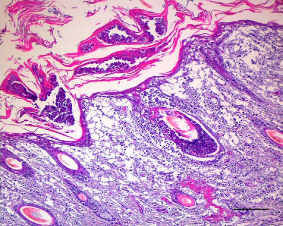
Photomicrograph of the skin of the bovine fetus shown in Figure 1. Diffuse severe orthokeratotic hyperkeratosis with neutrophilic infiltration and abscess formation in the stratum corneum. Note the moderate multifocal pyogranulomatous inflammatory infiltrate associated with superficial dermal necrosis and intravascular fibrin thrombi. Hematoxylin and eosin. Bar = 150 μm.
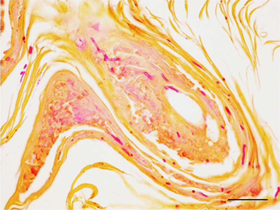
Photomicrograph of the skin of the bovine fetus shown in Figure 1. Note the hyaline, septate, and branched hyphae associated with the necrotic tissue of the hair follicle. Gridley stain. Bar = 70 μm.
Studies in other countries have reported a variable prevalence for mycotic abortion from 1% to 24.9%. 1 Aspergillus fumigatus is the species most commonly identified in cases of bovine mycotic abortion and is isolated in approximately 60–75% of cases. 1 Geotrichum candidum has been reported as a cause of systemic mycosis in dogs, cats, and human beings,2,7 gastrointestinal infections in gorillas, 3 clinical mastitis in cattle, and endometritis in cattle and buffalo. 12 However, abortions have rarely 11 been associated with this agent. Mycotic infections by G. candidum are considered opportunistic and are most likely related to immunosuppressive factors. 8 Factors associated with a greater susceptibility to fungal infections include previous immunosuppressive diseases, the prolonged use of antibiotics or hydrocortisone, and poor physical condition. In the current case, there was no evidence suggesting that the animal in question was immunocompromised. Mycotic abortions are generally associated with the ingestion of food products that have been inadequately preserved and exposed to high humidity, resulting in fungal contamination. 8 The case reported herein occurred in July, during the winter in southern Brazil, which is a period when rains are more frequent and the air humidity is higher; however, information about the feed consumed by this small herd was not available.
Mycotic abortion is characterized by fungal infection of the placenta and amniotic contamination and is associated with fetal infection occurring through contact with the skin and/or through aspiration of contaminated amniotic fluid, which can lead to fungal dermatitis and pneumonia. Partial alveolar expansion was observed in the present case, which indicates that the fetus was alive when it was expelled from the uterus. Most mycotic abortions occur during the final third of the gestation period, as was observed in the case of this fetus. In cases of mycotic abortion, the placenta is the main organ affected. In a study on bovine placentas, 9 it was observed that among the 55 cases in which the placenta contained fungal hyphae, only 7% of the fetuses exhibited some type of macro- or microscopic lesion. In the present case, however, the fetus was found to have lesions that strongly indicated mycotic infection, and the fungus was isolated from the fetal abomasal contents and skin, which facilitated the diagnosis. The cutaneous lesions observed were similar to those found in other cases of mycotic abortion 1 and were characterized by raised gray plaques that were rounded or confluent and appeared predominantly in the head and neck region. These characteristics are consistent with the results of the microscopic analysis, in which dermatitis and hyperkeratosis were observed. Lung and liver lesions are not often observed in cases of mycotic abortion, but when present, they are characterized by necrotic suppurative or granulomatous inflammation, which may be associated with fungal hyphae. 1
While other possible causes for this abortion cannot be excluded, the macroscopic and histological observation of lesions, together with the isolation of the fungus, indicated that infection with G. candidum might have caused this abortion. Serology could potentially rule out other causes of abortion because this type of analysis can identify antibodies against agents such as Leptospira sp., Campylobacter sp., and BVDV that could have led to abortion but were not detectable at the time of the fetal expulsion. Therefore, serologic tests could rule out other causes of abortion and complete the diagnosis. Although G. candidum was isolated from the clinical materials, instances in which the isolates were associated with tissue invasion are rare. 2 Information concerning bovine abortion due to G. candidum is scarce. 11 The current case highlights the fact that mycotic agents should be investigated as a potential cause of abortion, and the diagnosis of mycotic abortion should be reached only when lesions associated with mycotic elements compatible with cultured isolates are present.
Footnotes
Declaration of conflicting interests
The author(s) declared no potential conflicts of interest with respect to the research, authorship, and/or publication of this article.
Funding
The author(s) disclosed receipt of the following financial support for the research, authorship, and/or publication of this article: Conselho Nacional de Desenvolvimento Científico e Tecnológico (CNPq) of Brazil.